International Journal of
eISSN: 2381-1803


Clinical Case Volume 17 Issue 6
1SOLCA Cancer Institute, Cuenca, Ecuador
2School of Medicine, University of Cuenca, Ecuador
3Integral Medical Services (SERMEDIC), Cuenca, Ecuador
4Postgraduate in Pediatrics, Universidad Internacional del Ecuador (UIDE), Ecuador
5Center for Morphological and Surgical Studies (CEMyQ), Universidad de La Frontera, Chile
6D Program in Medical Sciences, Universidad de La Frontera, Chile
Correspondence: Enmanuel Guerrero, Hemato-oncology, SOLCA Cancer Institute, Cuenca, Ecuador
Received: October 31, 2024 | Published: December 4, 2024
Citation: Guerrero E, Alvarado R, Jaramillo D, et al. Pseudopapillary solid tumor of the pancreas: case report and literature review. Int J Complement Alt Med. 2024;17(6):251-253. DOI: 10.15406/ijcam.2024.17.00713
Solid pseudopapillary tumor of the pancreas (NSSP) is a rare tumor entity, of unknown cause and good prognosis. It mainly affects women between 25 and 35 years of age. Occasionally, it occurs in the pediatric age group and is characterized by the presence of abdominal pain and mass. Treatment is complete surgical resection. The following clinical case of a 10-year-old girl is shared in order to report the diagnostic and therapeutic experience of this disease. The patient debuted with pain and tumor mass at abdominal level, being initially diagnosed as Wilms tumor; however, due to the lack of response to chemotherapy, it was decided to perform an exploratory laparotomy, finding a mass confined to the body and tail of the pancreas. Pathological anatomy concluded that it was a NSSP. There is little documented evidence on this disease, especially in the pediatric population, so the case is important. It is a disease with a good prognosis and low mortality after surgical treatment.
Keywords: solid pseudopapillary neoplasm of the pancreas, wilms' tumor, pancreas
Solid pseudopapillary tumor of the pancreas (NSSP) or also called Frantz's tumor was first reported in 1934 by Lichtenstein, but it was not until 1959 that Virginia Frantz described the pathology in detail.1,2 It accounts for 1 - 2% of non-endocrine neoplasms of the pancreas.3 It is rare in children and usually appears in young women around the third or fourth decade of life (95%).3 It is characterized by the presence of pain, distension and abdominal mass. Complete surgical excision is the treatment of choice and the prognosis is good.4 The aim of this manuscript was to report the case of a 10-year-old patient diagnosed with NSSP at the Institute of the Society for the Fight Against Cancer (SOLCA) in the city of Cuenca (Ecuador). Initially, the patient presented a different clinical diagnosis; however, the pathological and surgical findings were decisive in establishing the definitive diagnosis and treatment. The CARE (Case Report Guildness) guidelines for clinical case reports were followed in the preparation of this article.5
We present a 10-year-old female patient with a history of left nephrectomy for hydronephrosis at the age of 4 years. Three months prior to care, she noticed the presence of an abdominal mass of progressive growth. One month ago, it was accompanied by pain in the epigastrium, colicky type, of intensity four out of ten on the visual analog scale. In addition, she reported constipation, hyporexia, anorexia and worsening abdominal pain, so she was brought to the Oncology Institute for treatment. During the physical examination, a solid abdominal mass of 15 cm in diameter, with lobular borders, non-mobile, painful to deep palpation, extending from the hypogastrium to the left flank, was palpated.
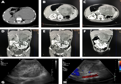
Blood tests did not show any alteration. Computed tomography (CT) of the abdomen showed a neoplastic lesion located in the left hemiabdomen measuring 14 x 12 cm, which displaced the pancreas and stomach posteriorly and showed extension to the pancreatic body and tail (Figure 1).
Several diagnostic suspicions were raised (Table 1); however, because of the clinical, epidemiological and radiological features, it was decided to treat the patient as a Wilms tumor.
|
Diagnostic suspicion |
Positive findings |
Negative findings |
|
Wilms tumor or nephroblastoma |
Epidemiology: Frequent in children under 15 years of age. Predominantly in girls. Clinical: Pain and firm abdominal mass, unilateral flank, hyporexia, anorexia and constipation. |
No hypertension, fever or anemia (<15%). |
|
Mesenteric cyst |
Epidemiology: Frequent in children under 15 years of age. Predominantly in children. Clinical features: abdominal pain, palpable mass, anorexia and constipation. |
No chronic picture (frequent form of debut). |
|
Neuroblastoma |
Epidemiology: Frequent in children under 1 year of age. Late diagnosis at 10 years of age (97%). Predominance in children. Clinical: Pain and abdominal mass, fixed, hard, located in the flanks, anorexia and constipation. |
No neurological symptoms (irritability, Horner's syndrome), nor clinical symptoms of genitourinary obstruction. |
Table 1 Differential diagnoses of NSSP with their epidemiological and clinical characteristics
Neoadjuvant chemotherapy based on weekly Vincristine (1.5 mg/m2) and Actinomycin (1.35 mg/m2) was started every three weeks, with the aim of reducing the tumor volume and making it resectable. After the fourth week of treatment, a new CT scan was performed, showing the absence of tumor regression (Figure 1).

Figure 1 Comparative computed tomography (CT) findings. A: CT scan of the abdomen performed before treatment, showing an abdominal mass of 14 x 12 cm and absence of the left kidney. B-F: CT scan of the abdomen after the fourth cycle of chemotherapy, showing an abdominal mass of 14.5 x 9.25 cm, without significant changes.
Surgical procedure and definitive diagnosis
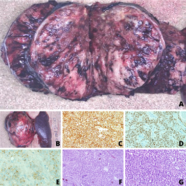
Due to the lack of response to the initial treatment, an exploratory laparotomy was planned, performing a complete resection of the tumor mass with splenectomy and distal pancreatectomy. A delimited tumor was resected, surrounded by a pseudocapsule measuring 19 x 11 x 10 cm, confined to the body and tail of the pancreas, which infiltrated the splenic vein and artery with solid, cystic and hemorrhagic areas (Figure 2: A, B).Anatomic pathology studies ruled out neoplastic involvement to other areas (lymph nodes or peritoneum) and confirmed the complete removal of the tumor. Microscopic findings included an epithelial neoplasm, consisting of monomorphous sheets with a solid growth pattern and polygonal cells in the midst of a hyalinized stroma.

Figure 2 Surgical specimen and immunostaining of solid pseudopapillary tumor of the pancreas. A) Tumor mass measuring 19 x 11 x 10 cm, dependent on the body and tail of the pancreas with hemorrhagic, solid and cystic areas. B) Distal splenopancreatectomy including a 12 x 7 cm purplish sple spleen and tumor mass. C) Vimentin. D) Prostagen receptor (PR). E) CD10. F) Hematoxylin - Eosin 10x. G) Hematoxylin - Eosin 20x.
The proliferative index was low, foamy macrophages and pseudopapillae with central nucleus surrounded by neoplastic cells were identified. Immunohistochemistry tests showed positivity for: CK cocktail, vimentin, prostaglandin receptor (PR), neural specific enolase (NSE), CD56 and CD10, thus concluding that it was an NSSP (Figure 2: C - G).
The patient had a good postoperative evolution, being discharged in good clinical condition. She did not need chemotherapy or radiotherapy and four years later, the patient is in complete remission.
NSSP or Frantz tumor is a rare neoplastic entity of unknown origin. According to the World Health Organization, it is defined as low-grade malignant tumors composed of loosely cohesive epithelial cells lacking a specific lineage of pancreatic epithelial differentiation.13 It mainly affects women between the ages of 25 and 35 years; however, the tumor may be present in pediatric age groups (20 to 25%), with an average age of 8 years old.4,6 As in this patient, the most common forms of presentation are pain (51.6%) and the presence of a palpable mass at the epigastric level (40.2%). As a consequence of compression and displacement of adjacent structures, patients may present with: nausea and vomiting (12.7%), diarrhea (7%), gastric fullness and weight loss. About 15% are usually asymptomatic and the diagnosis is usually incidental.3,4,6.7
The evaluation should be complemented with abdominal ultrasound, CT or MRI. Imaging studies describe NSSP as a mass at the level of the tail (35% to 44%), head (30% to 34%) and body (10% to 13%) of the pancreas well circumscribed.14 They measure between 0.5 to 34.5 cm in diameter, and are generally separated from the pancreatic parenchyma by a pseudocapsule with heterogeneous contrast uptake. They also present solid, necrotic, cystic and hemorrhagic areas, as in this clinical case.8,9 The confirmatory diagnosis is established by immunopathology studies, where solid nests of poorly cohesive cells forming a cuff surrounding the blood vessels are observed. In addition, the stroma shows various degrees of hyalinization and degeneration. Although NSSP is a well-circumscribed tumor, infiltration of the surrounding pancreatic tissue is also often found. There are no established morphological criteria for malignancy, although the latter is defined by the presence of metastases (5%-15%) in the liver, portal, spleen, lymph nodes, omentum, duodenum, colon, lung and retroperitoneum.15
Characteristically presents positivity for immunostains: SOX11 (100%), β-catenin (98%), CD56 (96%), alpha 1 anti-chymotrypsin (95%), vimentin (88%), alpha 1 antitrypsin (82%), androgen receptor (81%), CD10 (63%), among others.6,9 However, to establish a definitive diagnosis, β-catenin, CD10, synaptophysin and chromogranin are suggested.10,11 Differential diagnoses in pediatrics include renal tumors, neuroblastoma, cystic adenoma, cystoadenocarcinoma, lymphangioma, sarcomas, discogenic cyst, pseudocysts, hydatid cysts, among others.3,7,14
Definitive treatment is complete surgical resection.16 The prognosis is good, even in the presence of metastases, with a 10-year survival rate of 96%.9 Mortality is approximately 1.5% and the clinical factors associated with poor prognosis are male sex and advanced age.12–14 Long-term follow-up of patients is recommended, since 2% will present local recurrence and, of these, 20% will have metastases in the liver, lymph nodes, mesentery, omentum and peritoneum.3 Factors associated with recurrence are initial tumor size greater than 5 cm, vascular invasion, synchronous and lymph node metastases and positive margins.13 In this patient the surgical resection was complete, with negative margins, so her prognosis to date has been satisfactory.
Based on the clinical, epidemiological and radiological characteristics, the initial diagnostic suspicion of Wilms tumor was made. In these patients, biopsy is not indicated and the administration of 3 to 6 weeks of neoadjuvant chemotherapy is recommended in order to convert an initially unresectable tumor into a resectable one. However, due to the lack of response to treatment (since NSSP is a benign tumor that does not respond to chemotherapy), a surgical procedure was performed with the findings and favorable evolution described above.
NSSP is a low grade malignant neoplasm with a good prognosis. It should be suspected in any young female patient with abdominal pain and presence of solid or cystic pancreatic mass. This report is of importance to the medical literature because there is no clear evidence on the epidemiology and management of NSSP in pediatric patients.
Protection of persons
The authors declare that the ethical standards established by the World Medical Association and the Declaration of Helsinki were followed.
Data confidentiality
SOLCA Institute protocols regarding patient confidentiality were applied.
Right to privacy and informed consent
The authors obtained informed consent from the patient's father. This document is in the possession of the first author.
Thanks are due to the SOLCA-Cuenca Cancer Institute (Ecuador).
The authors declare that they have no conflict of interest.

©2024 Guerrero, et al. This is an open access article distributed under the terms of the, which permits unrestricted use, distribution, and build upon your work non-commercially.