International Journal of
eISSN: 2381-1803


Review Article Volume 4 Issue 4
1Medical doctor, Universal Medical Imaging Group, USA
2Naturopathic Practitioner, pH Miracle Center, USA
Correspondence: Robert O Young, ND, pH Miracle Center, 16390 Dia Del Sol Valley Center, California 92082, USA, Tel 818 987 6886, Tel 760 751 8321
Received: October 29, 2016 | Published: November 17, 2016
Citation: Migalko G, Young RO (2016) A Non Radioactive Alternative to Mammograms. Int J Complement Alt Med 4(4): 000130. DOI: 10.15406/ijcam.2016.04.00130
While a growing body of research now suggests that x-ray mammography is causing more harm than good in the millions of women who subject themselves to breast screenings, annually, without knowledge of their true health risks, the primary focus has been on the harms associated with over-diagnosis and over-treatment, and not the radiobiological dangers of the procedure itself.
In 2006, a paper published in the British Journal of Radiobiology, titled “Enhanced biological effectiveness of low energy X-rays and implications for the UK breast screening programme,” revealed the type of radiation used in x-ray-based breast screenings is much more carcinogenic than previously believed.
Recent radiobiological studies have provided compelling evidence that the low energy X-rays as used in mammography are approximately four times – but possibly as much as six times – more effective in causing mutational damage than higher energy X-rays. Since current radiation risk estimates are based on the effects of high energy gamma radiation, this implies that the risks of radiation-induced breast cancers for mammography X-rays are underestimated by the same factor.1
In other words, the radiation risk model used to determine whether the benefit of breast screenings in asymptomatic women outweighs their harm, underestimates the risk of mammography-induced breast and related cancers by between 4-600%.
The authors continued Risk estimates for radiation-induced cancer -principally derived from the atomic bomb survivor study (ABSS) - are based on the effects of high energy gamma-rays and thus the implication is that the risks of radiation-induced breast cancer arising from mammography may be higher than that assumed based on standard risks estimates.
This is not the only study to demonstrate mammography X-rays are more carcinogenic than atomic bomb spectrum radiation. There is also an extensive amount of data on the downside of x-ray mammography.
Sadly, even if one uses the outdated radiation risk model (which underestimates the harm done),* the weight of the scientific evidence (as determined by the work of The Cochrane Collaboration) actually shows that breast screenings are in all likelihood not doing any net good in those who undergo them.
In a 2009 Cochrane Database Systematic Review,** also known as the Gøtzsche and Nielsen’s Cochrane Review, titled “Screening for breast cancer with mammography,” the authors revealed the tenuous statistical justifications for mass breast screenings http://www.greenmedinfo.com/article/x-ray-mammography-every-woman-whose-life-prolonged-10-womens-lives-will-be-shortened-ie
Screening led to 30% over-diagnosis and overtreatment, or an absolute risk increase of 0.5%. This means that for every 2000 women invited for screening throughout 10years, one will have her life prolonged and 10 healthy women, who would not have been diagnosed if there had not been screening, will be treated unnecessarily. Furthermore, more than 200 women will experience important psychological distress for many months because of false positive findings. It is thus not clear whether screening does more good than harm.2
In this review, the basis for estimating unnecessary treatment was the 35% increased risk of surgery among women who underwent screenings. Many of the surgeries, in fact, were the result of women being diagnosed with ductal carcinoma in situ (DCIS), a “cancer” that would not exists as a clinically relevant entity were it not for the fact that it is detectable through x-ray mammography. DCIS, in the vast majority of cases, has no palpable lesion or symptoms, and some experts believe it should be completely reclassified as a non-cancerous condition.
A more recent study published in the British Medical Journal in 2011 titled, “Possible net harms of breast cancer screening: updated modeling of Forrest report,” not only confirmed the Gøtzsche and Nielsen’s Cochrane Review findings, but found the situation likely worse. This analysis supports the claim that the introduction of breast cancer screening might have caused net harm for up to 10 years after the start of screening.3
So, let’s assume that these reviews are correct, and at the very least, the screenings are not doing any good, and at worst, causing more harm than good. The salient question, however, is how much more harm than good? If we consider that, according to data from Journal of the National Cancer Institute (2011), a mammogram uses 4 mSv of radiation vs. the .02 mSv of your average chest x-ray (which is 200 times more radiation), and then, we factor in the 4-600% higher genotoxicity/carcinogenicity associated with the specific “low-energy” wavelengths used in mammography, it is highly possible that beyond the epidemic of over-diagnosis and over-treatment, mammograms are planting seeds of radiation-induced cancer within the breasts of millions of women***.
With the advent of non-ionizing radiation based medical diagnostic technologies, such as thermography and ultrasound, it has become vitally important that patients educate themselves about the alternatives to x-ray mammography that already exist. Until then, we must use our good sense – and research like this – to inform our decisions, and as far as the unintended adverse effects of radiation go, erring on the side of caution whenever possible.
In modern day oncology, surgeons biopsy the lymph nodes to determine how cancer is spreading or provide staging. Lymphocytes, a type of white blood cell that is found in these lymph nodes which are catch-basins for acidic waste and cancerous cells are responsible for breaking-down and removing cellular acidic waste and cancerous cells. Impaired lymphocytes or congested lymph nodes are at least one major factor in the many areas we test for functionality.
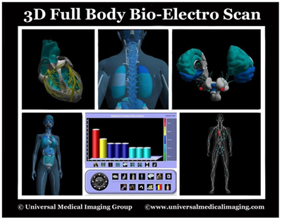
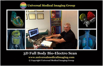
The lymphatic system, the lymph nodes and the lymphocytes themselves must be functional in preventing and reversing any cancerous condition. Using electrodes attached to the head, hands and feet we are able to test the functionality of the lymphatic system, circulatory system, muscular system, skeletal system, endocrine system, neurological system, reproductive system, vascular system, digestive system, and respiratory system, interstitial chemistry, interstitial pH for metabolic acidosis and the electro-conductivity of the cells to determine the state of health of ALL organs, glands and tissues in the prevention and reversal of any cancerous condition4 (Figures 1&2).

Figure 1 The patient is hooked up to electrodes to quantify pH, oxidative stress, mineral levels, hormone levels, and the state of health in all organs and organ systems.

Figure 2 Non-Invasive Bio-Electro Scan provides the quantitative measurements of interstitial pH to determine metabolic acidosis as a indicator of an inflammatory and/or cancerous condition.
We also test clinically for nutritional deficiencies and metabolic alkalosis or acidosis by measuring the interstitial chemistry, interstitial pH and the electro-conductivity. Measuring the pH of the interstitial fluids is more revealing of a cancerous condition since the blood is always trying to maintain its delicate alkaline pH of 7.365 and will not vary much. Based upon my theory that cancer is a compromised acidic environment of the interstitial fluids which may negatively affect the state of health of ALL body cells which make up the organs, glands and tissues. It is significantly more important to measure interstitial and intracellular fluids than blood fluids in order to obtain a correct chemistry and pH when making nutritional recommendations in the prevention and treatment of a cancerous condition.5–10
The following are quantitative measurements in healthy patients, without cancer, comparing Blood fluids with Intracellular and Interstitial fluids of the body compartments as a benchmark which I use to determine deficiencies in alkalizing minerals, protein and whether or not the patient is in metabolic acidosis or a pre-cancerous or cancerous condition (Note: all cancer patients are in interstitial metabolic acidosis, low in interstitial sodium and high in interstitial calcium and potassium).9,10
Venous blood: 130, Arterial blood: 137, Capillary blood: 135, Intracellular fluid: 10 and interstitial fluid: 135
Venous blood: 3.2, Arterial blood: 3.5, Capillary blood: 4, Intracellular fluid: 140 and interstitial fluid: 3.17
Venous blood: 2.5, Arterial blood: 2.2, Capillary blood: 2.3, Intracellular fluid: 0.0001 and interstitial fluid: 1.55
Venous blood: 0.64, Arterial blood: 0.62, Capillary blood: 0.60, Intracellular fluid: 58 and Interstitial fluid: 0.50
Venous blood: 104, Arterial blood: 101, Capillary blood: 103, Intracellular fluid: 4 and Interstitial fluid: 106
Venous blood: 22, Arterial blood: 24, Capillary blood: 23, Intracellular fluid: 10 and Interstitial fluid: 24
Venous blood: 2.5, Arterial blood: 2.3, Capillary blood: 2, Intracellular fluid: 75 and Interstitial fluid: 0.70
Venous blood: 0.8, Arterial blood: 0.6, Capillary blood: 0.5, Intracellular fluid: 2 and Interstitial fluid: 0
Venous blood: 1, Arterial blood: 1, Capillary blood: 1.01, Intracellular fluid: 0.20 and Interstitial fluid: 0.90
Venous blood: 0.66, Arterial blood: 0.630, Capillary blood: 0.676, Intracellular fluid: 0.2 and Interstitial fluid: 0.188
Venous blood: 80, Arterial blood: 90, Capillary blood: 89, Intracellular fluid: 20 and Interstitial fluid: 87.2
Venous blood: 46, Arterial blood: 40, Capillary blood: 42, Intracellular fluid: 50 and Interstitial fluid: 46
Venous blood: 7.36, Arterial blood: 7.4, Capillary blood: 7.38, Intracellular fluid: 7.2 and Interstitial fluid: 7.36
Venous blood: 72, Arterial blood: 74, Capillary blood: 73.7, Intracellular fluid: 68 and Interstitial fluid: 20.6
As we correct the deficiencies in the intracellular and interstitial fluids targeted with key alkalizing nutritional treatments, patients see the difference through follow-up tests using quantitative non-invasive 3-D Full Body Bio-Electro scanning. They also feel the difference physiologically and functionally.5–8
This is how we know proper alkalizing nutritional support in any cancerous condition is important in the prevention and treatment of cancer, the metastasis of cancer and the shrinking of a cancerous cyst or mass without chemotherapy and/or radiation. The best part about these alkalizing nutritional treatments is they are helpful in most, if not in all cancerous conditions.5–8
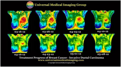
The following case study is with one of my patients who were diagnosed by biopsy with inflammatory ductal cell carcinoma who reversed her cancerous condition without chemotherapy, radiotherapy, and surgery (Figure 3).

Figure 3 An eight week treatment progress of a patient with medically diagnosed inflammatory ductal cell carcinoma with a 14.2 cm primary cancerous mass in the left breast. Note in the thermographs the increased temperature and size of mass reduced significantly over the eight week period following and alkaline lifestyle and diet.
Using breast thermography and tumor location and size measured by breast ultrasound you can see the week by week thermography progress of a 14.2cm tumor in the left breast reduce to less than 2cm in 8weeks of treatment using ANI protocol as outlined in this article and in Chapter 11 of the pH Miracle revised and updated book.5–8
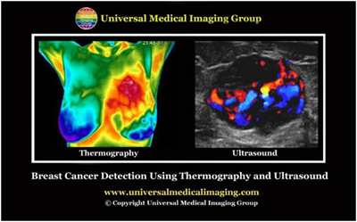
The safest, painless, non-invasive, affordable full body screening tests are a combination of a Medical Diagnostic Ultrasound and Thermography, which may give the Physician about 95% accuracy in detecting breast cancer (Figure 4).

Figure 4 Non-invasive thermograph showing the physiology of a cancerous mass in the left breast indicated in red from the increased temperature coming from an active cancer. Non-invasive ultrasound using dopler to see the blood supply to the mass confirms the anatomy of the cancerous mass and the possible malignancy.
Thermography is a physiological, non-invasive screening procedure that detects and records infrared heat emissions from the pre-cancerous or cancerous area, which can aid in the early detection of abnormal changes in body tissues, organs and glands. Thermography offers information that no other procedure can provide. The procedure is based on the principle that chemical and blood vessel activity in both pre-cancerous or cancerous tissue and the area surrounding a developing cancer is almost always higher in temperature than in the normal tissue.
Since pre-cancerous and cancerous masses are highly metabolic tissues, they need an abundant supply of nutrients to maintain their growth. The cells release substances that stimulate the formation of new blood vessels (neoangiogenesis). This process results in an increase in surface temperatures of the affected tissue, organ or gland.
The most promising aspect of medical diagnostic thermography is its ability to spot abnormalities years before the tumor is seen on any anatomical test. Since thermal imaging detects changes at the cellular level, this test can detect activity 8 to 10years before any other test. This makes it unique in that it affords the physician the opportunity to view changes before the actual formation of the cancerous tumor.
Studies have shown that by the time a tumor has grown to sufficient size to be detectable by physical examination or mammography; it has in fact been growing for about seven years achieving more than 25 doublings of the malignant cell colony. At 90days there are two cells, at one year there are 16 cells, and at fiveyears there are 1,048,576 cells–an amount that is still undetectable by a mammogram. Thermography has the ability to provide the patient with future risk assessment (Figure 5). If discovered, certain thermographic risk markers can warn the patient that she/he needs to work closely with their physician with regular checkups to monitor her health.4
Full-body ultrasound is an anatomical non-invasive, painless screening test without ionized radiation. Ultrasound, also known as sonography, uses sound waves to outline a part of the body. For this test, a small instrument called a transducer is placed on the skin (which is often first lubricated with ultrasound gel) and emits sound waves off body tissues. The echoes are converted by a computer into an image that is displayed on a computer screen.
Ultrasound imaging is “real-time,” meaning that it can show exactly what’s happening in the tissue, organ or gland at that moment, help to distinguish between cysts (fluid-filled sacs) and solid masses, detect increased vascularity around or within the mass, see the shape, exact size and location of the mass, cyst, calcification or dilated mammary ducts. These safe medical diagnostic tests can be done on early bases for a regular check up, or more often if the problem was detected, to monitor a noninvasive alkalizing nutritional treatment progress.
Early detection, which includes self examination and safe, painless, non-invasive medical diagnostic Full Body Bio-electro Scan (FBBES) Full Body Thermography (FBT) and Full Body Ultrasound (FBU) screenings with no ionizing radiation coupled with a supportive alkalizing nutritional diet and ANI whether or not the patient is receiving chemotherapy and/or radiation, I have found clinically that this approach in a precancerous or cancerous condition will saves lives.5–8
Is X-ray Mammography Findings Cancer or Benign Lesions?
The Dark Side of Breast Cancer Awareness Month
Does Chemo & Radiation Actually Make Cancer More Malignant?
None.
Author declares there are no conflicts of interest.
None.

©2016 Migalko, et al. This is an open access article distributed under the terms of the, which permits unrestricted use, distribution, and build upon your work non-commercially.