International Journal of
eISSN: 2574-9862


Research Article Volume 7 Issue 3
Club Auto Safari Chapín, Guatemala
Correspondence: M V Gustavo Adolfo González, Club Auto Safari Chapín, Guatemala
Received: September 27, 2023 | Published: October 9, 2023
Citation: Gustavo González MV, González Cifuentes MVLD. Retrospective study of the incidence of dental pathologies in big cats under human care in Guatemala, Central America. Int J Avian & Wildlife Biol. 2023;7(3):102-109. DOI: 10.15406/ijawb.2023.07.00198
Oral pathologies in both domestic and wild felids have gained importance in recent years and have been considered to be part of the most common and important pathologies in these species. A study carried out in different zoos in Belgium, the Netherlands and Germany showed that, of a total of 23 wild felids evaluated, 18 had dental lesions. The most affected teeth were the canines with fractures with and without pulp exposure, abrasion of the teeth and absence of teeth. Based on the aforementioned, it is intended that this research study provides useful, relevant and concrete knowledge that supports veterinary professionals in the diagnosis of dental injuries. The aim is to help improve the health and quality of life of large felids under human care in Guatemala.
Currently, veterinary medicine focuses on animal welfare, which is defined as compliance with the five freedoms or animal principles. These five freedoms seek to ensure that animals are free from pain, stress, hunger, suffering, and have normal behavior according to their species. As veterinary medicine focuses on animal welfare, untreated or non-diagnosed pathologies of the oral cavity acquire great relevance, since they violate the 5 principles. Because the oral cavity fulfills different functions such as feeding, defense and grooming, when the oral cavity is affected, the patient's general health is impacted, and can reach critical states. For this reason oral pathologies, in both domestic and wild felids, have gained importance in recent years and have been considered to be part of the most common and primary pathologies in these species.1 According to Brooks,1 the species where it has been most documented for dental pain to be extreme is human species. In humans, it has been determined that oral pathologies can cause sleep disturbances, productivity, and social and psychological disorders. Although the impact of dental pain in animals has not yet been determined exactly, it is considered that it may be similar to that of humans, considering that pain thresholds are similar. Diaz,2 reports that approximately 50% of domestic cats aged 5 years have dental lesions and that more than 80% of cats aged 8 years and older suffer from pathologies of the oral cavity. However, worldwide, in wild felids there is no data or references that indicate the percentage of prevalence of dental lesions. Studies have been carried out that report some common injuries observed in these species. In other countries, clinical cases have been reported, such as the case of a jaguar from the Bubalco Zoo in the town of Gral. Roca in the province of Río Negro.3 A study carried out in different zoos in Belgium, the Netherlands and Germany showed that, of a total of 23 wild felids evaluated, 18 had dental lesions. The most affected teeth were the canines (teeth numbers 104, 204 and 404) with fractures with and without pulp exposure, abrasion of the teeth and absence of teeth.4 This would be equivalent to 72% of the felids evaluated having dental problems.
Due to the aforementioned, it is intended that this research study provides useful, relevant and concrete knowledge that supports veterinary professionals in the diagnosis of dental injuries. The aim is to help improve the health and quality of life of large felids under human care in Guatemala.
The Felidae family belongs to the order Carnivora. Currently two subfamilies are known, which are Felinae and Pantherinae.5,6 The divisions are based on morphological, ethological and physiological considerations.6According to these divisions, the species have been grouped into: large felids or pantherids (subfamily Pantherinae), and small felids (subfamily Felinae). Members of the Felidae family are hypercarnivorous.7 This refers to the fact that these species consume more than 70% of their diet in meat, unlike other species of the Carnivorous order which consume 50-60% meat. Due to these eating habits, its anatomy has adapted to hunting, characterized by having a fairly flexible spine and a little modified axial skeleton.5,7 They are digitigrade animals with retractable claws, although these are not exclusive to them.5 This ability to retract their claws helps them when hunting as it reduces the noise when stepping and makes them extremely stealthy during hunting. According to Sánchez,7 these animals are equipped with “sensory adaptations” which facilitate the location of prey without being detected. Richard5 mentions that they have binocular vision, which allows them to have high visual quality. Sánchez7 talks about the vibrissae and the developed hearing level that allows them to capture tactile information, ultrasonic communications from their prey, facilitating hunting during the night hours. The skull is short and rounded with a notable reduction in teeth and great development of the carnivorous molars (formed by the first lower molar and the last upper premolar). We can say that the Felidae family has evolved to be hypercarnivorous, so the teeth must be in good condition so that their digestion and nutrition are not affected.
Anatomy of the oral cavity: The oral cavity extends from the lips to the pharynx, bordering laterally with the cheeks, dorsally with the palate, and ventrally with the tongue and mandibular tissues.8
It presents the following structures:
Generalities of dentition in felids
Felids are diphiodonts, which means that throughout their life they have two changes of teeth.4 The first teeth are known as deciduous teeth, which appear in puppies 2-6 weeks old. These are replaced by permanent teeth at approximately 3 months of age. According to Bollez,4 all Felidae species have a total of 26 deciduous teeth and 30 permanent teeth. To identify the teeth, the initials of the tooth are used, being I (incisors), C (canines), P (premolars) and M (molars). In Figure 1 we can see the dental formula of an adult cat.

Figure 1 Dental formula in adult cats.4
There are several dental nomenclatures, but the system internationally recognized by the International Dental Federation (FDI) and the World Health Organization is the modified Triadan nomenclature.9 In this nomenclature, each tooth is represented with a three-digit number, as seen in Figure 2. The first digit refers to the arch where the tooth is located, the second and third digits refer to the type of tooth, then remaining as follows:
Tooth anatomy
Díaz2 mentions that all felid teeth have the same elements regardless of shape and function. Those teeth that are multirooted (several roots) have additional structures, but their internal conformation does not change. The anatomical and histological conformation is presented below.
Anatomically, teeth are made up of three parts:
Histologically each tooth is made up of three tissues

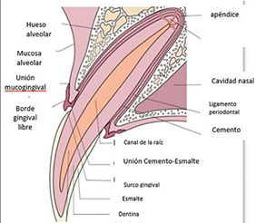
Figure 3 Anatomy and histology of the tooth. Fuente Díaz.2
Anatomy of the periodontium
Gorrel11 mentions that the periodontium is the structure that has the function of holding the tooth to the spindle of the mandible or maxilla. It provides a suspension apparatus to resist normal functional forces. As seen in Figure 4, the periodontium is made up of:

Figure 4 Anatomy of the alveolar bone. Fuente Díaz.2
Relationship between oral health and systemic conditions
Brooks,1 et al mentions that veterinary medicine is currently beginning to focus on the five freedoms of animals, which seek to minimize stress, fear, suffering and pain, as well as provide the animal with freedom to express normal behavior. Part of this animal well-being is the care of the oral cavity and its structures, since the pathologies of this cavity can trigger chronic pain and/or contribute to the appearance of both local and systemic diseases and prevent the expression of natural behaviors. In human medicine, the relationship between oral health and general health has been well described and documented in literature,4 while in veterinary medicine data is still being collected to confirm this relationship. The relationships between oral health and systemic health that have been found in veterinary medicine according to Bollez4 include periodontal disease and cardiovascular diseases such as endocarditis, myocarditis and atherosclerosis. In some cases, periodontal disease has also been associated with respiratory diseases. Bollez4 reports that some studies show that periodontal disease can be a risk factor for chronic kidney disease in cats. These data have been demonstrated in domestic cats and there is no literature or studies of this relationship in large felids, but they assume that it is a very similar situation in all mammal species.
Dental evaluation
In domestic cats, oral pathologies are common, and possibly in large felids this is very similar. There is not much information about it, since it has begun to be described and studied only recently. In domestic cats, dental evaluation should be carried out at least once a year.10 In these species the evaluation is carried out with the patient awake and the veterinarian is able to manipulate the patient completely. But, in the case of large felids, dental (or any type of evaluation) must be carried out under anesthesia, which is why many veterinarians have adapted to performing an evaluation without handling. For this, the veterinarian and the person in charge of caring for the animals evaluate behavioral patterns during feeding or, in some cases, witness the moment of dental trauma. Beers10 mentions that in most cases dental prophylaxis in large felids is ignored. Treatments in large felids are based more on curative medicine since oral cavity treatments focus on specific problems that are causing pain or are risk factors for local or systemic diseases.
Main dental pathologies
Due to the little information that exists on large felids, the focus of the information is mainly on pathologies in cats, which is the closest to them. According to Feline Dental Disease,13 pathologies of the oral cavity and teeth are quite common in cats. Approximately 50-90% of cats over four years of age suffer from some oral pathology. Calderon12 and Díaz2 consider that the most common dental pathologies in felines are: dental fractures, periodontal disease, loss or wear of teeth, and odontoclastic resorptions. As mentioned above, oral pathologies can cause pain and discomfort in the animal, which can affect its quality of life. In many cases, clinically dental disease presents itself as anorexia, which leads to a wide variety of health problems.
Periodontal disease
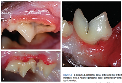
Periodontal disease is one that includes all disorders associated with the tissues that surround and support the dentition.14 In other words, this term refers to infection or inflammation of the periodontium.15 As seen in Figure 5, it affects the gum, cementum, periodontal ligament and alveolar bone. Calderón12 mentions that this disease is the most common pathology in adult cats, with the main causes being bacterial plaque, genetics, age, diet, chewing patterns and systemic health. Depending on the etiological origin it can be divided into:
Inflammatory that has four states

Figure 5 Periodontal disease in cats (a) Gingivitis; (b) Established periodontitis in the mandibular first molar; (c) Advanced periodontitis in the third and fourth premolars of the maxilla.8
Dystrophic: in which there is already gradual wear and tear of the size or function of the periodontium.
Tooth wear (abrasion/attrition): Calderón12 et al. defines this disease as progressive and abrasive loss of enamel and dentin. It can be classified according to the level of wear:
Regardless of whether it is physiological or pathological wear, the hypoplasia/hypomineralization that the enamel suffers weakens the tooth structurally, which can favor dental fractures. The teeth that suffer the most wear are the incisors (Figure 6).11 The presentation of these pathologies increases with age due to physiological wear, chewing patterns or diets, which result in loss of crown enamel and subsequently in possible exposure of the dental pulp.

Figure 6 Wear of incisor teeth without pulp exposure and wear of canines with exposure of dental pulp.11
Tooth mobility
Bellows8 mentions that, physiologically, the teeth have a certain mobility, which should be less than 0.2 mm in the incisor teeth and the rest of the teeth should not have any mobility. Also, he mentions that pathological mobility is in response to the forces of occlusion, trauma or reduction of the periodontium. Mobility is not diagnosed as a periodontal disease as such, but reflects a certain pathological adaptation of the periodontium. Mobility can be categorized into different degrees:
Dental fractures
Dental fractures can be a consequence of certain habits, games, fights or diets that end in trauma.12 Bellows8 mentions that, in dental fractures in felines, pulp exposure is more common than in other species because the pulp chamber is located just behind the dentin. In the other species, the dentin is found several millimeters behind the enamel to protect the pulp. Trauma that causes pulp exposure provides a gateway for oral bacteria which infect the pulp chamber.8 The resulting infection can continue along the pulp canal until it reaches the periodontal ligament, periapical tissue and, in severe cases, can reach the alveolar bone. When the infection has reached the alveolar bone it is common for fistulas to form which, being from the maxilla, tend to drain near the eye socket and, in cases where they affect the jaw, purulent drainage is observed below the chin. Dental fractures can be classified according to the complexity and the part of the tooth that is compromised.12 It is taken as a fracture with complications when there is pulp exposure, and without complications when there is no pulp exposure. The general classification of fractures is as follows:
Tooth resorption
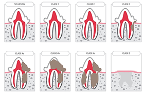
It is also known as feline odontoclastic resorptive lesion (FORL), “neck lesion” or “feline false caries”.16 "Feline Dental Disease"13 defines it as a process of destruction of hard dental tissue (Figure 7), which starts from the inside of the tooth and extends to other parts of the tooth and is caused by the activation of odontoclastic cells. It is estimated that between 30-70% of cats suffer from this pathology. Turini & Algorta17 report that it is a painful pathology that is characterized by odontoclastic resorption of dental tissue and its replacement by granulation tissue, and it is commonly associated with gingivitis. The most affected teeth are: the lower first molar, lower third premolar and upper fourth premolar, rarely affecting incisors. It can also manifest as a hyperemic line at the junction of the tooth and the gum ("Feline Dental Disease", 2019). However, when this sign appears the tooth has been damaged significantly. This type of injury can vary in severity from a small defect or injury in the gum to large defects or injuries in the root and/or crown of the tooth.

Figure 7 Classification of odontoclastic resorptive lesions in cats.16
Study design
Descriptive cross-sectional study based on clinical records from the years 2015 to 2019 in a private collection.
Location of area of study
The study area is located at a north latitude of 14°06'02.5'' and west longitude of 90°37'27.6'', the average altitude is 34 meters above sea level. It is located at km 87.5 of the CA2 highway in the department of Escuintla, which borders to the north with the municipality of San Juan Alotenango, Suchitepéquez; to the south with the municipality of Masagua; to the east with the municipalities of Palín, San Vicente and Guanagazapa; and to the west with La Democracia and Siquinalá.18 The climate in the study area is warm, ranging from 21-34°C. It has an average rainfall of 2,200 mm. and has 3 identified life zones: subtropical dry forest, warm subtropical humid forest and warm subtropical very humid forest.19
Study documents
The study was carried out based on the complete files of all the felid species (32 specimens) in the park, distributed as follows:
A review of the clinical records of the study population was carried out, which covered the years 2015-2019 to identify specimens with dental lesions. Data collection was carried out by preparing cards where the species, age, name and habitat number of the specimen under study were specified. The dental lesion that the specimen presented was identified and marked on an odontogram and the type or types of lesions that the specimen presented were classified using tables (Annex I). Upon completion of the review of all the files, tables were prepared where the number of affected animals, each dental lesion and the percentage it represented were calculated, to determine the most common lesion. Another table, contains data on the number of affected teeth, the number of animals that had an injury to this tooth and the percentage it represented (Annex II). Tables and graphs were used to represent the data obtained. Quantification of animals with dental injuries. Classification of dental injuries (Graphs 1–6) (Table 1–6).
|
Variable |
Conceptual definition |
Type and scale |
Unit of measurement |
|
Fractures without complications |
When there is partial loss of enamel and dentin without exposure of dental pulp. The information on the card about the damaged layers in the tooth was analyzed to determine the unit of measurement and place it in the record card boxes. |
Nominal qualitative |
of enamel |
|
crown |
|||
|
Coronary and root |
|||
|
Root |
|||
|
Fractures with complications |
When there is loss of enamel and dentin with exposure of dental pulp. According to the description of the clinical records, the affected layers were determined and classified in the registration form. |
Nominal qualitative |
Crown |
|
Coronary and root |
|||
|
Root |
|||
|
Physiological wear |
A result of chewing. It was recorded in drawings. |
Nominal qualitative |
Mild |
|
Advanced |
|||
|
Wear (abrasion/attrition) |
It is when there is loss of enamel or dentin due to an external object, such as metal cages, sticks, balls or bones. According to the data obtained from the documents, it was recorded in tables and graphs. |
Nominal qualitative |
Mild |
|
Advanced |
|||
|
Tooth mobility |
When there is mobility of the tooth axially or in another direction. A dental explorer is used to evaluate the level of mobility of the teeth. The data obtained will be recorded through tables on registration forms. |
Nominal qualitative |
Grade 0 |
|
Grade 1 |
|||
|
Grade 2 |
|||
|
Grade 3 |
|
Dental fractures |
Yes/No |
Tooth wear |
Yes/No |
Tooth mobility |
yes/No |
|
of enamel |
|
Mild physiological |
|
Grade 0 |
|
|
Crown without complications |
|
Advanced physiology |
|
Grade 1 |
|
|
Crown with complication |
|
Mild pathological attrition |
|
Grade 2 |
|
|
Coronary and root without complications |
|
||||
|
Uncomplicated root fracture |
|
Advanced pathological attrition |
|
Grade 3 |
|
|
Root fracture with complications |
|
Annex 2 Type of injury
Enter yes or no, as appropriate
|
Healthy animals |
Animals with injury |
Total |
|
14 |
18 |
32 |
Table 1 Quantification of animals with dental injuries
|
Type of injury |
No. of injuries found |
Percentage it represents (%) |
|
Dental fractures |
20 |
86.95 |
|
Tooth wear |
2 |
8.7 |
|
Tooth mobility |
1 |
4.35 |
|
TOTAL |
23 |
100 |
Table 2 General classification of dental injuries
|
Type of injury |
No. of affected animals |
Percentage it represents (%) |
|
of enamel |
0 |
0 |
|
Crown without complications |
0 |
0 |
|
Crown with complication |
18 |
78.26 |
|
Coronary and root without complications |
0 |
0 |
|
Uncomplicated root fracture |
1 |
4.35 |
|
Root fracture with complications |
1 |
4.35 |
|
TOTAL |
20 |
100 |
Table 3 Quantification of dental fractures
|
Type of injury |
No. of affected animals |
Percentage it represents (%) |
|
Mild physiological |
1 |
4.35 |
|
Advanced physiology |
0 |
0 |
|
Mild pathological attrition |
1 |
4.35 |
|
Advanced pathological attrition |
0 |
0 |
|
Mild physiological |
0 |
0 |
|
Advanced physiology |
0 |
0 |
|
TOTAL |
2 |
8.7 |
Table 4 Qualification of dental wear
|
Type of injury |
No. of affected animals |
Percentage it represents (%) |
|
Grade 0 |
0 |
0 |
|
Grade 1 |
1 |
4.35 |
|
Grade 2 |
0 |
0 |
|
Grade 3 |
0 |
0 |
|
TOTAL |
1 |
4.35 |
Table 5 Quantification of tooth mobility
|
No. of affected tooth piece |
No. of animals affected |
Percentage it represents (%) |
|
103 |
1 |
2.17 |
|
104 |
5 |
11 |
|
105 |
1 |
2.17 |
|
106 |
1 |
2.17 |
|
107 |
2 |
4.35 |
|
108 |
1 |
2.17 |
|
204 |
9 |
19.55 |
|
205 |
1 |
2.17 |
|
206 |
1 |
2.17 |
|
207 |
3 |
6.51 |
|
208 |
1 |
2.17 |
|
304 |
9 |
19.55 |
|
305 |
1 |
2.17 |
|
306 |
1 |
2.17 |
|
401 |
1 |
2.17 |
|
404 |
6 |
13 |
|
405 |
1 |
2.17 |
|
406 |
1 |
2.17 |
|
TOTAL |
46 |
100 |
Table 6 Quantification of affected tooth
The lesions presented by the specimens were categorized and placed in tables. The variables that were used are qualitative. The data obtained is presented through tables and graphs. The information included is the type of injury and the number of specimens that presented it with percentages.
The study was carried out on all the large felids (32) in the collection, which is made up of 10 African lions (Panthera leo), 8 jaguars (Panthera onca) and 14 pumas (Puma concolor). Of these, 56% (18) have some dental injury. Of this population with dental injuries, 86.95% have dental fractures, of which the pieces most affected are pieces 204 and 304, corresponding to the upper and lower left canines with a 19.55% prevalence each, followed by the tooth 404 (lower right canine) with 13%. According to the results, dental wear and mobility have a prevalence of 8.7% and 4.35% respectively. Studies have been carried out in which it has been determined that the incidence of dental injuries in large felids increases with age, although fractures, especially of canines, can occur at any stage of life. It should be considered that, as mentioned Gorrel,10 one of the main causes of the most common pathology being canine fractures, which are the most prevalent in the study, may be due to pathological attrition, that is, wear of the enamel due to trauma. In the case of the study, the most probable cause of attrition is the wear of the pieces due to bites on the cages in times of prolonged confinement, which weakens the piece and favors fracture, mainly of the canines since in these species these teeth are large and can get stuck in the material of the bedroom/cage more easily than the other teeth. As there is friction between the tooth and the metal/concrete, it causes gradual loss of enamel and weakening of the tooth until it ends in fracture.
There is a high frequency of dental pathologies in large cats under human care in Guatemala and therefore it is important to evaluate and have the basic knowledge to offer animals an adequate diagnosis and treatment.
None.
The authors declared that there are no conflicts of interest.

©2023 Gustavo, et al. This is an open access article distributed under the terms of the, which permits unrestricted use, distribution, and build upon your work non-commercially.