International Journal of
eISSN: 2574-9862


Mini Review Volume 7 Issue 2
Fundación Refugio Salvaje, El Salvador, Centro América
Correspondence: MV Gustavo Adolfo González, Fundación Refugio Salvaje, El Salvador, Centro América
Received: March 31, 2023 | Published: May 3, 2023
Citation: González MVGA. Handrearing of a hippopotamus (Hippopotamus amphibius) at “Fundación Refugio Salvaje”. Int J Avian & Wildlife Biol. 2023;7(2):40-44. DOI: 10.15406/ijawb.2023.07.00188
The Nile hippopotamus is a semi-aquatic mammal with populations that have decreased in the last 10 years, which is why it has been placed in CITES Appendix II, and as vulnerable on the IUCN red list. The artificial breeding of hippos is poorly documented, possibly due to the complexity that this species presents. The Wild Refuge Foundation (FURESA) located in El Salvador, C.A. has kept a pair of Nile hippos (Hippopotamus amphibius) in its facilities. Both specimens have enjoyed good health and have procreated three pups, which died between 3 and 5 days after birth, considering little maternal attention as the main cause of deaths. Due to the above situations, artificial breeding was decided for this occasion. Housing conditions, medical history, feeding techniques and diet, which were used in this breeding, are described.
Keywords: hand‐rearing, captive hippopotamus, Hippopotamus, amphibius
The Nile hippopotamus is a semi-aquatic mammal that inhabits shallow lakes, rivers and swamps in the Ethiopian region of the world, with populations that have decreased in the last 10 years, which is why it has been placed in CITES Appendix II and as vulnerable, for the IUCN red list.1,2 The artificial breeding of hippos is poorly documented, possibly due to the complexity that this species presents.3-5 In 1970, Weber reports the rearing of a calf at the Melbourne Zoo.6 Another report of hippopotamus calf rearing is the one described in 1978 by Wilson and Littlewood, starting rearing from the 10th day of birth.5 That same year, the Ouwehands Zoo, Netherlands proceeded to raise a calf from the 2nd day of birth.7 Recently, in 2017, a premature female hippopotamus was bred by the Cincinnati Zoo, USA.3,4
“Fundación Refugio Salvaje” (FURESA) located in El Salvador, C.A. (altitude 600 AMSL, 13°42'42.2" N 89°27'0.6 W), has kept a pair of Nile hippos (Hippopotamus amphibius) in its facilities. Both specimens have enjoyed good health and have procreated three calves, two of which died between 3 and 5 days after birth, considering little maternal attention as the main cause of death. Due to the above situations, artificial breeding was decided for the third calf, the subject of this article. After 7 months of gestation and having taken the mating date, it was decided to separate the male from the female one month before delivery, towards a lock with a swimming pool and patio, although smaller than the exhibition area. After 8 months of gestation, the delivery was reported on January 11, 2020 at 6:00 a.m. After delivery, the calf was separated to be transferred to the veterinary hospital. At 10:00 a. m. the calf was finally separated from his mother. He was removed from the premises, and transferred, at 11:00 a.m.
Accommodation
The calf, baptized with the name of "Kibo", (from the Japanese meaning Hope and Kiboko from the Swahili in a diminutive form, which corresponds to hippopotamus), was placed in a 4 x 4 meter dormitory, using hay as a substrate for bedding and infrared lights forming a lamp, to keep warm, especially at night (Image 1).
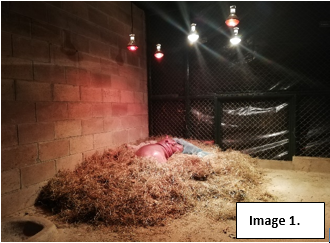
Image 1
From the first day of birth, the calf was accompanied day and night by its caregivers, since it showed stress when he was left alone. Likewise, stuffed animals were placed around him, which gave him comfort These stuffed animals were disinfected daily (Image 2 and 2a). A continuous bedroom, of the same size, housed the pool, which was made of plastic, round, 1.83 meters x 51 cm, with a capacity of 880 liters, for use by young children (Intex®, Easy Set®, China). The water of the pool was renewed in a daily basis. The wáter was heated to 28°C via a tankless electric heater (Aqua Power®, Thailand). At the end of the day, the pool was emptied, washed with powdered soap and disinfected with chlorine, letting it drain overnight. In this pool, he defecated the meconium the first day, and continued to defecate there for the rest of the breeding. During the first 4 days of life, he entered the pool for 1-hour periods in the morning and 1-hour in the afternoon. The rest of the day he liked to sleep in his bedroom. After the first week of age he already liked to stay and play in the pool for longest periods of time, during which he learned to get in and out of it by himself (Image 3).
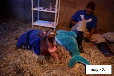
Image 2
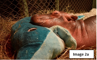
Image 2a
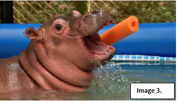
Image 3
Feeding schedule
From day one, at 3:30 p.m., the oral feeding of replacement milk for bovine calves (Fortamilk® Lactalis Feed, France) began, at a rate of 250 ml orally, every two hours until 12:00 a.m. On the second day, feeding began at 06:00 until 24:00, with the same volume and frequency (250ml every two hours). During the initial two days of feeding, the calf was fed with a bottle, but the sucking réflex was not normal, so it was uncertain that the intake amounted for the total of mililiters stipulated. By day 3, there still wasn’t any presence of sucking reflex. At that momento, the veterinarian team decided to feed him with a 122cm, 16Fr/Ch double-lumen gastric tube (Salem Sump®, Covidien, MA, USA) Via this tube the calf was fed 500ml of formula, per feeding, 7 times a day, starting at 06:00 and ending at 24:00, every 3 hours.
The offered quantity corresponds to 10% of its live weight. The Fortamilk® formula was prepared at a rate of 250gm of powdered formula diluted in one liter of water for the first few days. From day 20, 250gm of powdered formula were diluted in 900ml of water and, after a month, in 800ml of water. This formula provided 22% crude protein, 20% fat, 7.5% ash, 0.3% fiber, 0.70% calcium, 0.55% phosphorus, 0.55% sodium, 50,000 IU/kg of vitamin A, 10,000 IU/ kg vitamin D, 160mg/kg vitamin E, 5mg/kg vitamin B1, 10mg/kg vitamin B2, 500mg/kg vitamin C, 2.50mg/kg vitamin K3, 50mg/kg iron, 0.15mg/kg kg of iodine, 7mg/kg of copper, 35mg/kg of manganese, 60mg/kg of zinc and 0.20mg/kg of organic selenium. A capsule of lactase equivalent to 9000 FCC (Lacto Digestomen® Richam Internacional, S.A. Guatemala) was added to each dose. Around day 20 of age, he began to suck and take the bottle by himself.
The adjustments of the amount of formula offered, were made based on, weight and daily consumption, with the best consumption being 6.25% of its live weight, divided into 5 doses a day, every 4 hours, starting at 06:00 a.m. And ending at 10:00 p.m. Table 1 summarizes the rearing diet.
|
Age in days |
Amount |
No. of shots |
% of V.P. |
Observations |
|
1 |
250ml |
5 |
||
|
2 |
250ml |
10 |
||
|
3 |
500ml |
7 |
10 |
with probe |
|
5 |
600ml |
7 |
10 |
with probe |
|
8 |
800ml |
6 |
11 |
with probe, suction attempt |
|
18 |
800ml |
6 |
11 |
probe and bottle |
|
23 |
800ml |
6 |
11 |
Take in a bottle |
|
38 |
900ml |
4 |
5.25 |
|
|
45 |
950ml |
5 |
5.25 |
|
|
50 |
1200ml |
5 |
6.5 |
Quite an appetite, Take in a bottle |
|
100 |
1300ml |
5 |
6.5 |
|
|
150 |
1400ml |
3 |
||
|
180 |
1400ml |
2 |
3 |
start weaning |
|
200 |
0 |
0 |
4 |
fully weaned |
Table 1 Food
Weaning
Weaning began around day 180 of age. By that time he began to show interest in eating solid food. Therefore, he was presented with a tabulated diet based on 3% of his live weight (320 pounds)- The diet consited of 30% concentrate (concentrate of dairy cow 16%® Tecnutral, El Salvador) and 70% vegetables (spinach, carrot, lettuce and green grass). 25% of the diet was given in the morning, and the other 75% in the afternoon. Likewise, during the day he received two doses of formula.
At 185 days of age, he was only consuming the diet offered in the afternoon, and continued with two feedings of formula during yhe day. It wasn’t until day 200 that he began to consume both diets: the one in the morning and the one in the evening. Therefore, formula feedings were suspended. During this phase, the daily weight gain was very unstable and he had many ups and downs, so by day 215 of age, a new diet adjustment was made. This change was based on 4% of live weight following the same characteristics of the previous diet. On day 230, the diet was adjusted to 5% of its live weight, changing the distribution, since it was divided into 50% in the morning and 50% for the afternoon. At 300 days of age, the food consumption continued to increase and, therefore, a diet based on 8% of their live weight (400 pounds) was recalculated, and distributed in 20% dairy cow concentrate and 80 % vegetables.
Upon reaching 3 years of age, a decrease in food consumption was observed, so the diet was tabulated based on 6% of its live weight (estimated at 2000 pounds), distributing the total diet into 10% concentrate and 90 % vegetables.
Growth
The weight was taken daily in the early morning hours, before his first feed, from the day it was born until the 424th day of age, as presented in table 2 & 3.
|
Date |
Days of birth |
Weight (pounds) |
Weight (kg) |
|
11-01-20 |
0 |
87.4 |
39.73 |
|
12-01-20 |
1 |
87.4 |
39.73 |
|
13-01-20 |
2 |
87.4 |
39.73 |
|
14-01-20 |
3 |
91.4 |
41.55 |
|
15-01-20 |
4 |
95.6 |
43.45 |
|
16-01-20 |
5 |
98 |
44.55 |
|
17-01-20 |
6 |
98.4 |
44.73 |
|
18-01-20 |
7 |
98.6 |
44.82 |
|
19-01-20 |
8 |
98.2 |
44.63 |
|
20-01-20 |
9 |
101 |
45.9 |
|
21-01-20 |
10 |
104.6 |
47.55 |
|
22-01-20 |
11 |
106.4 |
48.36 |
|
23-01-20 |
12 |
110.4 |
50.48 |
|
24-01-20 |
13 |
114 |
51.82 |
|
25-01-20 |
14 |
115.4 |
52.45 |
|
26-01-20 |
15 |
117 |
53.18 |
|
27-01-20 |
16 |
117.4 |
53.36 |
|
28-01-20 |
17 |
115 |
52.27 |
|
29-01-20 |
18 |
115.2 |
52.36 |
|
30-01-20 |
19 |
118.2 |
53.73 |
|
31-01-20 |
20 |
117.4 |
53.36 |
|
01-02-20 |
21 |
116.2 |
52.82 |
|
02-02-20 |
22 |
120.2 |
54.64 |
|
03-02-20 |
23 |
126.2 |
57.36 |
|
04-02-20 |
24 |
125.2 |
56.9 |
|
05-02-20 |
25 |
130.2 |
59.18 |
|
06-02-20 |
26 |
137 |
62.27 |
|
07-02-20 |
27 |
139 |
63.18 |
|
08-02-20 |
28 |
140.8 |
64 |
|
09-02-20 |
29 |
141.6 |
64.36 |
|
10-02-20 |
30 |
141 |
64.09 |
|
11-02-20 |
31 |
141.8 |
64.45 |
Table 2 Daily weight
|
Date |
Days of birth |
Weight (pounds) |
Weight (kg) |
|
20-02-20 |
40 |
154.2 |
70.09 |
|
01-03-20 |
50 |
166.2 |
75.54 |
|
11-03-20 |
60 |
173 |
78.64 |
|
21-03-20 |
70 |
189.2 |
86 |
|
31-03-20 |
80 |
205.2 |
93.08 |
|
10-04-20 |
90 |
223.4 |
101.33 |
|
20-04-20 |
100 |
243.8 |
110.82 |
|
30-04-20 |
110 |
252 |
114.55 |
|
10-05-20 |
120 |
278.2 |
126.45 |
|
20-05-20 |
130 |
300 |
136.64 |
|
30-05-20 |
140 |
304 |
138.18 |
|
09-06-20 |
150 |
316 |
143.63 |
|
19-06-20 |
160 |
316 |
143.64 |
|
29-06-20 |
170 |
310 |
140.91 |
|
09-07-20 |
180 |
321 |
145.91 |
|
19-07-20 |
190 |
326.2 |
148.18 |
|
29-07-20 |
200 |
315 |
143.18 |
|
08-08-20 |
210 |
301.6 |
137.09 |
|
19-08-20 |
220 |
316 |
143.63 |
|
29-08-20 |
230 |
316 |
143.64 |
|
08-09-20 |
240 |
325 |
147.72 |
|
18-09-20 |
250 |
343.4 |
156.09 |
|
29-09-20 |
260 |
355.2 |
161.45 |
|
08-10-20 |
270 |
369.2 |
167.82 |
|
21-10-20 |
283 |
390.4 |
177.27 |
|
30-10-20 |
292 |
388.2 |
176.45 |
|
08-11-20 |
300 |
396.8 |
180.36 |
|
18-11-20 |
310 |
389.4 |
177 |
|
15-12-20 |
338 |
437 |
198.63 |
|
18-12-20 |
341 |
442 |
200.9 |
|
06-01-21 |
360 |
472 |
214.55 |
|
10-02-21 |
395 |
533 |
242.27 |
|
11-03-21 |
424 |
600 |
272.73 |
Table 3 Weight (every 10 days)
Expected weight gain
Average daily weight gain for the first week was 2.25 pounds; second week was 2.68 pounds; third week was 3 pounds; and, the fourth week had an average growth of 4.36 pounds. By the second month of age he had an average growth of 5 pounds per day; In the third month, 7 pounds per day; and, for the fourth month 9 pounds per day was reported.
At one year of age, the daily growth average was 22.48 pounds. Graph 1, shows the comparative development of Milly (raised in the Melbourne Zoo, Australia)6 and Kibo (raised in “FURESA”, El Salvador).
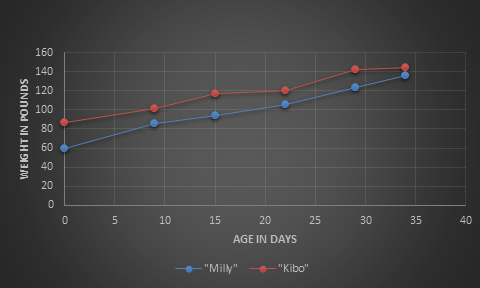
Graph 1 Comparative Kibo vs Milly.
Medical history
At physical evaluation on the first day of birth, he appeared to be normal, with a weight of 87.4 pounds (39.73 kilograms). The umbilicus was disinfected with chlorhexidine. On day one, he was feeded orally with 100 ml of a colostrum supplement based on immunoglobulins, probiotics, vitamins and energy components. (Sprinter®, Anupco, England). On the second day after birth, the veterinarian team decided to adminístrate preventive antibiotic therapy using ceftiofur free crystalline acid (Excede® Zoetis Inc. Kalamazoo, MI.) at a rate of 6mg/kg subcutaneously.
From day one through day 11, the physiological constants were taken according to Table 4.
|
Age (in days) |
Body temperature °C |
Frequency Cardiac (bpm) |
Oxygen saturation % |
|
1 |
36.3 |
||
|
2 |
35.9 |
||
|
3 |
37 |
102 |
99 |
|
4 |
37 |
78 |
97 |
|
5 |
37 |
75 |
98 |
|
6 |
37.2 |
93 |
98 |
|
7 |
37 |
80 |
100 |
|
9 |
37 |
77 |
100 |
|
10 |
37 |
80 |
100 |
|
11 |
36.4 |
85 |
100 |
Table 4 Physiological constants
The navel cleaning was performed from the first day of birth with chlorhexidine. On the third day of the calf's life, the appearance of liquid from the navel was observed, determining that it was urine. Therefore, it was established that he presented persistence of the urachus (Image 4). Conservatively, the treatment was decided based on the application of a solution of iodine and alcohol (50:50) by means of gauze compresses, (three times a day) keeping said compress for a period of 5 minutes. This treatment was carried out until day 30, at which time the navel was healed, and the calf began to urinate only through the penis, although with some difficulty. It wasn't until 70 days that he urinated normally. During this period of time, two ultrasonographic studies of the urinary bladder and navel were performed (study carried out and interpreted by M.V.Z. Silvia Arévalo, “Escalón Veterinary Clinic”, El Salvador). The first study showed a cranial portion of the urinary bladder immersed in the patent umbilical foramen (patent urachus. See image 5). The second study showed abundant soft tissue, and high vascular activity was observed. This activity was evaluated using Doppler mode, and although the urachal stenosis persisted due to concentric hyperechogenic material, the pseudourethra attached to the navel urachus was observed with greater distension, compared to the last study. A cranial portion of the urinary bladder was observed immersed in the pseudourethra, very well defined with the umbilical urachus. The other portion of the urinary bladder ended in a urinary vesicle prior to the urethra that had been formed by the great distension of the latter (see images 6,7 and 8).
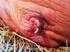
Image 4 Umbilical veins and arteries are observed.
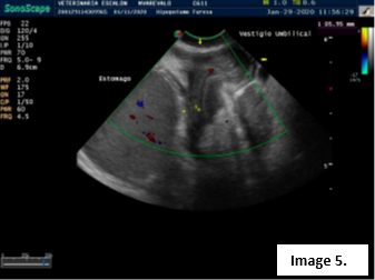
Image 5
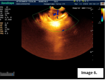
Image 6
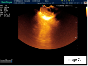
Image 7
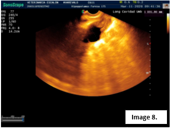
Image 8
On the second day of birth, the presence of a heart murmur was heard on cardiac auscultation, more frequently on the right side, which remained audible until 6th day of age. An echocardiography was performed on day 2 of age, the result of which determined that the murmur was suggestive of postvalvular aortic stenosis. Later, at one week of age, the murmur was no longer audible, possibly because the thickness of the skin no longer allowed it. The calf never showed any clinical signs of heart disease (Image 9).
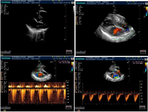
Image 9 Echocardiographic study. Studies conducted and interpreted by MVZ Silvia Arévalo. “Escalón Veterinary Clinic”, El Salvador.
Exercise
From the second day after birth, in the morning hours it was taken out for a walk for a period of 5 minutes, the calf always following its caregiver, a routine that was gradually increased and which, by the 10th day of birth, already had a duration of 15 to 20 minutes per day. By the age of one month, this routine was continued, but by then, he was already jogging and his physical strength was greater. On each walk he liked to touch his caregiver's heels with his mouth or head, considering that it was a way to feel safe and guided.
During the first three months of rearing, he had long periods of sleep during the day and slept even more at night. As he got older, he slept more in the morning and was more active in the afternoon and twilight.
Tooth development
The dentition was developing from the first day of birth, presenting mainly the 2 lower canines and much less the 2 lower incisors. At 6 months of age, the eruption of the lower incisors was completed and the eruption of the premolars began. At one year of age, the eruption of the upper canines began.
Medical problems
At one year and ten months of age, he presented a small skin lesion on the right side, close to the hip, which grew noticeably within a week. Said lesion was manifested especially by depigmentation and erythema and, later, vesicles with serosanguinous discharge, skin necrosis and ulceration. The lesion reached a diameter of approximately 15 cm. Based on the lesions observed, it was established that the condition was the product of dermatitis, possibly due to Streptococcus sp.8 It was treated locally with chlorhexidine-based cleaning, twice a day, with the addition of amoxicillin (Inovo Amoxi 50%®, Innovaciones Nutricionales, S.A de C.V El Salvador) at 20mg/kg, in the food, twice a day per-15 days). The case was resolved after 10 days of treatment (Image 10). Suspecting that this condition could be influenced by the temperature of the water in the pond in which he lived, the branches of the trees that surrounded it were cut in order to allow more sunlight to enter and increase the temperature of the water (which remained generally cold), considering an acceptable range of pond temperatura to be between 24 and 27°C.8
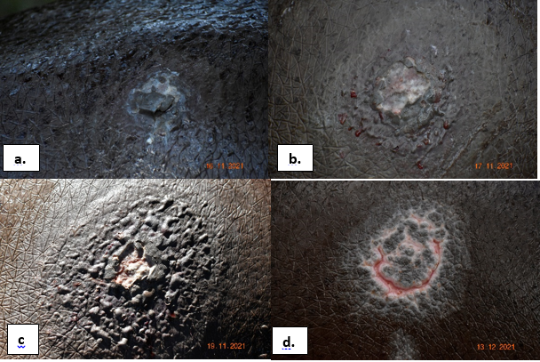
Image 10 Dermatitis (a. beginning, b. and c. intermediate, d. scarring.
A coproparasitological examination (centrifugation/flotation) was performed at two and a half years of age, and it was oserved the presence of: oocysts of Eimeria spp. The established treatment was based on toltrazuril at a rate of 20mg/kg in a single dose and repeated after 30 days. He also presented Strongyloides type eggs. The treatment this time was based on ivermectin in oral paste at a dose of 0.20mg/kg in a single dose, repeated after 30 days. One month after the second dose, another coproparasitological test was performed with a negative result for parasites.
Based on the achievement obtained with Kibo’s breeding, I consider it important to mention the following conclusions:
I thank God for giving me the opportunity to take care of Kibo, who taught me a lot with his love and joy, and despite the adversities, it was achieved! To the zookeepers at FURESA, who were always willing to take care of him and generate enthusiasm. To Luis Martínez for his support in care. To Eng. Elisa Rodríguez for her great support and to Dr. Silvia Arévalo for her contributions in the ultrasounds performed.
Author declare that there is no conflict of interest.

©2023 González. This is an open access article distributed under the terms of the, which permits unrestricted use, distribution, and build upon your work non-commercially.