International Journal of
eISSN: 2574-9862


Case Report Volume 2 Issue 2
1Nandankanan Zoological Park, India
2Department of Pathology, Orissa Veterinary College, India
Correspondence: Rajesh Kumar Mohapatra, Nandankanan, Bhubaneswar, India. PIN-754005, Bhubaneswar, India
Received: April 20, 2017 | Published: July 5, 2017
Citation: Sahu SK, Panda SK, Mohapatra RK. Gout in Egyptian vulture (neophron percnopterus): a case report. Int J Avian & Wildlife Biol. 2017;2(2):46-48. DOI: 10.15406/ijawb.2017.02.00014
A case of articular and visceral gout is reported in Egyptian vulture (Neophron percnopterus). The vulture was suffering from lameness and died after living more than 40 years in captivity. Urate crystals in the periarticular tissue and kidney were found. The presence of semi-fluid chalky-white granular substance in the swollen hock joints were confirmed by De Galantha’s stain as urate crystals.
Gout is a metabolic disorder characterized by retention of uric acid and urates in the body tissue. It usually occurs in two separate syndromes: visceral gout and articular gout, which may occur together or independently.1,2 Visceral gout is the accumulation of uric acid tophi on serosal surfaces of the pericardium, liver capsule, air sacs and within the kidney but may be found in any tissue.3 Articular gout is characterized by accumulation of urates in the synovial capsules and tendon sheath of the joints particularly in the foot and hock regions.4 Visceral gout was reported to be a common finding of vulture post-mortem examination in India.5–9 Reports on articular gout in vulture are limited. Egyptian vultures (Neophron percnopterus) are endangered birds belonging to the Family Accipitridae. The present study reports a case of articular and visceral gout in an Egyptian vulture.
The Egyptian vulture under study was received at Nandankanan Zoological Park, Odisha, India on 23.03.1972. At receipt the bird was already adult with typical white plumage and black flight feathers. It was housed in a naturalistic soil substrate enclosure with existing trees. The vulture was provided with rats/two day–old chicks every day and 300gm of buffalo meat six days a week (except Mondays).
On 22.05.2012, the vulture was observed with swelling on its right leg and exhibiting limping. It was able to take minced meat but could not grasp chick or rat, which was its normal feed earlier. It had preferred to sit on the ground but not perched on the trees may be due to its inability to bend the toes. Physical examination revealed painful swelling of both hock joins which bulged through the skin with an opening, discharging creamy and pasty contents. Along with this all articulation of the toes of both limbs were swollen. Based on the clinical signs it was diagnosed as articular gout. It was treated with antibiotics and anti inflammatory drugs along with local dressing of wound with antiseptics. Common salt was added to drinking water to increase water intake. Multi-vitamin and minerals added to the meat given was well accepted. But the health condition did not improve and the vulture died on 20.07.2012. Post mortem examination revealed the death was due to senility associated with articular and visceral gout. The swollen hock joints of the said bird (Figure 1-4) revealed the presence of semi-fluid chalky-white granular substance suggestive of urate crystals, which were confirmed by De Galantha’s stain showing blackish deposits.10

Figure 1 Articular gout in Egyptian vulture and the vulture under study with swelling on articulation of limb.
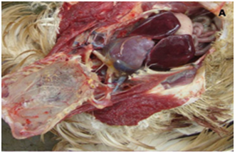
There was gross enlargement of Liver with haemorrhagic patches (Figure 5). The cortical surface of kidney revealed presence of gouty deposits (Figure 6). Cut surface also showed presence of Gouty deposits in parenchyma of kidney. Sections of kidney when stained with Haematoxylin and Eosin stain revealed degeneration and necrosis of tubular epithelial lining of kidney and interstitial congestion (Figure 7). Similarly, De Galantha’s stain of sections of kidney revealed degeneration and necrosis of epithelium of proximal convoluted tubules (Figure 8). Basement membranes also revealed presence of black deposits of urates (Figure 9). The presence of urates also found in tuft of glomeruli with increased Bowman’s space (Figure 10).

Figure 5 Affection of liver and histopathology of gouty kidney A. Liver enlargement with haemorrhagic patches.

Figure 8 Degeneration and necrosis of tubular epithelial lining of kidney and interstitial congestion (40X, Haematoxylin and Eosin stain).
The cause of avian gout is unknown, but vitamin A deficiency, pyelonephritis, renal neoplasia, high protein diet and incorrect amino acid balance are predisposing factors.2,8 In particular gout which is less common than the visceral form, urates are deposited around the joints, tendon sheaths, ligaments and periosteum.2 Similar cases of articular gout had been reported in chicken and turkey.11-13 Though, visceral gout had been reported in Himalayan griffon vulture, White backed vulture,5,8,9 no records of articular gout in vulture could be found from the available literature. Therefore, it seems that the present case is the first report of articular gout and visceral gout in Egyptian vulture.
There were reports describing death of vultures due to Diclofenac Sodium (Non Steroidal Anti-inflammatory Drug) associated with visceral gout. But in the present case the buffalo meat provided to the vultures were procured from buffalo slaughter house managed by the park, Here the buffaloes are screened for a considerable time period and allowed to slaughter. Therefore, there is limited of NSAID contamination of meat. So, it can be inferred that, the presence of gout may be due to senility as the said vulture lived up to 40 years, 3 months and 28 days in the park, which is the highest longevity report for this species.14
None.
The author declares no conflict of interest.

©2017 Sahu, et al. This is an open access article distributed under the terms of the, which permits unrestricted use, distribution, and build upon your work non-commercially.