International Journal of
eISSN: 2574-9862


Review Article Volume 3 Issue 3
Faculty of Agriculture and Natural Resources, Islamic Azad University, Iran
Correspondence: Mahrad Nassirkhani, Entomology Department, Faculty of Agriculture and Natural Resources, Islamic Azad University, Arak branch, Arak, Iran, Tel 9891 3141 6830
Received: May 20, 2018 | Published: June 11, 2018
Citation: Nassirkhani M. A redescription of pselaphochernes scorpioides (Hermann) (Pseudoscorpiones; Chernetidae). Int J Avian & Wildlife Biol. 2018;3(3):233-236. DOI: 10.15406/ijawb.2018.03.00092
This paper provides a redescription of widely distributed pseudoscorpion Pselaphochernes scorpioides (Hermann, 1804) based on the newly collected specimens from Tehran and Fars Provinces, Iran.
Keywords: arachnida, pseudoscorpions, faunistic, the middle east, Iran
eb, external basal; esb, external sub–basal; ib, internal basal; isb, internal sub–basal; ist, internal sub–terminal; est, external sub–terminal; it, internal terminal; et, external terminal; t, terminal; b, basal; sb, sub–basal; st, sub–terminal
The genus Pselaphochernes Beier, 1932 currently contains 17 species, of which only two species were found in Iran: Pselaphochernes anachoreta (Simon, 1878) reported from Fars Province-southern Iran by Beier1 and Pselaphochernes scorpioides (Hermann, 1804) recorded from Guilan Province-northern Iran by Mahnert.2 In this contribution, P. scorpioides distributing around Europe, northern Africa, and the Middle East and Central Asia3 is redescribed and illustrated based on the specimens collected from central north and southern Iran, as new provincial records for the species.
The materials used in this study were examined and illustrated with an Olympus BH-2 compound microscope and drawing tube attachment. The specimens are lodged in Collection of the Acarology Laboratory, Islamic Azad University of Arak, Iran. The morphological terminology and mensuration follow.4-7
Genus Pselaphochernes Beier, 1932
The members of the genus Pselaphochernes can be separated from the other genera of the family by the following combination of characters: female spermathecae relatively short and T–shaped, each branch terminated to a huge vesicle; posterolateral corner of coxa IV normal; cheliceral rallum with three blades; and tarsus IV with a long tactile seta situated medially.
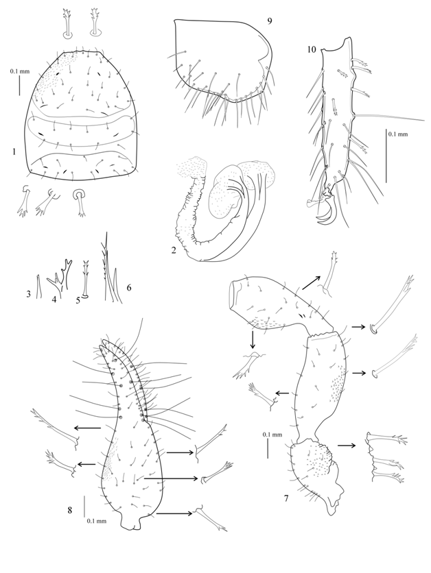
Pselaphochernes scorpioides (Hermann, 1804) (Figure 1–11), Pselaphochernes scorpioides (Hermann), Beier, 1932: 131.
Material examined
IRAN: Fars Province: 4♀, Ardakan [30°15′43″N, 51°58′59″E, altitude 2200m], Sepidan, leaf litter, July 13 2015, leg. M. Nassirkhani. Tehran Province: 1♂, 2♀, Lavizan [35°46′15″N, 51°30′46″E, altitude 1600m], Shemiran, decayed leaf litter, September 20 2013, leg. M. Nassirkhani (IAUA).
Redescription
Adults. Body length: 1.75mm (♀2.12–2.32mm)
Carapace: reddish brown; heavily granulate, not coarsely; L/W 1.12–1.21 (♀1.00–1.10); 2 transverse furrows present; with five pairs lyrifissures (Figure 1).
Tergites: reddish brown, intensively sclerotized, completely granulate; I–X with median suture line, XI not divided; half–tergites I–III without discal setae, IV with one lateral discal seta, V–X with one lateral and one central discal seta; tergites X without long tactile setae; XI with four slightly long dentate setae; all setae short and stout with terminal denticulations; chaetotaxy 10:10:10:12:12:12:12:12:11–12:10–12:9–10:2 (♀8–10:10:10:10–11:12–13:12–13:12:12:12–13:10–12:8:2).
Sternites: brown, lighter in colour than tergites; lightly sclerotized; less granulate than tergites; IV–X with median suture line, XI undivided; females with T–shaped spermathecae, each arm end to a huge vesicle (Figure 2); XI with four relatively long setae; chaetotaxy 22–26:(1)8(1):(2)6(2):18:18:19:16:17:14:8:2 (♀14:(1)10(1):(2)4(2):15:20:20:18:18:16:8:2).
Pleural membrane: longitudinally roughly striate.
Chelicera: light brown; galea short with indistinct rami in males (Figure 3), and in females long, with 6 rami (divided medially to two branches, each with 3 terminal rami) (Figure 4); fixed finger with 4 retrorse teeth; movable finger with galeal seta situated distally, with one point and large sub–apical tooth; hand weakly sclerotized, with 5 setae, b, sb and es dentate (Figure 5); rallum with 3 blades, distal blade longest and denticulate (Figure 6) serrula exterior and interior present; serrula exterior with 17–19 blades.
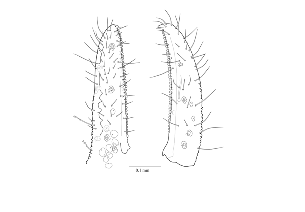
Pedipalps: reddish brown, darker in colour than carapace; intensively sclerotized; completely granulate, chelal fingers finely granulate; coxa with 19–21 setae, a few setae denticulate, with two distinct lyrifissures, manducatory lobe with 2 mariginal and 3 discal simple setae; trochanter swollen, with 2 dorsal rounded humps (Figure 7), L/W 1.81–1.87 (♀1.82–2.05); femur L/W 2.50–2.61 (♀2.75–2.83); patella with 2 lyrifissures situated basally (Figure 7), L/W 2.04–2.20 (♀2.27–2.40); chela (with pedicel) L/W 3.20–3.33 (♀3.13–3.29); chela (without pedicel) L/W 2.96–3.04 (♀2.93–2.96); hand (with pedicel) L/W 1.64–1.75 (♀1.73–1.81); movable finger slightly shorter than hand (with pedicel); hand (with pedicel) 1.02–1.05 (♀1.08–1.13) times longer than movable finger; fixed finger with 8 and movable finger with 4 trichobothria (Figures 8,11); fixed finger with 35–36 and movable finger with 36–38 cusped teeth (♀ 36–39 with and 36–41 with teeth); movable finger with 4–5 exterior accessory teeth, and fixed finger with 5–7 exterior accessory teeth (Figure 11); fixed finger with 7–9 sensory spots, mostly located basally (Figure 11); movable finger with two sensory spots located between trichobothria sb and st; venom apparatus present only in movable finger, nodus ramosus situated at same level as st.
Legs: brown; granulate; sub–terminal setae curved and simple; arolia simple and shorter than claws; claws smooth and symmetrical; coxal setae arranged: 12:12–14:16–18:24–28 (♀12–13:12–14:15–18:28–37); coxal setae simple and acute; coxa IV with normal shape (Figure 9); prolateral margin of all segment with simple setae, retrolateral margin with denticulate setae; leg I: tibia L/D 3.33 (♀3.43–4.00); tarsus L/D 4.40–4.60 (♀5.40–5.60); leg IV: tibia+patella L/D 3.90–4.18 (♀3.84–4.00); tibia without tactile seta, L/D 3.55–4.00 (♀3.90–4.62);tarsus with a long tactile seta situated medially (TS =0.5), retrolateral margin with one bulge situated proximal to tactile seta (Figure 10 ), L/D 4.33–4.66 (♀5.00–5.16).

Figure 1–10 Pselaphochernes scorpioides (Hermann, 1804), ♀: 1. carapace, dorsal view (setae magnified, granulation pattern shown partly); 2. spermatheca; 3. galea (♂); 4. galea, female; 5. dentate seta on cheliceral hand; 6. rallum; 7. basal segments of pedipalp, dorsal view (setae magnified, granulation pattern shown partly); 8. right chela, dorsal aspect; 9. left coxa I; 10. tarsus IV.

Figure 2 Pselaphochernes scorpioides(Hermann, 1804), ♀: left chelal fingers, lateral view (showing trichobothriotaxy, chelal teeth, and position of nodus ramosus).
Dimensions in mm
♂Carapace: 0.58/0.48. Pedipalp: trochanter 0.29/0.16; femur 0.47/0.18; patella 0.43/0.21; chela (with pedicel) 0.80/0.24; chela (without pedicel) 0.73; hand (with pedicel) L.0.42; movable finger L. 0.40. Leg I: femur 0.14–0.17/0.09–0.10; patella 0.20–0.25/0.08–0.09; tibia 0.20/0.06; tarsus 0.22–0.23/0.05. Leg IV: femur 0.16–0.17/0.10; patella 0.31–0.34/0.11; Femur+patella 0.43–0.46; tibia 0.32/0.08–0.09; tarsus 0.26–0.28/0.06. ♀Carapace: 0.60–0.65/0.60. Pedipalp: trochanter 0.31–0.35/0.17–0.19; femur 0.51–0.55/0.18–0.20; patella 0.48–0.50/0.20–0.22; chela (with pedicel) 0.87–0.94/0.27–0.30; chela (without pedicel) 0.80–0.88; hand (with pedicel) L.0.48–0.52; movable finger L. 0.45–0.47. Leg I: femur 0.17–0.18/0.10–0.11; patella 0.24–0.26/0.09; tibia 0.24–0.26/0.06–0.07; tarsus 0.27–0.28/0.05. Leg IV: femur 0.17–0.18/0.11; patella 0.34–0.36/0.12–0.13; Femur + patella 0.48–0.50; tibia 0.35–0.37/0.08–0.09; tarsus 0.30–0.31/0.06.
The pedipalps of Pselaphochernes scorpioides (Hermann, 1804) collected from central north and southern Iran and from Turkey [8] are slightly longer and thinner than the specimens collected from Europe e.g. the pedipalpal hand (without pedicel) size is 0.43/0.29 mm (♂) and 0.48/0.31 (♀) for the European specimens.9–10 The pedipalp of the female from Pakistan, the only other described specimen from the Middle East, is distinctly longer than that of the previously studied specimens, e.g. the pedipalpal femur size is 0.69/0.22 mm, chela (with pedicel) is 1.20/0.38mm, chelal hand (with pedicel) is L. 070 mm and the movable chelal finger L. is 0.61mm.11 In due attention to the morphometric characters, the type of the cheliceral setae [see 12: Figure. 17], the position of a tactile seta on tarsus IV [see 12: Figure. 16], and the tergal setae arrangement [see 12: Figure 16], the specimen from Pakistan is very close to Pselaphochernes macrochaetus Redikorzev, 1949 which was synonymized with P. scorpioides by Schawaller.13 Based on the type of the cheliceral setae (b and sb dentate in the female from Pakistan), numbers of accessory teeth on the chelal fingers (the fixed chelal finger of the Pakistani female bears five and the movable chelal finger only two exterior accessory teeth), the shape of spermathecae (see 11: Figure. 148), and the position of a tactile seta on tarsus IV (slightly distal to middle in the types and in the female from Pakistan [see 11: Figure. 149], the Pakistani female differs from P. scorpioides. Total of these differences raises the possibility of revalidation of P. macrochaetus, but still the types must be reexamined to clarify.
Pselaphochernes anachoreta (Simon, 1878) the only other species found in Iran can be recognized from P. scorpioides by the Pedipalpal size, e.g. femur is 0.69/0.30mm, chelal hand (without pedicel) is 0.64/0.37 mm, and movable finger is 0.62mm (♂) (♀0.62/0.25mm, 0.60/0.37mm, and 0.50mm).9,10 Pselaphochernes scorpioides is probably more widespread throughout the country than the present records indicate. It favors rich decaying organic material and is often found in synanthropic habitats such as compost, manure, dung and straw debris. This species has also been found in leaf litter, dead woods and the nests of the red–ant Formica rufa and is commonly phoretic on flies.14
The author is very grateful to Mr. Mahmoud Nassirkhani for his assistance.
The author declares that he has no competing interest and has not a financial relationship with the organization that sponsored the research.

©2018 Nassirkhani. This is an open access article distributed under the terms of the, which permits unrestricted use, distribution, and build upon your work non-commercially.