International Journal of
eISSN: 2574-9862


Research Article Volume 4 Issue 1
Division of Arachnology, Department of Zoology, Sacred Heart College, India
Correspondence: Aneesh V Mathew, Division of Arachnology, Department of Zoology, Sacred Heart College, Thevara, Cochin, Kerala, India
Received: December 18, 2018 | Published: January 10, 2019
Citation: Mathew AV, Joseph M. A redescription of Paratemnoides plebejus (with) (Pseudoscorpiones; atemnidae). Int J Avian & Wildlife Biol. 2019;4(1):1-3. DOI: 10.15406/ijawb.2019.04.00142
Widely distributed species Paratemnoides plebejus Carl With1 is redescribed in this paper by observing newly collected specimens from the Western Ghats and other reserve forest areas of Kerala, India.
Keywords: morphology, taxonomy, variation, Western ghats
eb, exterior basal; es, external setae; esb, exterior sub-basal; est, exterior sub-terminal; et, external terminal; gs, galeal setae; ib, interior basal; is, internal setae; isb, interior sub-basal; ist, interior sub-terminal; it, interior terminal; ls, lateral setae; sb, sub-basal; sbs, sub-basal setae; st, sub-terminal; t, terminal
The pseudoscorpiones genus Paratemnoides Harvey2 belonging to the subfamily Atemninae Kishida, 1929 of the family Atemnidae Kishida, 1929. The genus is cosmopolitan in distribution having 31 nominal species Harvey,3 including five representatives from India, which all were originally described under Chelifer Geoffroy, 1762 and Paratemnus Beier,4 Paratemnoides indicus (Sivaraman, 1980), Paratemnoides laosanus Beier3 Paratemnoides mahnerti (Sivaraman, 1981), Paratemnoides pallidus (Balzan, 1892) and Paratemnoides plebejus Carl With,1 Beier.2 Paratemnoides is characterised by the trichobothrial pattern of fixed chelal finger: the tactile hair it of the fixed finger in or proximal of the finger and always far farther from the fingertip than the distance between isb, and ist and st of movable finger closer to sb than to t Beier.5
The specimens used for the present study were collected from Western Ghats, one of the biodiversity hot spots of the world, and Kanjirapally in the Kerala region of southern India. The specimens were preserved in 70% ethanol and studied under Leica M205C stereomicroscope (at the highest possible magnification) and Labovision AXL compound microscope. Drawings were made by the aid of a drawing tube attached to the microscope. Scanning electron micrographs were taken using the Scanning Electron Microscope (JEOL Model JSM–6390 LV) available at Sophisticated Test & Instrumentation Centre facility of Cochin University of Science and Technology. The specimens were examined by preparing temporary slide mounts by immersing the specimen in 75% lactic acid at room temperature for several days, and mounting them on microscope slides with 18 mm cover slips. Permanent slide mounts were prepared by clearing the specimen in clove oil and by mounting using Canada balsam. All specimens were examined by using Leica S8AP0 stereomicroscope and Labovision AXL compound microscope. All measurements are in millimeters (mm) and were executed with the aid of Leica DFC 295 digital camera mounted on Leica M205C stereomicroscope (at the highest possible magnification) using the measurement module of the software package Leica Application Suite (LAS), version 4.3.0. Microphotographs were taken using Leica DMC 2900 digital camera mounted on Leica M205A stereomicroscope with the software package LAS, version 4.5.0. using LAS montage facility.
INDIA: 4 m#, 3 f# (ADSH PS0004) Kerala, Kottayam, Pazhayidom [9º 29’45”N 76º 47’22”E, 60m alt], 27 December 2017, leg. M.V. Aneesh, under the bark of Heavea brasiliensis, by hand. 3 m#, 2 f# Kerala, Kottayam, Pazhayidom [9º 29’45”N 76º 47’22”E, 60m alt], 27 December 2017, leg. Merin Tom, under the bark of Artocarpus heterophyllus, by hand. 6 m#, 4 f# Kerala, Kottayam, Paippad [9º 25’25”N 76º 35’31”E, 30m alt], 16 October 2017, leg. M.V. Aneesh, Kerala, Ponmudi [8º 44’22”N 77º 7’10”E, 380m alt], 21 February 2018, leg. M.V. Aneesh. 2 m#, 1 f# Kerala, Pathanamthitta, Punnackadu [9º 19’12”N 76º 42’36”E, 40m alt], 18 October 2018, leg. M.V. Aneesh, under the bark of Artocarpus heterophyllus, by hand, 7 m#, 3 f# Kerala, Trivandrum, Karyavattom Campus [8º 33’36”N 76º 52’48”E, 20m alt], 23 March 2018, leg. M.V. Aneesh, under the bark of Artocarpus heterophyllus, by hand.
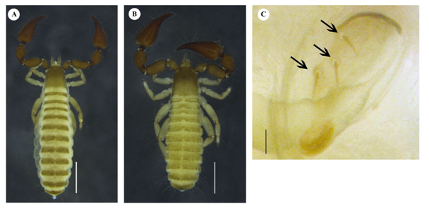
Adults (Figure 1A–1C). Chitinized regions reddish brown, rest of pale–brown.

Figure 1 Paratemnoides plebejus. male (A) and female (B) paratypes: A–B Dorsal; C Spiracle IV, ventral. Scale bars: A–D, 0.5mm. Arrows 1, 2 & 3 indicate spiracle setae.
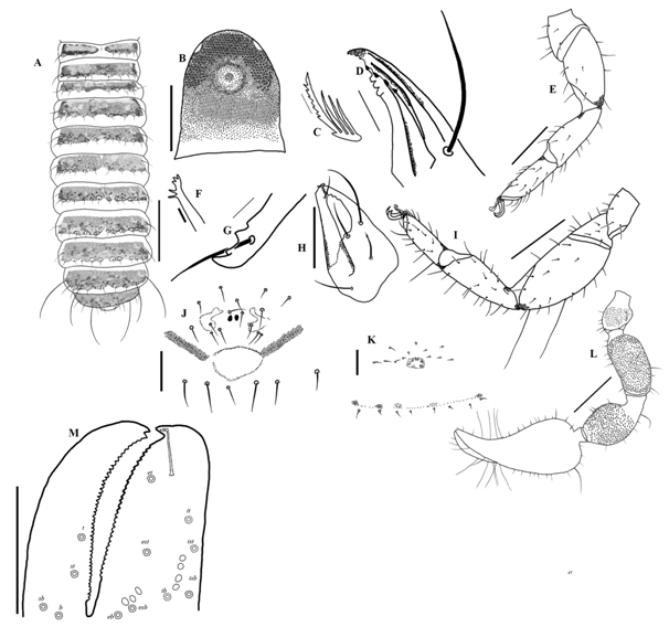
Chelicera (Figure 2): palm with rasp-like ornamentation, 4 setae; ls and is longer than fixed finger, bs dentate (Figure 3). Finger with three triangular teeth (Figure 2G). Lamina interior with four apically dentate lobes. Fixed finger with three apical lobes followed by four triangular serrations (Figure 2D). Serrula exterior with 23 blades in both males and females; rallum with 4 blades, distal blade with seven serrations (Figure 3C). Galea long, 3 terminal and 2 sub terminal rami in males (Figure 2F), 2 terminal and 3 sub terminal rami in females.6-9

Figure 2 Paratemnoides plebejus. A–D Male paratype. A Habitus, dorsal; B Areolum Bothridium. C Vestitural setae. D Right chela, dorsal.

Figure 3 Paratemnoides plebejus. Female paratype (A, K) male paratype (B–L) and nymphs (M–O). A Tergites, dorsal; B Carapace, dorsal; C Flagellum; D Lamina interior; E Left leg I, dorsal; F Galea; G Fixed finger of left chelicera, dorsal; H Left chelicera, dorsal; I Left leg IV, dorsal; J Genital area, ventral; K Genital area, ventral; L Left chela, retrolateral; M Left pedipalp; Scale bars: A–D, F–K, 0.5mm E, L–P, 0.2mm.
Pedipalps (Figure 3L–M): Trochanter and femur pale reddish-brown; patella and chela dark reddish; trochanter, femur, patella and chela finely granulated. Dorsal tubercle of trochanter well developed, with 3 setae at apex; trochanter 1.33 (m#), 1.63 (f#) x longer than broad. Femur 2.11 (m#), 2.0 (f#) x longer than broad; 3 pseudotactile setae. Patella 1.81 (m#), 1.90 (f#) x longer than broad, with stout pedicel; 2 pseudotactile setae at base. Chela 2.62 (m#), 2.43 (f#) x longer than broad; fixed finger with 32 (m#f#) teeth, movable finger with 46(m#), 49 (f#) teeth. Nodus ramosus not extending to et (Figure 3L). Trichobothrial pattern (Figure 3L): t distal to middle of movable finger; one pseudotactile seta present laterally near tip of finger; one pseudotactile seta laterally between t and st; st closer to sb than to t; sb closer to b than to st; et near to the tip of fixed finger, est distal to the middle, et, est, esb, eb near to the inner margin; it farther from the fingertip, it, ist, isb, ib far away from inner margin; three sense spots present above esb, four sense spots between ist and ib. Palm with short dentate setae, 5 tactile setae near base.
Carapace: 1.01–1.25 (m#), 1.05–1.40(f#) x longer than broad; more strongly sclerotized in anterior region and thus darker, smooth and glossy, with two distinct raised eyespots, without transverse furrow with ca. 59 (m#), 61 (f#) setae, including 4 (m# & f#) near anterior margin, 9 (m#), 10(f#) near posterior margin. Vestitural setae dentate (Figure 1C).
Coxal region: Chaetotaxy m#, 9:6:8:16, f#, 9:8:9:16;
Legs: golden yellow, with acuminate setae, articulation between femur and patella oblique. Leg I (Figure 2E): femur 1.06, patella 2, tibia 2.56, tarsus 2.83 x longer than broad. Leg IV: femur + patella 2.32, tibia 2.75, tarsus 2.84 x longer than broad. Tactile seta situated at base of tarsus IV. Femur IV with 2 tactile setae, 1 at apex, other more proximal (Figure 2I).
Opisthosoma: Width constant from tergite 1 to 11. Tergites 2–5 undivided; tergites 1 and 6–11 partially divided. Sternites with faint medial suture, with small dentate and moderately long, simple sternal setae. Anterior spiracle with 2 (m#), 3 (f#) setae, posterior spiracle with 1 (m# & f#) seta. Tergal chaetotaxy: m#, 9:10:10:13:16:14:16:15:14:14 (including 4 tactile setae):13 (including 2 tactile setae): 2, f#, 12:12:11:13:16:15:17:15:15:17 (including 4 tactile setae):14 (including 2 tactile setae):2. Sternal chaetotaxy: m#, 13:11:13:19:17:21:14:16:16 (including 4 tactile setae):15 (including 4 tactile setae):2, f#, 28:12:17:17:17:17:17:16:14 (including 4 tactile setae):14 (including 4 tactile setae):2.
Measurements males: Holotype followed by other males in parentheses (where applicable): body length 3.3(3.7–4.2). Carapace (0.823–0.937/0.729–0.809).
Pedipalps: Trochanter 0.255/0.254 (0.379–0.391/0.284–0.295), trochanter pedicel 0.088/0.160 (0.084–0.089/0.144–0.154), femur 0.632/0.328 (0.580–0.687/0.325), femur pedicel 0.067/0.137 (0.065–0.093/0.140–0.150), patella 0.566/0.400 (0.674–0.697/0.371–0.378), patella pedicel 0.114/0.174 (0.099–0.110/0.147–0.152), chela (with pedicel) 1.249/0.544 (1.296–1.319/0.503–0.520), chela (without pedicel) 1.179/0.544 (1.240–1.274/0.503–0.520), hand 0.716 (0.686–0.769), movable finger 0.532 (0.545–0.577). Leg I: femur 0.180/0.169 (0.213–0.216/0.172–0.203), patella 0.396–0.198 (0.370–0.444/0.193–0.202), tibia 0.346/ 0.135 (0.372–0.380/0.145–0.149), tarsus 0.292/0.103 (0.304–0.311/0.110). Leg IV: femur + patella 0.738/0.317 (0.674–0.823/0.285–0.300), tibia 0.549/0.199 (0.518–0.530/0.190–0.198), tarsus 0.338/0.119 (0.332–0.348/0.112–0.124).
Female: Paratype: body length 4.5 (4.0–5.7). Carapace 0.966/0.686 (0.817–1.062/0.774–1.003).
Pedipalps: Trochanter 0.241/0.254 (0.231–0.346/0.245–0.371), trochanter pedicel 0.066/0.138 (0.062–0.065/0.140.145), femur 0.645/0.321 (0.533–0.808/0.312–0.441), femur pedicel 0.077/0.139 (0.065–0.080/0.135–0.194), patella 0.707/0.371 (0.682–0.799/0.378–0.488), patella pedicel 0.121/0.155 (0.106–0.150/0.134–0.223), chela (with pedicel) 1.284/0.528 (1.331–1.663/0.524–0.686), chela (without pedicel) 1.203 (1.192–1.530), hand 0.778 (0.746–0.967), movable finger 0.535 (0.522–0.798). Leg I: trochanter 0.157/0.140 (0.151/0.133), femur 0.206/0.160 (0.190–0.319/0.154–0.276), patella 0.407/0.193 (0.350–0.475/0.188–0.251), tibia 0.369/0.142 (0.379–0.489/0.138–0.178), tarsus 0.314/0.101 (0.316–0.406/0.099–0.158). Leg IV: trochanter 0.284/0.174 (0.202–0.286/0.156–0.163), femur + patella 0.780/0.315 (0.744–1.124/0.332–0.403), tibia 0.603/0.196 (0.565–0.770/0.158–0.195), tarsus 0.366/0.134 (0.318–0.445/0.128–0.158).
Morphological characters show intraspecific variation. The arrangement and chaetotaxy shows variation. But the number of tactile setae remains the same. The setae in the posterior region of the carapace shows much variation from 9 - 11 in males. The carapace is highly sclerotised in the anterior region, thus the colour lightens to the posterior from middle. Uncsclerotised circular region is present around every setae in the tergite. There are unsclerotised small patches in every tergite. The longitudinal division of tergites shows variation. Arrangement of setae in each segment shows variation.
Our gratitude to Rev. Fr. Prasanth Palackappillil (Principal, Sacred Heart College, Thevara, Cochin) for providing all the facilities for completing this work. Thanks to Mr. Pradeep M. Sankaran (Division of Arachnology, Sacred Heart College, Thevara, Cochin) for assistance. We extend our heartfelt thanks to Dr Mark S. Harvey (Western Australian Museum, Perth, Australia) for providing relevant literature.
Author declares that there is no conflict of interest.

©2019 Mathew, et al. This is an open access article distributed under the terms of the, which permits unrestricted use, distribution, and build upon your work non-commercially.