eISSN: 2469-2778


Case Report Volume 11 Issue 4
1Master in Cytopathology, first and second degree specialist in pathological anatomy, second degree specialist in oncology, university of medical sciences of Camagüey. Manuel Ascunce Domenech Provincial Teaching Hospital, Camagüey, Cuba
2Master in Clinical Nutrition, first and second degree specialist in internal medicine, second degree specialist in intensive care and emergency medicine, university of medical sciences of Camagüey. Manuel Ascunce Domenech Provincial Teaching Hospital, Camagüey, Cuba
Correspondence: Pedro León Acosta, Master in Clinical Nutrition, first and second degree specialist in internal medicine, second degree specialist in intensive care and emergency medicine, university of medical sciences of Camagüey. Manuel Ascunce Domenech Provincial Teaching Hospital, Camagüey, Cuba
Received: November 01, 2023 | Published: November 16, 2023
Citation: Torres PR, Acosta PL. Lymphomatoid granulomatosis with infrequent manifestations: case report. Hematol Transfus Int. 2023;11(4):105-108. DOI: 10.15406/htij.2023.11.00315
Introduction: lymphomatoid granulomatosis is a very rare disease with few reports in the medical literature.
Objective: to present the case of a lymphomatoid granulomatosis that constitutes a variety of primary lymphoproliferative neoplasms of the lung and constitutes the first case reported in our hospital.
Case presentation: 44-year-old male, veterinarian with a history of health without toxic habits that began with respiratory symptoms, dermatological lesions, constitutional syndrome and flaccid paraplegia. After performing the essential complementary tests which showed pancytopenia; an image suggestive of pulmonary metastasis was found in the chest X-ray, which was corroborated in the pulmonary tomography; also performing magnetic resonance imaging that showed lesion compression at the dorsal level. Pathology studies of the skin and lungs revealed lymphomatoid granulomatosis with spinal cord invasion. The patient received chemotherapy treatment and died abruptly a month after it began.
Conclusions: lymphomatoid granulomatosis is a rare disease currently considered as a lymphoid neoplasm associated with Epstein-Barr virus and large B-cell lymphoma. We present the case of a patient with said pathology, accompanied by neurological injury and whose diagnosis was only possible by pathological studies; making it impossible to carry out a necropsy due to family refusal.
Keywords: lymphomatoid, granulomatosis, diagnosis, treatment
Lymphomatoid granulomatosis (LG) was first described by Liebow et al.1 in 1972. Initially it was considered a disease caused by a proliferation of T-cell lymphoid cells; today it is considered a rare type of lymphoma. Large B associated with Epstein-Barr virus (EBV) infection with almost exclusive extranodal involvement and characterized by an angiocentric and angiodestructive growth pattern.2 It is a rare disease, unknown not only by a large number of clinicians, but also by other medical specialties that are related to this entity; whose treatment and prognosis are not defined. Its diagnosis is difficult and complex due to its similarity to vasculitic processes, which is why clinical suspicion is important.3 Cases have been separated into three histological grades based on the relative amount of tumor cells and reactive cells; a grade is assigned to the disease between 1 and 3. Grade 1 is characterized by a net predominance of small lymphocytes without cytological atypia, occasional EBV-transformed lymphoid cells (less than 5 EBV-infected cells in a high-power field) and the absence of necrosis; grade 2 is characterized by a variable number of lymphoid cells transformed by EBV (between 5 and 20 infected cells) and grade 3 presents a large number of lymphoid cells transformed by EBV (more than 50 infected cells accompanied by few lymphocytes of small size and extensive areas of necrosis),4,5 this patient showed grade 2. The case of a patient affected by this disease is presented, which represents the first case reported in our hospital in four decades.
Male patient, white, 44 years old, veterinary doctor by profession, with health history, without family or work history of interest. No toxic habits. It begins with a fever of 38 ˚C and 39˚C in the afternoon and respiratory distress not related to effort, associated with a predominantly nocturnal dry cough; all of these symptoms were accompanied by constitutional impairment expressed by asthenia; anorexia and mucosal paleness. He went to the doctor's office where he was medicated with bronchodilators and steroids, showing slight improvement. Ten days later he presented non-pruritic skin lesions on the trunk and right arm. He went to the emergency service of the Manuel Ascunce Domenech Provincial Hospital in the City of Camagüey where his admission was decided.
Compliance with the ethical component of the clinical research
The institution's research ethics committee accepted the publication of the case report, after approval by the family members by signing the informed consent to disclose the studies carried out on the patient, which included permission to publish the photos. Compliance was maintained in removing identifying information from all patient-related data.
Patient perspective
Despite the patient's fatal outcome, close communication was maintained with the patient and his family; constantly informing those of the results of the clinical and complementary evaluations, leaving them satisfied with our work.
Clinical findings
The physical examination revealed pallor of the mucous membrane and skin lesions in the form of red-purple, painful papules measuring between 5 mm and 1 cm approximately confluent, located mainly on the trunk and right upper limb. Fill capillary 4s. Respiratory system: Fr 27 rpm Vesicular murmur decreased. Crackling rales scattered in both lung fields. Not voiceless pectoriloquy. Cardiovascular System: Central HR: 103 bpm BP: 100/70 mmHg. Rhythmic heartbeat. Don't blow. Digestive System: Mouth without alterations. Globules, depressible, non-painful abdomen, no visceromegaly. No abdominal murmurs. Normal RHA Hemolymphopoietic System: no splenomegaly, no lymphadenopathy, non-painful bone percussion. Rumpel Leede test negative. Genitourinary System: No nephromegaly. Without modifications.
Nervous System: Without alterations.
Diagnostic evaluation
Multiple complementary exams are performed Hb 7.5 g/L; leuco 2.5×10⁹ /L; platelets 35×10 9/L. Cultures of blood, urine, feces and serological tests for multiple bacterial and viral microorganisms were all negative. Immunological studies such as: antinuclear antibodies, ANCA, anti-phospholipids and protein electrophoresis were all normal. Medullogram, medullary biopsy and medullary culture ruled out lymphoproliferative, neoplastic or infectious processes; hypercellularity was found without maturation alterations of the different hematopoietic series. Fundus without alterations. Electrocardiogram: sinus tachycardia. No ST-T alterations. Abdominal ultrasound: normal.
Therapeutic intervention
Given the diagnosis of systemic GL, treatment with the CHOP regimen (cyclophosphamide, adriamycin, vincristine and methylprednisolone) is imposed, adding allopurinol, omeprazole in cycles and transfusion of 2 units of red blood cells, significantly improving all the patient's symptoms and signs except paraplegia.
Approximately thirty days later, he began to experience dyspnea, tachycardia, progressive worsening of his clinical condition, and he died. It was not possible to perform an autopsy due to family refusal. A chest x-ray was performed (Figure 1), revealing bilateral radiopaque images with a metastatic appearance, which was confirmed by computed axial tomography (CAT) (Figure 2). A biopsy of the lesions was performed, which resulted in angiocentric lesions of the superficial and deep plexus of the dermis, accompanied by necrosis and granulomatous reaction (Figure 3A & 3B).

Figure 1 PA chest radiology. Bilateral radiopaque images located in hilar.
(a) Peripheral
(b) Basal
(c) Regions of both lungs

Figure 2 Lung CT. Presence of isodense images in posterior segments that show multiple metastases, some with a tendency to nodulation.

Figure 3A Macrophotography. Complex exophytic maculopapulonecrotic lesion located on the left flank.

Figure 3B Microphotography. Histological section of skin. Angiocentric lesions of the superficial and deep plexus of the dermis, accompanied by necrosis and granulomatous reaction. H/E-20x.
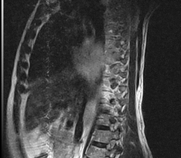
The day after the skin biopsy was performed, he began to experience pain in the thoracolumbar spine that was not relieved with analgesics, requiring the use of opiates, and with a neurological physical examination compatible with flaccid paraparesis; Therefore, magnetic resonance imaging (MRI) was performed (Figure 4) which revealed: diffuse tumor infiltration with extension to the spinal cord and lytic lesions of D4 and D5 vertebral bodies due to possible metastatic infiltration of a lung neoplasm. FNAC of the lung was performed (Figure 5), obtaining pulmonary infiltrate with the presence of atypical Stemberoides cells accompanied by abundant inflammatory cells, histiosites and plasma cells, in addition, small granulomas and lymphophagocytosis were observed. Subsequently, immunohistochemistry confirmed the diagnosis of the disease (Figure 6).

Figure 4 MRI of the dorsal column. Observe diffuse tumor infiltration with extension to the spinal cord and lytic lesions of D4 and D5 vertebral bodies due to possible metastatic infiltration of a lung neoplasm.
GL is currently considered a rare lymphoproliferative process in which the tumor cell is a B lymphocyte infected and transformed by EBV.5 These tumor cells are accompanied by a variable number of plasma cells, lymphoid cells with immunoblastic morphology, small and reactive T lymphocytes that are usually CD4+ and histiocytes.6 The involvement is extranodal and presents angiocentric and angiodestructive growth, causing vascular damage by different mechanisms; Therefore, the symptoms of the disease are those derived from vascular injury in the different affected organs.7 It is a rare entity and whose knowledge is still scarce, its incidence being highest in the fifth decade of life and in men; although it is reported that there are no differences between both sexes.2,3 It is rare in children and adolescents.5 The main organ affected is the lung in 50% of cases, also affecting the skin, gastrointestinal tract, liver, kidneys, heart and central nervous system (CNS),6–8 as seen in this patient. GL usually presents in relation to immunosuppression, with documented cases associated with organ transplants, Wiskott-Aldrich syndrome, human immunodeficiency virus (HIV) and X-linked proliferative syndrome.3,4 Although GL is a lymphoproliferative process, it is striking that it usually shows respect for secondary lymphoid organs such as the spleen and lymph nodes.6,9 Very rarely, the disease can affect the bone marrow, causing hematological cytopenias, as reported in the medical literature4,6,9 and observed in this case. Cases of extramedullary hematopoiesis associated with GL have been described without detecting evidence of marrow involvement.9
This patient did not present splenomegaly or lymphadenopathy. GL is an entity histologically similar to a lymphoma, but due to the characteristic pleomorphic lymphoid infiltrate, the inflammatory component and the absence of involved lymph nodes, it was not designated as a lymphoma despite its poor prognosis and systemic progression in some cases.10 Several studies of pulmonary large cell lymphomas have identified cases indistinguishable from GL. Histologically, it is characterized by a pleomorphic cellular infiltrate composed of atypical large lymphoid cells mixed with small lymphocytes, plasmocytes, epitheloid histiocytes, and other inflammatory cells. Small non-caseating granulomas can be seen, with vascular invasion being the hallmark of GL that shows transmural infiltration of large and medium arteries and veins that can produce necrosis of the vessel wall. Occlusion of the lumen with extensive ischemic necrosis can produce infarcted areas with necrotic tumor cells. No caseous or fibrinoid necrosis is observed in GL.1,3,5,7 GL immunophenotype studies show that the majority of cells in the infiltrate are B lymphocytes and show a positive reaction for CD 79, CD 30 and CD 43 in some cases; CD 15 negative and reaction for EBER ‒1 and PML positive.10
The majority of cases correspond to B lymphoproliferative lesions rich in T cells (1‒2). Atypical large cells stain with B markers such as CD 20 and CD 79 and coexpress PML (EBV).5,7 Recent studies have shown that the majority of GL cases contain EBV DNA.5,6 The differential diagnosis is with vasculitis such as: Wagener granulomatosis and Churg-Strauss disease given the involvement of the vessels. Other important differential diagnoses are with primary pulmonary nodular lymphoreticular hyperplasia, classic variant Hodgkin lymphoma, extranodal nasal type T/NK lymphoma, peripheral T lymphoma, tumors such as lymphoepithelioma type lung carcinoma,11 as well as Langerhans cell histiocytosis. The treatment of GL is varied and describes bone marrow transplantation, the use of interferon alfa-2a, and treatment with cyclophosphamide and steroids. Other authors prefer to use the CHOP system12,13 which was used by us. Due to the progress in recent years in the field of monoclonal antibodies and the known effect of rituximab in hematological malignancies such as B-cell lymphoma, the prognosis of these patients has been considerably improved. Rituximab has also been used with the CHOP regimen, but with not very encouraging results.13 Due to the progress in recent years in the field of monoclonal antibodies and the known effect of rituximab in hematological malignancies such as B-cell lymphoma, the prognosis of these patients has been considerably improved. Rituximab has also been used with the CHOP regimen, but with not very encouraging results.13 Due to the progress in recent years in the field of monoclonal antibodies and the known effect of rituximab in hematological malignancies such as B-cell lymphoma, the prognosis of these patients has been considerably improved. Rituximab has also been used with the CHOP regimen, but with not very encouraging results.13
Lymphomatoid granulomatosis is a rare entity unknown to many clinicians and specialists in other branches of internal medicine. It is found in both sexes, being more common in the fifth decade of life and very rare in children and adolescents. The lung is the main organ affected, although it can damage other organs causing various clinical manifestations. In this patient, skin, bone marrow, and dorsal column involvement occurred, along with fever, which is not common in these cases. The diagnosis is made by clinical manifestations, histopathology and immunohistochemistry. The disease must be studied in an interdisciplinary manner.
None.
The authors declare that there is no conflicts of interest.
None.

©2023 Torres, et al. This is an open access article distributed under the terms of the, which permits unrestricted use, distribution, and build upon your work non-commercially.