eISSN: 2469-2778


Research Article Volume 9 Issue 3
1Department of Anatomy and Physiology, Kansas State University, Kansas
2Cellphire, 9430 Key West Ave, Rockville, MD 20850, Kansas
3Department of Anatomy and Physiology , Midwest Institute of Comparative Stem Cell Biology, Kansas State University, Kansas
Correspondence: Mark L Weiss, Department of Anatomy and Physiology, Kansas State University, 1600 Denison Ave, Coles 17 Hall room 105, Manhattan, KS 66506; Kansas
Received: May 10, 2021 | Published: May 20, 2021
Citation: Wright A, Zheng YY, Lee A, et al. Doxorubicin delivery via novel lyophilized/reconstituted platelet-product has anti-cancer activity. Hematol Transfus Int J. 2021;9(3):41-51. DOI: 10.15406/htij.2021.09.00251
Background: Cancer is a global health concern with millions of people diagnosed annually. Chemotherapy remains the primary treatment for many cancers despite its toxic side effects. Currently, there is focus on developing nanoparticle systems to enhance the specificity and efficiency of chemotherapeutics. Biological drug delivery methods, e.g., platelets, offer advantages compared to traditional, synthetic nanoparticles and may enhance therapeutic potency by avoiding immune interactions. Platelets are a potential source of biological nanoparticles for drug delivery but, their short shelf-life limits their therapeutic potential. Lyophilization has been used to increase the shelf-life and stability of platelets.
Methods: Here, we evaluate a modified version of Cellphire’s proprietary freeze-dried platelet product loaded with the cytotoxic drug, doxorubicin, for delivery to hemangiosarcoma cells.
Results: We demonstrate that 1-10% volume/volume (v/v) addition of doxorubicin-loaded Lyophilized Human Platelets (DOX-LHP) to hemangiosarcoma cultures have potent anti-cancer activity by inhibiting proliferation, metabolism, and promoting apoptosis. Further, hemangiosarcoma cells exposed to 5% and 10% v/v DOX-LHP contained 2.2x and 4x more intracellular doxorubicin compared to cells treated with media containing 5 µM free doxorubicin.
Conclusions: These results suggest that lyophilized, DOX-LHP overcome the storage limitations of platelets and once reconstituted they function as effective drug delivery vehicles for cytotoxic compounds.
Keywords: drug delivery, platelets, cancer cell targeting, biological nanoparticles, lyophilization
DOX, doxorubicin; LHP, lyophilized human platelets; DOX-LHP, doxorubicin-loaded lyophilized human platelets; TGA, thrombin generation assay; T-TAS, total thrombus-formation analysis system; CAT, calibrated automated thrombogram; DHSA-1426, canine hemangiosarcoma cell line; HSA media, standard hemangiosarcoma cell culture media; AOPI, acridine orange/propidium iodide; MF, mean fluorescence intensity; ANOVA, analysis of variance; EMF, enhanced metafiles; WST-1, water soluble tetrazolium salts; LCP, lyophilized canine platelets; DOX-LCP, doxorubicin-loaded lyophilized canine platelets
Cancer is the leading cause of death before the age of the 70 in the United States and it is expected to become the leading cause of death in all countries by the end of the 21st century.1 For many cancers, the use of cytotoxic drugs remains the leading therapy.2 Doxorubicin (DOX), an anthracycline type of cytotoxic drug, inhibits cancer cell proliferation by targeting topo-isomerase IIα. DOX is typically administered intravenously. Once administered, the chemotherapeutic is systemically distributed where it can damage cancers and healthy cells and tissues. In addition, many cancers develop multidrug resistance to chemotherapeutics during treatment rendering the drugs ineffective.3–5 There has been interest in the development of nanoparticles to deliver chemotherapeutic drugs to improve therapeutic stability and efficacy.2,3,6,7
Traditional nanoparticle systems used in drug delivery include liposomes, polymeric nanoparticles, polymer-drug nano-conjugates, dendrimers, and inorganic nanoparticles.3 Interactions of synthetic nanoparticles with the immune system can cause antibody production against the nanoparticle or the cargo.8 For this reason, biologicals, e.g., red blood cells, platelets, and nanoghosts, have been explored as drug delivery systems.9–12 Platelets are an ideal choice for chemotherapy drug delivery because they are rapidly replenished by the body, providing ample supply for extraction and loading, and express receptors that interact with tumor cells, thus improving specificity for cancer.11,13 Platelets are not without their own challenges since they have specific handling requirements and a limited shelf-life of 5-7 days.14,15 Efforts to extend the shelf-life of platelets are critical to minimize loss of units.16–20 On method to extend platelets’ shelf-life is lyophilization, and Cellphire (https://www.cellphire.com/) has developed proprietary methods for producing freeze-dried platelets for application as a hemostatic agent.21,22 Cellphire expanded their technology platform by developing techniques to load therapeutic cargo into platelets. For example, for testing in this short report, human platelets were loaded with DOX prior to lyophilization (called DOX-lyophilized human platelets, DOX-LHP). Here, we characterize DOX-LHP, and evaluated its stability and dose-response relationship on canine hemangiosarcoma cells in vitro.
Preparation of DOX-LHP
Three apheresis units (Blood Center: Atlanta Blood Services Cobb Location) were pooled, acidified with ACD to pH of 6.6 -6.8, and counted via AC·T Diff Coulter (per manufacturer’s instructions) prior to centrifugation at 1470 x g for 20 minutes at 21°C (Beckman Coulter). Following centrifugation, platelet poor plasma was removed then the platelet pellet was resuspended at 2500 x 103 cells/µL in Cellphire proprietary loading buffer® with 2 µM of each platelet aggregation inhibitors PGE1 and GR144053 (Tocris, Catalog No. 1620 and 1263, respectively). Platelets were incubated at 37°C for 10 minutes to allow for platelet activation inhibition. At the end of this incubation, an equal volume of Cellphire proprietary loading buffer® with or without DOX (Sigma Aldrich, Catalog No. D1515 – 10MG) was added for loading or negative control, respectively. These sublots were incubated at 37°C on a rocker for 3 hours in the dark to allow for drug loading. Antiplatelets (1 µM each) were re-supplied every hour. Loaded or unloaded platelets were each diluted 12 – fold with Cellphire proprietary loading buffer® then centrifuged at 1470 x g for 20 minutes at 21°C (Beckman Coulter). Free DOX was removed with the supernatant, and the remaining loaded or unloaded platelets were resuspended at 2000 x 103 cells/µL in Cellphire proprietary loading buffer® with 6% polysucrose. 2 mL of loaded or unloaded suspensions were aliquoted into 5 mL amber vials for lyophilization according to Cellphire’s proprietary method to generate DOX-LHP and unloaded LHP, respectively. After lyophilization, the vials were baked at 80°C for 24 hours to anneal.10
Quantification of DOX Load
DOX-loaded platelets pre- and post-lyophilization were assessed for drug retention and platelet functionality. Drug retention was evaluated by flow cytometry to quantify percentage of DOX-loade platelets and a fluorescent plate reader was used to quantify DOX load in fg/particle. For flow cytometry, the following conjugated antibodies or protein were used: anti – CD41 – Alexa Fluor 700 (Biolegend, Catalog No. 303728), anti – CD62P – APC – Cy7 (Biolegend, Catalog No. 304944), Annexin V – Pacific Blue (BD Biosciences, Catalog No. 9169526). Isotype control for anti – CD62P is mouse anti-human IgGk (Biolegend, Catalog No. 400128). Sizing beads, 0.5 µm (Beckman Coulter, Catalog No. 6602336) and 2.5 µm (Bangs Laboratories, Catalog No. 833), were used to define platelet sized particles 0.5 – 2.5 µm. Platelets were stained with primary antibody or isotype control for 20 minutes at room temperature before washing with HBS buffer (150 mM NaCl, 10 mM HEPES, pH 7.4 with 1 M NaOH). Thirty thousand events per sample was acquired by a NovoCyte ACEA Biosciences cytometer and analyzed by NovoExpress software (Agilent Technologies, Santa Clara, CA). DOX is a fluorescent compound, detectable by flow through the PE–Cy5 channel. Flow evaluation of DOX-load examined % of CD41+ DOX+ platelets in loaded sample compared to unloaded control. To quantify DOX loading, 1 mL aliquot of loaded or unloaded platelets were washed three times with HMT buffer (1X HEPES – Tyrode’s Buffer Salts, 5 mM Dextrose, pH 6.6 – 6.8 with 1 M HCl) via centrifugation at 845 x g for 10 minutes at room temperature followed by resuspension. The pellet was resuspended in 1 mL HMT buffer and the particle count determined using the AC·T Diff Hematology Analyzer (per manufacturer’s instructions), then sonicated three times at 26kHz for 30 seconds with 2 – 5 minutes interval of rest at room temperature to release intracellular DOX into the aqueous solution. These samples were then centrifuged (Eppendorf Centrifuge 5424, 18,000 x g, 20 minutes, room temperature) to remove membrane debris from suspension. Quantification of DOX was achieved by comparison of the relative fluorescent emission of the sample with a standard curve after 500 nm excitation and 600 nm emission using the TECAN Infinite M200 PRO (Mannedorf, Switzerland). A 96-well polystyrene, half area, non-treated, black with clear flat bottom plate (Corning) was used in which 50 µL of sample was plated per well in triplicates. DOX load per platelet (fg/particle) was calculated based on standard curve generated from serial dilution of platelet lysate and the platelet count of the sample prior to sonication.
Functional characterization of DOX-LHP
Functionality of DOX-LHP was assessed by Thrombin Generation Assay (TGA) and Total Thrombus – Formation Analysis System (T–TAS). TGA was performed with loaded- or unloaded-platelets resuspended at 4.8 x 10³ particles/µL in 30% Octaplas (Octapharma, Catalog No. 8 – 68982 – 955 – 01) diluted (v/v) in Cellphire-proprietary control buffer® and PRP reagent (Diagnostica Stago, Catalog No. 86196) to assess thrombin generation. Cephalin (BIO/DATA Corporation, Catalog No. 32300013), diluted 1 to 50 in Cellphire proprietary control buffer®, and 30% Octaplas were used as positive and negative assay controls, respectively. Evaluation of thrombin generation was conducted automatically by the Calibrated Automated Thrombogram (CAT) software by comparison of the sample with a known thrombin calibrator (Diagnostica Stago, Catalog No. 86192) measured in tandem with each sample. The substrate for TGA reaction was 5 prepared as instructed per FluCa–Kit (Diagnostica Stago, Catalog No. 85197). For T – TAS, DOX- or unloaded-LHP was centrifuged at 3900 x g for 10 min at room temperature for removal of Cellphire- proprietary control buffer. Washed platelets were resuspended at 350 x 103 cells/µL in George King Pooled Normal Plasma (George King Bio-Medical, Inc., Catalog No. 0010) then assessed for occlusion in the presence of CaCTI (Zacros, Catalog No. TR0101) on the AR Chip (Zacros, Catalog No. TC0101).
Endotoxin testing
Endotoxin testing was performed for both unloaded- and DOX-LHP. Samples were rehydrated and diluted 50-fold in LAL reagent water (Endosafe W130) prior to endotoxin analysis. Measurement of endotoxin was completed per manufacturer’s instructions using Charles River Endosafe® nexgen – PTSTM (Charles River, Wilmington, MA, Equipment ID: 1 – 365 – 0000).
Cell culture
A canine hemangiosarcoma cell line (DHSA-1426), derived from the splenic tumor of a 10-year old neutered male Golden Retriever, was purchased from Kerafast (Catalog No. EMN017; Boston, MA, USA). Cells were thawed and cultured according to the manufacturer’s protocol. Cell culture media (HSA media) consisted of Ham’s F12 (ATCC, Catalog No. 30-2004), 10% fetal bovine serum (FBS, GE Healthcare Life Sciences, Catalog No. SH30071.03), 0.05 mg/mL endothelial cell growth supplement (ECGS, Thermo Fisher, Catalog No. CB-40006b), 0.01 mg/mL heparin (1000 USP units/mL), 10 mM HEPES buffer (Sigma Aldrich, Catalog No. H4034-100G), and 100 μg/mL Primocin (Invivogen, Catalog No. ant-pm-1), as suggested by the manufacturer. Initially, cells were plated at a fixed density of 10,000 cells/cm2 on tissue culture-treated plastic dishes, then expanded and cryopreserved to build a stock. Cells were grown at 37°C, 5% CO2, condensing humidity in a Heracell 150i incubator. Half of the volume of media was replaced every 2 days until cells reached 80-95% confluence. To passage, DHSA-1426 cells were lifted via incubation in 1.75% Nattokinase (Bulk Supplements, Catalog No. NATT100) for 30 minutes at 37°C. Cells were detached by gentle tap agitation applied to the culture dish, then rinsed with an equal volume of HSA media. Cells were collected and pelleted by centrifugation (5 minutes at 200 x g, room temperature). The supernatant was discarded and cells were resuspended in 1 mL warm, fresh HSA medium for cell count analysis. 6 Cryopreservation of DHSA-1426 cells was achieved by suspension of cells in a 1:1 volume/volume (v/v) ratio of DHSA cell culture medium and freezing medium (Human Embryonic Stem Cell Cryopreservative, MTI-GlobalStem). These cells were kept on ice prior to immediate transfer to a controlled-rate freezing container (Mr. Frosty) and stored at -80°C overnight. After 24 hours, the vials were transferred to the vapor phase of the liquid nitrogen tank for long-term storage. To seed plates for experiments, stocks were thawed and plated at the specified density. Thus, all experiments were performed on cells from the same passage.
Rehydration of LHP
The lyophilized platelet product was reconstituted according to Cellphire’s instructions. The reconstituted product was used within 1 hour from rehydration. The rehydrated DOX-LHP product was pelleted via centrifugation to remove the free-DOX in supernatant before being re-suspended in medium for use in cell culture with DHSA-1426 cells. Treatments were prepared by adding unloaded- or DOX-LHP product in 1- 10% v/v ratio in HSA cell culture media.
Cell count and population doubling
To determine population doubling, cells were plated at a density of 10,000 cells/cm2 in T-25 flasks in triplicate and allowed to grow overnight. The following morning, the media was removed and the cells were exposed to treatment or control for 24 hours. Treatment groups were: 1%, 5%, and 10% v/v LHP-containing media, either unloaded- or DOX-loaded. Standard DHSA cell culture media and media containing 5 µM free DOX (Sigma Aldrich, Catalog No. D1515-10MG) served as the negative and positive controls, respectively. Cells were passed and analyzed after 48 hours or 96 hours post-exposure. For analysis, a live/dead cell count was obtained using acridine orange/propidium iodide (AOPI) viability staining solution (Nexcelom Bioscience, Catalog No. CS2-0106-5ML) on a Nexcelom Auto 2000 Cellometer (immune cells, low RBC standard program) (Lawrence, MA, USA). Technical triplicates were averaged for each treatment or control group. Population doubling time was calculated using the following formula:
Water soluble tetrazolium salts (WST-1) assay
An experimental schema is shown in Figure 1. Cell proliferation and viability were quantified using a WST- 1 assay (Sigma Aldrich, Catalog No. 5015944001) per manufacturer’s instructions. As shown in the experimental chematic (Figure 1), cells were plated at densities of 3500 and 7000 cells/well of four wells on a 96 well plate/dose/treatment (USA Scientific, Catalog No.CC7682-7596) (e.g., technical quadruplicates), at time point designated as -24 hours. After incubation at 37°C, 5% CO2, in HSA medium for 24 hours, the media was removed and replaced with a treatment or control media, this is designated time 0. Treatment groups included 1%, 5%, and 10% v/v LHP-containing media, either unloaded- or DOX- loaded. Standard HSA cell culture media and media containing free DOX at 4 different concentrations (0.2, 1, 2, and 5 μM) served as the negative and positive controls, respectively. At 24 hours, the treatment or control medium was removed by gentle aspiration, the plates were washed once with HSA medium, then replenished with fresh medium. Plates were analyzed by WST-1 assay at time points 0, 24 hours, 48 hours, and 96 hours according to the manufacturer’s instructions. Briefly, plates were incubated with WST-1 reagent for 2 hours and absorbance was read at 460 nm using a SpectraMax i3X plate reader. The absorbance readings were compared between unloaded- and DOX-loaded LHP products and analyzed by analysis of variance (ANOVA) with factors being treatment, dose, and time. The absorbance readings for different DOX concentrations were analyzed by ANOVA with the factors being dose and time.
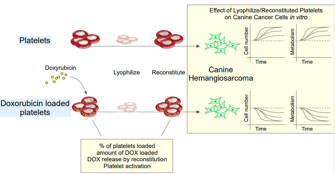
Here, we evaluate a modified version of Cellphire’s proprietary freeze-dried platelet product loaded with the cytotoxic drug, doxorubicin, for delivery to hemangiosarcoma cells. Platelets could be efficiently loaded with doxorubicin. Doxorubicin was retained following reconstitution of the lyophilized platelet product. We found that 1-10% volume/volume (v/v) addition of doxorubicin-loaded platelets (DOX-loaded) to hemangiosarcoma cultures had potent anti-cancer activity by inhibiting proliferation, metabolism, and promoting apoptosis. Further, hemangiosarcoma cells exposed to 5% and 10% v/v DOX-loaded platelets contained 2.2x and 4x more intracellular doxorubicin compared to cells treated with media containing 5 µM free doxorubicin. These results suggest that lyophilized, DOX-LHP overcome the storage limitations of platelets and once reconstituted they function as effective drug delivery vehicles for cytotoxic compounds.
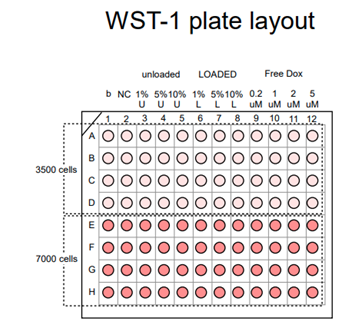

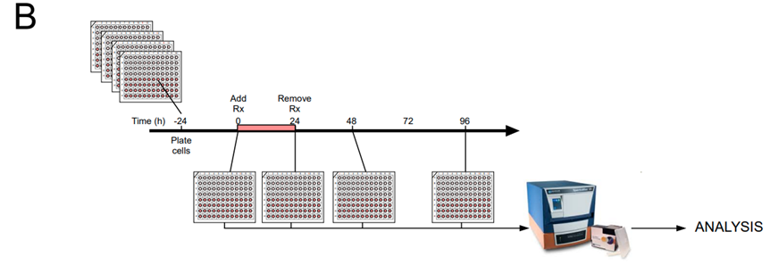
Figure 1 Experimental schematic. A. DHSA-1246 cells were plated in four technical replicate 96-well plates at two densities (3500 and 7500 cells/well) in quadruplicates at time -24 hours. B. At the indicated times (0, 24, 48, and 96 hours) an assay, for example, WST-1 or Annexin-V assay was performed. Time 0 correlates to time of treatment (Rx). Times 24, 48, and 96 correlates to time post-exposure to treatment or control media. At the indicated time point, a plate is incubated in reagent according to the manufacturer’s protocol for WST-1 or Annexin V and the readout is obtained using a SpectraMax i3X plate reader, and then analyzed.
Annexin-V/7-AAD assay
An experimental schema is shown in Figure 1. Cells were characterized for apoptosis using the annexin- V/7-AAD apoptosis detection kit for flow cytometry analysis (eBioscience, Catalog No. 88-8006-72). Briefly, cells were plated at a density of 10,000 cells / cm2 in T-25 (treatment groups) or T-75 (control groups) flasks. After 24 hours, the cells were exposed to treatment (1%, 5%, or 10% v/v LHP-containing media, 8 unloaded- or DOX-loaded) or controls (negative control DHSA medium or positive control 5 μM DOX) and incubated overnight. Following 24 hours of treatment, media was removed by gentle aspiration, cells were washed once, and fresh HSA media was added. At 24 hours, 48 hours, and 72 hours cells were harvested and stained with Annexin-v/7-AAD for flow cytometric analysis per manufacturer’s protocol. Unstained cells and single stains were used to establish background fluorescence and used for compensation. Heat- shocked cells were used as an additional positive control. Cells were analyzed using an LSR Fortessa X- 20 Special Order Research Product flow cytometer with 405, 488, 561, and 640 nm excitation lasers with a total of 13 parameters plus forward scatter and side scatter. The BV421 filter was used to collect the Annexin-V label, the PerCP-Cy5.5 filter was used for the 7-AAD stain, and the PE-TxRed filter was used for the DOX fluorescence. Samples were blindly analyzed and at least 10,000 gated data points were recorded for all samples.
DOX content was detected using the PE-TxRed channel in flow and measured as mean fluorescence intensity (MFI). The MFI for the negative control, cells treated with HSA media alone, was used to normalize to zero for each time point analyzed. The range was calculated relative to cells exposed to 5 μM free DOX media.
Statistical analysis
ANOVA assumptions were tested. If they were met the analysis proceeded and the results are presented as least square means estimates and standard error of the least square mean. If ANOVA assumptions were not met, the data was transformed using standard mathematical transformation, and again ANOVA assumptions were tested. Three-way repeated measures ANOVA was used with factors: treatment, dose, and time, or two-way ANOVAs with the factors being dose and time. The data for 3500 and 7000 cells/well cell densities was analyzed using separate ANOVAs. Following significant main effects or interactions, post hoc analysis was conducted with p value correction for multiple comparisons. In all cases, p < 0.05 for two tailed analysis was considered significant. Graphs were prepared using Sigmaplot for Windows version 14.0 (build 14.0.3.192, Systat Software) and saved as enhanced metafiles (EMF). The EMF files were edited and assembled into final figures using Canvas X version 19 (build 133, Canvas GFX, Inc). 9
Generation of unloaded- and DOX-LHP
Platelets, either unloaded or DOX-loaded were lyophilized using Cellphire’s proprietary technology. After lyophilization, unloaded- and DOX-LHP were rehydrated with sterile water in the same volume used for aliquoting (2 mL) and analyzed. Figure 2A shows that 83-87% of the platelets were loaded with DOX. Lyophilization and rehydration caused significant increases in platelet α-granule release (labeled by CD62P) and membrane flipping (labeled by Annexin V) for both unloaded- and DOX-LHP (Figure 2B). Lyophilization and rehydration did not change the percentage of LHP loaded with DOX, as shown in Figure 2C, but it did cause a significant loss of approximately 40% of the DOX-loading as shown in Figure 2D. Thus, while the percentage of DOX-loaded platelets does not appreciably change after lyophilization (Figures 2A & C), Figure 2D shows a significant release of DOX from the LHP after rehydration. When considered with the Annexin V staining shown in Figure 2B, the Dox-loss may be due to membrane fracture or flipping, although studies to confirm this hypothesis are ongoing. Therefore, to eliminate potential confounding effects of DOX release following rehydration, the DOX-LHP were centrifuged and resuspended prior to every experiment to wash out released DOX. Unloaded-LHP were similarly washed prior to analysis for consistency.
Lyophilization and rehydration did not impact hemostatic activity shown by the ability to occlude a simulated vessel coated with collagen and tissue thromboplastin (Figure 2E), nor did it impact the ability to generate thrombin in response to an initiating reagent (Figure 2F). Loading with DOX, lyophilization and rehydration had minimal impact on platelet function. LHP retain their capacity to occlude capillaries coated with collagen and thromboplastin, with both unloaded- and DOX-LHP rehydrated samples occluding the capillary at around 15 minutes (Figure 2E). Also, both unloaded- and DOX-LHP produce similar endogenous thrombin potential (ETP) of 1227 and 1218 nM*min and reach similar thrombin peak heights of 69 and 75 nM, respectively (Figure 2F).
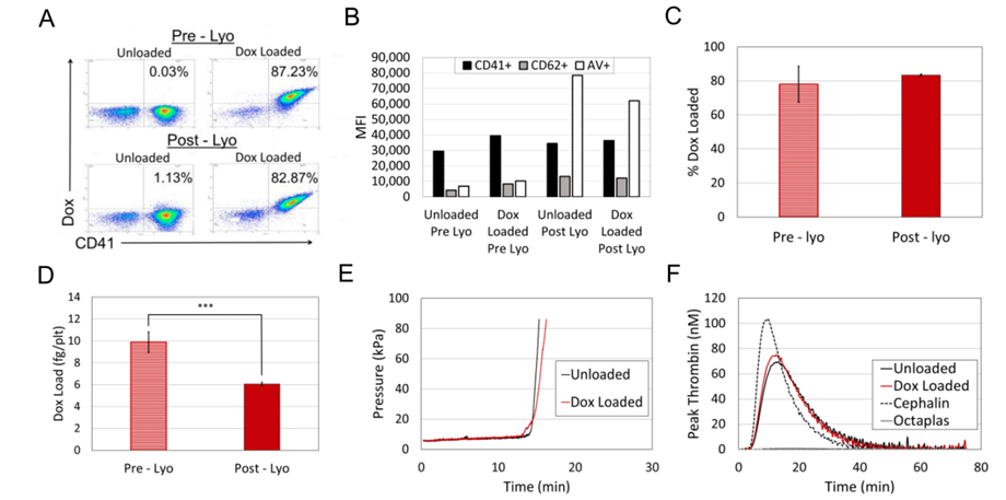
Figure 2 Characterization of Unloaded- and DOX-LHP. A. DOX-loading of platelets and LHP. Flow cytometry revealed that between 83 and 87% of the platelets contained DOX (e.g., double-stained for DOX and CD41) pre – and post – lyophilization, respectively. B. Effect of DOX loading and rehydration on LHP. Platelets or LHP were identified using anti-CD41 (CD41+). Platelet activation was assessed using anti- CD62P (CD62P+) and Annexin V (AV+). DOX loading caused only minor increases in platelet activation. Both unloaded and DOX loaded samples showed significantly increased activation after lyophilization and rehydration as demonstrated by increased MFI values for CD62P and AV. LHP C. Effect of LHP rehydration on DOX-loading. Rehydration did not change the % of DOX-loaded LHP. D. Effect of LHP rehydration on DOX-content. Using TECAN Infinite M200 PRO measurement, there was a 40% loss of DOX-content after reconstitution. E. Function of LHP. Rehydrated LHP were functional, as assessed by T – TAS to evaluate capacity for thrombus formation. DOX-loading did not affect thrombus formation. F. Function of LHP. Rehydrated LHP were functional as assessed by thrombin generation (using thrombin generation assay). There was an approximate 8% increase in peak thrombin generation after DOX-loading. Bars in C and D are averages of 3 replicates with 1 standard deviation. ***p<0.001 (Student’s t – test).
Lyophilized/Reconstituted DOX-LHP inhibit canine hemangiosarcoma cell growth
Preliminary results showed inhibited cell expansion following exposure to free DOX or DOX-LHP-containing media. In contrast, cells exposed to unloaded-LHP-containing media or standard HSA cell culture media continued to grow over 5 days (data not shown). The effect of unloaded and DOX-LHP product on live cell counts is shown in Figure 3A or population doublings shown in Figure 3B. Note that the live cell count data was transformed to meet ANOVA assumptions (Supplemental Table 1 for ANOVA table). Factors (Treatment, Dose, Time) and interaction terms (Treatment x Dose, Treatment x Time, and Treatment x Dose x Time) were significant. As shown in Figure 3A, cells exposed to either DOX-LHP (1-10%) or media containing 5 µM free DOX did not expand while cells exposed to unloaded-LHP or standard cell culture media continuously proliferated over the 96-hour experiment. All doses of DOX-LHP reduced cell growth significantly, and a dose-response relationship was observed. Specifically, higher doses of DOX-LHP had a greater reduction in cell growth, with 10% DOX-LHP having the greatest reduction in cell count. The highest dose of DOX-LHP was not different from the positive control (5 µM free DOX). In contrast, unloaded- LHP treatment allowed for uninhibited cell growth over time. There was a significant difference in cell growth between all doses of unloaded-LHP compared to the negative control (HSA cell culture media); this suggests that unloaded-LHP (1-10% v/v) impact cell growth. A similar trend is observed when the data is plotted as population doublings (Figure 3B).

Figure 3 (A) Live-cell count of canine hemangiosarcoma cells 48 hours and 96 hours after exposure to treatment or control media. Cells exposed to 5 µM free doxorubicin (DOX, positive control) or 1, 5 or 10% DOX-LHP did not continue to grow at 96 hours post-exposure. In contrast, cells exposed to 1, 5, or 10% unloaded-LHP or standard cell culture media (negative control) continued to grow over the 96 hours. No significant differences were found in the cell count among treatment groups at 48 hours. At 96 hours post- exposure, DOX-LHP significantly reduced the cell count and all 3 treatment groups were significantly different from one another. Note: Data was transformed by square root function to meet ANOVA assumptions and transformed data is plotted here. Note: ANOVA least squares mean estimates and SE of the least squares means are plotted. (B) Population doublings of cells 48 hours and 96 hours after exposure mto treatment or control media. Cells exposed to free DOX or DOX-LHP had less than 1 population doubling 96 hours after treatment. Cells exposed to unloaded-LHP or standard cell culture media had more than 2 population doublings 96 hours after exposure to treatment. *p < 0.05 in two tailed testing.
WRIGHT A et al. DOX LHP paper.
|
Source of variation |
DF |
SS |
MS |
F |
P |
|
treatment |
1 |
6141472.679 |
6141472.679 |
1427.102 |
<0.001 |
|
dose |
2 |
284310.190 |
142155.095 |
33.033 |
<0.001 |
|
time |
1 |
758563.946 |
758563.946 |
176.268 |
<0.001 |
|
treatment x dose |
2 |
180684.973 |
90342.486 |
20.993 |
<0.001 |
|
treatment x time |
1 |
1592867.330 |
1592867.330 |
370.137 |
<0.001 |
|
dose x time |
2 |
93772.096 |
46886.048 |
10.895 |
<0.001 |
|
treatment x dose x time |
2 |
45744.818 |
22872.409 |
5.315 |
0.012 |
|
Residual |
24 |
103282.978 |
4303.457 |
||
|
Total |
35 |
9200699.009 |
262877.115 |
Supplemental Table 1
ANOVA on live cell counts
Dependent Variable: Transformed live cell counts (sqrt)
Normality Test (Shapiro-Wilk): Passed (P = 0.359)
Equal Variance Test (Brown-Forsythe): Passed (P = 0.285)
WRIGHT A. et al. DOX LHP paper
In summary, the experiment shows that DOX-LHP inhibits cell proliferation. The highest dose of DOX-LHP (10% v/v) inhibited cell proliferation equally well compared to 5µM free DOX (added to the medium). In contrast, while cells continued to expand when exposed to unloaded-LHP, there was reduced proliferation compared to the negative control (HSA medium).
DOX-LHP product impacts canine hemangiosarcoma cell metabolism
To quantify canine hemangiosarcoma cell metabolism, WST-1 assay was used. In pilot data not shown, there were large variations in absorbance readings. To address this, the experiment was designed that averaged technical quadruplicates. Cells were plated at two different densities and analyzed at four different time points. In separate plates, the effect of five different free DOX concentrations on cell metabolism was evaluated. Here, raw data (absorbance values at 480 nm) were transformed by first normalizing and then by Log scaling in order to meet the ANOVA assumptions (ANOVA table is provided in supplemental Figure 2). Transformed data are plotted in Figure 4. ANOVA main effect [TREATMENT, TIME] and interaction term [TREATMENT, TIME] were significant, but main effect [DOSE] and the [DOSE x TIME] interaction effect were not significant. In Figure 4, both low plating density (A) and high plating density (B), cells exposed to unloaded-LHP continued to metabolize over time, as indicated by a significant increase in absorbance over the 96-hour time course. In contrast, cells exposed to DOX-LHP did not continue to grow, as indicated by decreased absorbance over the 96-hour time course.
As shown in Figure 4A, at the 3500-plating density, cells exposed to the DOX-LHP product had reduced metabolism at 24, 48, and 96 hours compared to the 0-hour reading. As shown in Figure 4B, at the 7000- plating density, cells exposed to DOX-LHP product had reduced absorbance at 48 and 96 hours compared to the 0-hour reading. Pilot data suggested a dose-response between unloaded and DOX-LHP, but no significant dose-response was observed here. Specifically, 1% DOX-LHP were equally effective at reducing cellular metabolism as 5 and 10% v/v doses of DOX-LHP. As expected, the negative control increased metabolism over the 96-hour time course and the positive control decreased metabolism over the 96-hour time course (data not shown).

Figure 4 Doxorubicin (DOX)-loaded lyophilized human platelets (LHP) inhibit metabolism of canine hemangiosarcoma cells as measured by WST-1 assay. Initial plating density of 3500 cells per cm2 (A) and 7000 cells per cm2 (B). Note raw data (absorbance values at 480 nm) were transformed by first normalizing and then by Log scaling in order to meet the ANOVA assumptions. The ANOVA least square mean estimates and standard error of LS means are plotted here. ANOVA found no significant (Dose) main effect or interactions; all doses were pooled for presentation (see supplemental data for ANOVA table).
(A) When plated at lower density (3500 cells per cm2), exposure to DOX-LHP (black symbols) significantly reduced metabolism over the 96 h experiment, e.g., cells had lower absorbance values than those exposed to unloaded-LHP (white symbols) at all time points after exposure (e.g., 24, 48, and 96 hours). In contrast, unloaded-LHP continue increase metabolism, as indicated by increasing absorbance over the 96 h experiment. (B)Cells plated at high density showed the same effect: DOX-LHP significantly reduced metabolism over the 96 h experiment, and cells exposed to unloaded-LHP continued to increase their metabolism over time, as indicated by an increase in absorbance. Significant differences between unloaded- and DOX-LHP treatment groups are indicated by an asterisk. *p-values < 0.05 post hoc comparisons.
To establish whether free DOX produced a dose-response relationship for comparison with the DOX-LHP, canine hemangiosarcoma cells were exposed to five different concentrations of free DOX (0, 0.2, 1, 2, and 5 μM) and cell metabolism was assessed at 0, 24, 48, and 96 hours using WST-1 assay. These results are shown in Figure 5. Raw data (absorbance values at 480 nm) were transformed by first normalizing and then by Log scaling in order to meet the ANOVA assumptions. ANOVA found significant main effect (DOSE) and interaction term (DOSE x TIME, supplemental data for ANOVA tables). In Figure 5, least square mean estimates and standard error of the least square means is plotted to reflect the DOSE x TIME interactions (transformed data is plotted). As shown, at both plating densities cells exposed to the highest concentration of free DOX (5 μM) decreased metabolism over the 48-hour and 96-hour time course. Cells exposed to 0.2-5 μM free DOX tended to have decreased metabolism at 96 hours in a dose-response manner. Plating density affected response to 0.2-2 µM free DOX since high density increased metabolism at 24 hours and low plating density did not. Also, significant decreases in metabolism after exposure to 2 μM DOX were noted at 48 and 96 hours at low density, however only 96 hours showed a significant response at high density. In summary, 0.2-5 µM free DOX tended to decreased metabolism of canine hemangiosarcoma cells as measured by the WST-1 assay 96 hours after exposure, independent of plating density. However, when evaluating 24- and 48-hour time points, dose and plating density affect results. To quantify apoptotic cells after treatment with DOX-LHP, cells were stained with Annexin-V/7-AAD and analyzed via flow cytometry. Cells were analyzed at time points 24, 48, and 72 hours after exposure to treatment or control conditions. ANOVA for cell death found significant main effects [Treatment] and [Dose], but main effect [TIME], and their interactions were not significant (Supplemental Table 3 for ANOVA table). Figure 6 shows the significant treatment effect of DOX-LHP. In contrast to cell death, ANOVA for apoptosis found significant main effect [Dose] and [TIME], and main effect [Treatment] was not significant, but a trend was observed for apoptosis to increase after exposure to DOX-LHP (data not shown, Supplemental Table 4). As expected, exposure to the positive control, 5 µM free DOX, increased both death (sin transformed data: 0.9903 on day 1, 0.8910 on day 2, and 0.9976 on day 3) and apoptosis (31% on day 1, 60% on day 2, and 62% on day 3) over the three days. These results, together with the results shown in Figure 3, indicate that exposure to 5 and 10% DOX-LHP produce cell death, similar to that of 5 µM free DOX.

Figure 5 Effect of free doxorubicin (DOX) treatment on the metabolism of canine hemangiosarcoma cells measured by WST1 assay. Initial plating density of 3500 cells per cm2 (A) and 7000 cells per cm2 (B). Note raw data (absorbance values at 480 nm) were transformed by first normalizing and then by Log scaling in order to meet the ANOVA assumptions. The ANOVA least square mean estimates and standard error of LS means are plotted here. Cells exposed to 0 μM DOX (black circles, standard cell culture media) had an increase in absorbance over 96 hours at the lower density (A) but no change in absorbance at the higher plating density (B). The highest dose of DOX (5 μM) caused monotonic decreases in absorbance for both plating densities. Significantly lower absorbance for 5 μM treatment was noted at 48 and 96 hours at both plating densities. In contrast, DOX doses 0.2-2 μM all showed decreased absorbance compared to 0 h at 96 h in both plating densities.
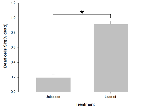
Figure 6 Effect of Doxorubicin (DOX)-loaded (Loaded) versus unloaded (Unloaded) lyophilized human platelets (LHP) on canine hemangiosarcoma cells death. The percent dead cell data from the Annexin V assay was transformed with a sine function. Treatment with DOX-LHP significantly increased the number of dead cells.
|
Source of variation |
DF |
SS |
MS |
F |
P |
|
treatment |
1 |
3.319 |
3.319 |
107.007 |
<0.001 |
|
dose |
2 |
0.0601 |
0.0300 |
0.969 |
0.385 |
|
time (h) |
3 |
0.424 |
0.141 |
4.553 |
0.006 |
|
treatment x dose |
2 |
0.116 |
0.0579 |
1.868 |
0.162 |
|
treatment x time (h) |
3 |
3.830 |
1.277 |
41.162 |
<0.001 |
|
dose x time (h) |
6 |
0.146 |
0.0243 |
0.783 |
0.586 |
|
treatment x dose x time (h) |
6 |
0.345 |
0.0576 |
1.856 |
0.100 |
|
Residual |
72 |
2.233 |
0.0310 |
||
|
Total |
95 |
10.473 |
0.110 |
Supplemental Table 2
ANOVA on 3500 plating density WST assay
Dependent Variable: Transformed log (normalized Abs 460 nm)
Normality Test (Shapiro-Wilk): Passed (P = 0.444)
Equal Variance Test (Brown-Forsythe): Passed (P = 0.252)
|
Source of variation |
DF |
SS |
MS |
F |
P |
|
treatment |
1 |
2.236 |
2.236 |
109.058 |
<0.001 |
|
dose |
2 |
0.0180 |
0.00901 |
0.440 |
0.646 |
|
time (h) |
3 |
0.913 |
0.304 |
14.845 |
<0.001 |
|
treatment x dose |
2 |
0.0619 |
0.0310 |
1.511 |
0.228 |
|
treatment x time (h) |
3 |
2.253 |
0.751 |
36.628 |
<0.001 |
|
dose x time (h) |
6 |
0.262 |
0.0437 |
2.131 |
0.060 |
|
treatment x dose x time (h) |
6 |
0.0580 |
0.00967 |
0.472 |
0.827 |
|
Residual |
72 |
1.476 |
0.0205 |
||
|
Total |
95 |
7.278 |
0.0766 |
ANOVA on 7000 plating density WST assay
Dependent Variable: Transformed log (normalized Abs 460 nm)
Normality Test (Shapiro-Wilk): Failed (P < 0.050)
Equal Variance Test (Brown-Forsythe): Passed (P = 0.473)
WRIGHT A. et al. DOX LHP paper
|
Source of variation |
DF |
SS |
MS |
F |
P |
|
treatment |
1 |
2.331 |
2.331 |
119.546 |
<0.001 |
|
dose |
2 |
0.167 |
0.0835 |
4.281 |
0.040 |
|
time |
2 |
0.0513 |
0.0256 |
1.315 |
0.305 |
|
Residual |
12 |
0.234 |
0.0195 |
||
|
Total |
17 |
2.783 |
0.164 |
Supplemental Table 3
ANOVA on Annexin V dead cell %
Dependent Variable: Transformed sin(% dead cells) in Annexin V assay
Normality Test (Shapiro-Wilk): Passed (P = 0.588)
Equal Variance Test (Brown-Forsythe): Passed (P = 1.000)
|
Comparison |
Diff of means |
p |
q |
P |
P<0.05 |
|
loaded vs. unloaded |
0.720 |
2 |
15.463 |
<0.001 |
Yes |
All Pairwise Multiple Comparison Procedures (Tukey Test):
Comparisons for factor: treatment
|
Comparison |
Diff of means |
p |
q |
P |
P<0.05 |
|
10.000 vs. 1.000 |
0.229 |
3 |
4.010 |
0.037 |
Yes |
|
10.000 vs. 5.000 |
0.0639 |
3 |
1.120 |
0.715 |
No |
|
5.000 vs. 1.000 |
0.0639 |
3 |
2.890 |
0.144 |
No |
Comparisons for factor: dose
Power of performed test with alpha = 0.0500: for treatment: 1.000
Power of performed test with alpha = 0.0500: for dose: 0.511
Power of performed test with alpha = 0.0500: for time: 0.0891
Least square means for treatment:
Group Mean
unloaded 0.195
loaded 0.915
Std Err of LS Mean = 0.0465
Least square means for dose:
Group Mean
1.000 0.424
5.000 0.588
10.000 0.652
Std Err of LS Mean = 0.0570
WRIGHT A et al. DOX LHP paper.
|
Source of variation |
DF |
SS |
MS |
F |
P |
|
treatment |
1 |
589.389 |
589.389 |
3.428 |
0.089 |
|
dose |
2 |
1835.111 |
917.556 |
5.337 |
0.022 |
|
time |
2 |
1413.444 |
706.722 |
4.111 |
0.044 |
|
Residual |
12 |
2063 |
171.917 |
||
|
Total |
17 |
5900.944 |
347.114 |
Supplemental Table 4
ANOVA for Apoptosis results from Annexin V experiment
Dependent Variable: Apoptosis
Normality Test (Shapiro-Wilk): Passed (P = 0.208)
Equal Variance Test (Brown-Forsythe): Passed (P = 1.000)
Here, we developed a novel anti-cancer therapy which combines the platelet’s special affinity for cancer cells with the antitumor efficacy of DOX. Four important observations encapsulate our findings. First, Cellphire’s lyophilization technology produces functional LHP. Upon rehydration, LHP have pro-coagulation activity as indicated by thrombin release and thrombus formation upon stimulation. Second, about 83% of the platelets could be loaded with up to 10 fg/platelet of DOX prior to lyophilization, and DOX-LHP retained approximately 60% of the DOX upon rehydration. Third, DOX-LHP had anti-cancer activity on canine hemangiosarcoma cells in vitro. This was demonstrated by dose-dependent inhibitory effects of DOX-LHP on canine hemangiosarcoma proliferation and metabolism in vitro, and significant accumulation of dead hemangiosarcoma cells over the 96-hour observation period after exposure compared to either unloaded- LHP or control medium. Furthermore, the highest “dose” tested, 10% v/v, inhibited cell proliferation and caused cell death equally as well as 5 µM free DOX while the lowest dose tested 1% v/v also had significant effects on sarcoma cells. Fourth, application of DOX-LHP produced specific drug effect as indicated by two observations: first, a dose-dependent inhibition was caused by DOX-LHP, and second, application of unloaded-LHP enhanced rather than inhibited cancer cell growth. Taken together, these observations suggest that this platform holds promise in treating cancer and should be advanced into animal preclinical cancer models. Our observations are supported by other work showing that platelets or platelet membrane formulations can be engineered to specifically deliver DOX to cancers.10,13,23,24 The work by others indicates that further modification of platelet membrane, perhaps by adding TRAIL with DOX (e.g., as done by Hu Q, Sun W et al.23), might offer further optimization of anti-cancer action. Nevertheless, different from prior publications, our drug formulation was lyophilized and reconstituted prior to testing. This addition which enables extended shelf-life and off-the-shelf application is a significant advancement in the field.
One limitation of the present work was the lack of testing in cancer-bearing animals. Based upon these promising in vitro data presented here, DOX-LHP should be advanced into animal preclinical testing perhaps into mouse models or into canines with naturally occurring hemangiosarcoma, since this model is considered as an excellent large animal model of the human disease.25,26 This is an important next step in translation to the clinic. However, due the relative dearth of canine platelets, compared to human platelets, it would have been difficult to produce DOX-loaded lyophilized canine platelets (LCP) for these preliminary studies. Nevertheless, a Cellphire subsidiary does produce a clinical product based on LCP and future studies investigating canine hemangiosarcoma treatments will seek to overcome the source limitations and produce DOX-LCP that are species-matched to prevent xenograft complications. The use of human cancer cell lines is another option, and mice models are well-established for many different cancers, however, xenograft complications in mice models using LHP will likely again be prohibitive. Compared to previous studies employing platelets as a drug-delivery vehicle, our work has the advantage of extended shelf life afforded by lyophilization. Shelf-life is a disadvantage for any platelet-derived therapies owing to the issues surround their sensitivity to storage conditions and short shelf-life.14,16,18,22 Multiple attempts have been made to extend the shelf-life of platelet storage.14,16,18 That the Cellphire process enables DOX retention and platelet activation following rehydration highlights advantages of our novel agent.
In conclusion, a novel platelet-based doxorubicin nano-formulation for the specific delivery of drug to cancer was described. By taking advantage of the known affinity platelets have for cancer cells, we speculate that this technology can efficiently deliver DOX into cancer cells in vivo, to stimulate cell apoptosis and death. Our work revealed enhanced cancer cell death and apoptosis following DOX-LHP incubation in a dose- dependent manner, and specificity indicated by comparison to effects of unloaded-LHP (which had the opposite effect on cancer growth, metabolism, and cell death). Therefore, we suggest that this platform have significant advantages over prior platelet-based nano-formulations that supports further translational testing.
We greatly thank Kaori Knights and Nora Springer of the Kansas State University Flow Cytometry laboratory for providing experimental assistance. We thank Dr. Hong He, MD, PhD for providing technical assistance. We acknowledge the use of the KSU Department of Anatomy and Physiology shared equipment which is 18supported by the State of Kansas. We acknowledge Marie Aponte for her work on TGA and endotoxin analysis at Cellphire. MLW acknowledges BGW for her life-long support and counsel.
The authors declare no conflicts of interest.
The datasets used and analyzed during this study are available by materials transfer agreement. Please contact corresponding author. Please direct inquires to Keith Moskowitz at Cellphire regarding LHP.
YYZ, AL, BI, MA, DS and KM are employed by Cellphire. The sponsors had no impact on the collection data or the interpretation of these results. AW and MLW report no competing interests.
This research was supported by a sponsored contract with Cellphire for in vitro testing of LHP. Other support was provided by the Terry Johnson Center for Basic Cancer Research, and the State of Kansas to the Midwest Institute for Comparative Stem Cell Biotechnology.
AW collected all experimental data involving DHSA-1426 cells with LHP and DOX-LHP and was a major contributor in writing the manuscript. YYZ contributed in the flow and T-TAS experiments, data analysis and interpretation, and edits of the final manuscript. AL and BI collected flow and T-TAS experiments on LHP. DS and KM designed the experiments, were involved with data analysis and interpretation, and contributed to the final manuscript. MLW designed the experiments, performed most of the data analysis and interpretation, and was a major contributor in writing the manuscript. All authors read and approved the final manuscript.

©2021 Wright, et al. This is an open access article distributed under the terms of the, which permits unrestricted use, distribution, and build upon your work non-commercially.