eISSN: 2576-4497


Case Report Volume 5 Issue 2
1Médico General, Hospital México, Costa Rica
2Médico Residente de Neurología, Hospital México, Costa Rica
Correspondence: Muñoz Murillo Juan Pablo, Médico Residente de Neurología, Hospital México, Costa Rica, Tel (506)87080142
Received: September 14, 2022 | Published: September 22, 2022
Citation: Rodolfo CZ, Pablo MMJ. Joint tuberculosis: a diagnosis that should be kept in mind. Hos Pal Med Int Jnl. 2022;5(2):29-32. DOI: 10.15406/hpmij.2022.05.00204
Tuberculosis is an infectious disease of high prevalence in developing countries. Factors such as underestimation of the disease and difficulty in diagnosis have generated delay in the diagnosis and with it significant morbidity and mortality.
With regard to the joint location, most of them are born and immigrated from endemic areas and also with immune compromise. Its usual presentation is a monoartritis of large joints and the most frequent locations with spine, hip and knee. Radiological findings may suggest the picture, however, the diagnosis is made by identifying the germ.
We present the case of a 29-year-old patient with systemic lupus erythematosus who underwent immunosuppressive treatment, who presented with a chronic condition of joint pain in the right hip and progressive functional limitation who, after several studies, documented a tuberculous arthritis. The patient receives antitubercular agents and is followed up by external consultation.
Keywords: tuberculosis spinal, hip, antitubercular agents
Tuberculosis (Tb) remains a public health problem worldwide, with developing countries being the most prevalent sites. Overcrowding, poor hygiene and immune compromise are risk factors to consider. Others such as injection drug use, diabetes, malignancies, aging, and pharmacological treatment, especially tumor necrosis factor alpha (TNF-α) inhibitors, have also been linked.1–3
Extrapulmonary manifestations occur in approximately 20% of all patients with Tb. Regarding tuberculous arthritis (TA), this represents 2 to 5% of all cases of Tb, as well as 11 to 15% of cases of extrapulmonary Tb, and is located behind lymph node Tb in frequency and urogenital.4 In 90% of cases, its presentation is monoarticular and is characterized by being paucibacillary. Disseminated forms have been described, but these are related to the analyzed population and diagnostic methods.1,4,5
There are 3 microorganisms related to AT. Mycobacterium africanum, which is endemic in certain African countries, Mycobacterium bovis in relation to unpasteurized milk or the application of bacillus Calmette-Guérin (BCG) in bladder cancer and the main one is Mycobacterium tuberculosis, an aerobic bacillus, strict, small and immobile.3,4
The case of a 29-year-old female patient is presented. He denies the use of tobacco, alcohol and drug addiction, reports allergy to paracetamol and NSAIDs, has not received transfusions. As pathological history, she is a carrier of systemic lupus erythematosus (SLE) with adequate control, diagnosed in 2015, for which she is treated with azathioprine 200 mg every day, plaquinol 1 tab every day, prednisone 7.5 mg/d, MTX 25 mg/d, folic acid, calcium 600 mg/d, Vit D 3 drops every day, the monitoring of his disease takes him to the Rheumatology service of Hospital México.
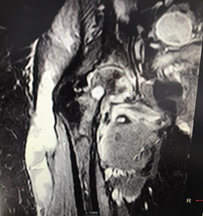
The patient reports pain in the right hip that began in May 2017, with an evolution of approximately 10 months, with functional limitation that progressively increased until she first felt the need to use a cane to walk until she was unable to walk. Therefore, on an outpatient basis, a magnetic resonance imaging (MRI) of the affected joint was requested (Figure 1); the report of said study indicates septic arthritis of the hip as the first possibility. However, due to the insidious evolution of the disease, the diagnosis is questioned and the patient is admitted through the outpatient clinic of the rheumatology service to complete studies.

Figure 1 Magnetic resonance imaging of the right hip showing bone edema, morphological alteration of the right femoral head, large amount of peripheral fluid (hyperintense) that affects the pectineus, hamstring and obturator muscles, extending through the tensor facia lata caudally for 16 cm. Collection suggestive of an inflammatory process (indicated by a red arrow). Source: MRI of the patient's right hip.
On admission, reports occasional feverish sensation, not quantified, pain in the right hip 10/10 that relieves being in a sitting position, does not tolerate the supine position, or the active or passive mobilization of the coxofemoral joint. He denies genitourinary and respiratory symptoms, denies bleeding, psychosis, seizures. He refers to being well controlled of his underlying disease.
In the physical examination at the time of admission, BP 121/75mmHg, MAP 92, HR 102 beats/min, Temp 36.5°C, Sat 02 97% AA is reported. Alert patient, oriented, cooperative. Not IY at 45°. Oral mucosa without lesions. Rhythmic heart, without murmurs. Lung fields with conserved vesicular murmur without added sounds. Soft and depressible abdomen, peristalsis present, not painful on palpation. No foot edema. Normal, symmetrical pedal and radial pulses. No skin lesions, no bleeding data. Preserved strength, no sensitivity alterations, intact cranial nerves, non-assessable gait, normoreflexic, preserved higher mental functions. With limitation in range of motion of the right hip, without erythema, slight edema, no increase in local temperature. The patient also has a recent echocardiogram that reports no heart valve disease, ejection fraction of 60%, normal pericardium, no signs of pulmonary arterial hypertension, no masses or thrombi, preserved diastolic and systolic function of the left ventricle, dimensions of cardiac structures in normal range. Table 1 shows the patient's admission laboratories, which remained similar during hospitalization, in addition, serologies were performed for hepatitis B, C, HIV, as well as VDRL, protein electrophoresis, and microproteinuria, all of the above normal.
Blood cell count |
Liver function |
Kidney function (mg/dl) |
Coagulation |
Electrolytes |
|||||
Hemoglobin |
9.8 g/dl |
AST |
15 IU/L |
Cr |
0.49 mg/dl |
TP |
75% |
Na |
139 mg/dl |
Hematocrit |
32.30% |
ALT |
12 IU/L |
NU |
14.7 mg/dl |
TPT |
31.2 s |
Cl |
108 mg/dl |
Platelets |
326000 |
GGT |
73 IU/L |
INR |
1.19 |
K |
4.1 mg/dl |
||
VAW |
94 pg |
BT |
0.3 mg/dl |
Ca |
9.1mg/dl |
||||
Leukocytes |
3100 |
BD |
0.04 mg/dl |
P |
3.1mg/dl |
||||
Segmented |
2300 |
FA |
79 IU/L |
||||||
Bands |
1% |
ALB |
3.6g/dl |
||||||
Table 1 Laboratory data of a patient with SLE undergoing studies for joint pain in the right hip
Upon admission, the orthopedic service is consulted, where the MRI images are evaluated, which at first are impressed by incipient avascular necrosis of the femoral head, which at that time did not require surgical management, without intra-articular collections that indicate arthritis. septic, extra-articular collection close to the subtrochanteric bursa is observed. They recommend taking a culture of it and requesting a computerized axial tomography (CAT) of the right hip, due to being a more sensitive study to assess bone pathology.
Subsequently, sampling of the collection by a radiologist guided by ultrasound (US) is coordinated. A previous exploration with US revealed an image compatible with a peritrochanteric collection, which extends caudally in the anterolateral aspect of the thigh to the junction between the middle third and the distal third, in the plane between the subcutaneous fat and the muscular structures. 5 cm deep, with a volume of not less than 150 cc. Due to the density of the material, it was possible to extract only approximately 1 cc, with a frankly purulent appearance, which was sent for smear and culture. Likewise, on 2/21/18, a CT scan of the right hip was performed without contrast medium, pending its official report.
The studies are taken to a general orthopedic session where a CT scan is evaluated that shows a decrease in the joint space of the right coxofemoral joint, with changes of osteoarthrosis, which does not show avascular necrosis of the femoral head. Regarding the "collection" observed in the MRI in the extra-articular space, it is determined that it must correspond to a probable chronic bursitis, given that the final culture report shows 1+ leukocytes and is negative for bacteria, she is discharged. for orthopedics patient, and without a clear diagnosis, it was decided to request a new US where only the anterior and partial lateral face of the thigh could be assessed due to the patient's pain; slight loss reported the sphericity of the right femoral head, slight increase in the echogenicity of the ileopsoas as well as possible inflammatory changes, possible edema. Iliopsoas bursitis is not observed. The well-known collection of soft tissue is observed, with dense peritrochanteric content that extends caudally in the anterolateral aspect of the thigh until the union between the middle and distal thirds. Minimal joint effusion. Collection 160cc.
In addition, taking into account the elevation of the erythrocyte sedimentation rate (ESR) and C-reactive protein (CRP) during hospitalization (Table 2), in the context of the known intra-articular collection, the infectology service is consulted, which recommends performing an MRI of lumbosacral spine plus psoas, culture of joint fluid for mycobacterium, PCR for TB, glucose, cellularity, culture for fungi and fastidious bacteria, in addition to purified protein derivative (PPD) skin test, which is performed on 5/3/ 18, with an induration greater than 10 mm. This result makes it more important to consider an open biopsy, which had been deferred initially by the orthopedic service due to considering a higher risk of contamination, since it was a collection without having had previous trauma or communication with the outside.
Fecha |
3/1 |
15/2 |
19/2 |
26/2 |
3/3 |
6/3 |
7/3 |
VES mm/h |
102 |
78 |
113 |
129 |
- |
92 |
|
PCR mg/l |
36.4 |
29.3 |
56.7 |
75.4 |
75 |
93 |
|
PCT ng/ml |
- |
0.1 |
0 |
0 |
0.1 |
0.1 |
0.1 |
Table 2 Count of inflammatory markers (ESR, PRC and PCT) of the patient during hospitalization
Finally, the case with orthopedics is discussed again with the recent findings and it is coordinated to take the patient to the surgery room, where a sample of lumpy, non-fetid whitish liquid, soft tissue and bone (greater trochanter) is taken for culture and biopsy. On 3/7/18, a positive culture for Mycobacterium tuberculosis complex was reported, and it was decided to isolate the patient. The soft tissue biopsy showed no alterations.
Infectology reassesses the case and suspends the isolation, since the patient does not have an open wound, for which there is a low probability of dissemination and she does not present respiratory symptoms. Two months of tetra-associated treatment (Isoniazid, rifampin, pyrazinamide, ethambutol and pyridoxine) and 10 months of bi-associated treatment are given, with confirmed sensitivity to all drugs. The patient after surgical drainage presents complete ranges of motion with moderate assistance in the lower limb law. Radiology sends the official report of the previously requested hip CT where they report, in addition to the already known pathology, vertebral destruction of the body of L5 with increased perivertebral soft tissue that contacts the ventral aspect of the dural sac and extends towards the left psoas where it is identified a collection of approximately 20 cc, in addition to hypodensity of the lower half of the body of L4. Findings in possible relationship with his known TB. It should be noted that the rest of the spine is not evaluated in this study since it was a CT scan of the right hip. Through the external consultation, follow-up is given, pending MRI of the spine and psoas, in addition, follow-up is given by physical therapy, and referral to the TB program.
Mycobacteria in general are characterized by an early transition to the intracellular space, thereby managing to evade the attack of other components of the immune system. This and other strategies at the cellular level have been used by these microorganisms in order to persist and spread. One is the production of enzymes to quench reactive oxygen species produced by macrophages. Even if intracellular mechanisms fail, TNF-α-mediated macrophage apoptosis can occur, however, many virulent strains compromise this step and thus continue infection. Interleukin 12, TNF-α, and interferon gamma are potent mechanisms of cellular immunity. States of compromised immunity, whether acquired or hereditary, or the result of pharmacological effects at that level, generate a predisposition to the activation of tuberculosis6, a risk factor presented by the patient in the exposed case.
In the case of AT, it is the result of activation of the primary focus and secondary hematogenous spread, or contiguous or lymphatic spread of Tb infection can occur after percutaneous inoculation.3,6 Cases of inoculation have been reported by thorns, contaminated objects, needles, prostheses and in relation to aesthetic procedures,6 this route should be rescued as a possible, although not as frequent, source of inoculum, since the portal of entry of the microorganism in the case presented is unknown, since an infection respiratory infection was never documented, nor was it found in any other objective source, for which other studies had to be completed, and seeds in different organs had to be ruled out, considering his underlying immunosuppressive condition.
In the musculoskeletal system, this microorganism is phagocytosed by mononuclear cells, then the tubercle with the caseous center is formed when these are arranged in a ring around epithelioid cells. The final liquefaction would lead to the formation of cold abscesses,3 data that appeared in the clinical evolution of the patient and probably without giving it sufficient importance, which could have led to an earlier diagnostic suspicion.
Initially, synovitis and granulation tissue formation develop, leading to the development of effusion with fibrin deposition, forming the so-called "rice" bodies. This granulation tissue begins to destroy the cartilage, leading to bone demineralization and caseous necrosis. The cartilage is destroyed peripherally, preserving the joint space for a considerable period of time, which has important clinical implications.2
TA usually presents insidiously with chronic joint pain and variable inflammation, associated with loss of function and local muscle atrophy. In 15% there is fistulization to the skin and a satellite adenopathy should be sought.2,4 Constitutional symptoms such as weight loss, night sweats and fatigue, although they are infrequent if they are present, the diagnosis should be considered.7
In approximately 50% of cases, there are no respiratory symptoms or radiological evidence of Tb, and the differential diagnosis should be made with chronic osteomyelitis, chondroma, osteoid osteoma, villonodular synovitis, sarcoidosis, Paget's disease, hyperparathyroidism, and brucellosis.2,8
Microscopic confirmation and culture is necessary, however, as it is a paucibacillary picture, false negative results are frequent and are positive from 3 to 10 weeks. A synovial aspirate sample is an excellent specimen to perform a Tb PCR in order to reach the diagnosis or detect resistance genes.1–3
Other noninvasive tests, including PPD testing and IFN-gamma release assays (IGRAs), may be helpful but do not differentiate latent from active disease. With regard to elevated ESR and CRP in the clinical evolution of the patient (Table 2), they help to suspect infection at that level, however, they are normal in 10 to 20% of cases.4,6,7
The joint fluid, in case of effusion, will almost constantly reveal inflammation with leukocytes of 5,000 to 50,000 elements per cubic mm and in 60 to 80% neutrophils predominate and lymphocytes in 20 to 30%.4,5 In plain radiography, the ¨Phemister¨ triad has been described, which suggests TA in some cases (periarticular osteoporosis, peripherally located bone erosion and progressive decrease in joint space), although the images do not have the findings. described if they were documented in the patient's CT scan and hip X-ray. Ultrasound can help identify effusions. However, it is very nonspecific. CT and MRI help in the diagnosis, especially in determining the extension, with MRI being the best diagnostic imaging method since it manages to document the "rice" bodies that suggest involvement by Tb.2,4,6,9
Regarding treatment, it will be medical according to the latest WHO consensus and surgical treatment is reserved for special cases or complications.4,8 The cornerstone of treatment is 3 to 4 drugs based on sensitivity profiles and its duration varied from 12 to 18 months. The most commonly used are rifampin (R), isoniazid (H), pyrazinamide (Z), ethambutol (E), and streptomycin (S). You can have a good response with antifungal therapy and early mobilization. Severe cases of peripheral joint involvement can be successfully treated surgically.2,8
In the case of our patient, a young woman with the risk factor due to her background disease and immunopressure and the monoarticular, chronic clinical picture, with associated edema and without According to the literature, greater local inflammation is initially very suggestive of Tb.
The absence of pulmonary Tb data should not lead to the diagnosis being dismissed, as it was reviewed, they are absent in 50% of the cases. With respect to the studies carried out, the CT showed clear initial data of this clinical picture and the MRI showed more non-specific findings, however, it is described that the imaging study par excellence is magnetic resonance imaging.
The other tomographic findings of injury to lumbar vertebrae (Pott's disease) suggest that the joint involvement could be due to local dissemination. The microbiological studies for negative pyogenics, the density of the material and the finding in the operating room of lumpy, whitish, non-fetid material, retrospectively mean that our case has a lot of association with the final diagnosis.
It is of the utmost importance to suspect this clinical entity since the results largely depend on prompt antifungal therapy in order to reduce the risk of sequelae and complications. Unfortunately, this condition, due to its non-specific and insidious nature, leads to a late diagnosis on many occasions.
None.
The authors declare that there are no conflicts of interest.

©2022 Rodolfo, et al. This is an open access article distributed under the terms of the, which permits unrestricted use, distribution, and build upon your work non-commercially.