eISSN: 2576-4497


Case Report Volume 5 Issue 1
Department of Urology, Academic Hospital Habib Bourguiba, Sfax, Tunisia
Correspondence: Mohamed Trigui, Urology Department, Tunisia, Tel + 27711449
Received: May 10, 2022 | Published: May 25, 2022
Citation: Kahla AB, Trigui M, Majdoub B, et al. Cutaneous metastases from an upper urinary tract carcinoma: A unusual entity and a poor prognosis. Hos Pal Med Int Jnl. 2022;5(1):1-2 DOI: 10.15406/hpmij.2022.05.00197
We report the case of a 58-year-old male with a history of diabetes who presented with a macroscopic hematuria. On physical examination, he was afebrile. Investigations revealed an anemia. A CT scan showed a tumor of the bladder and upper excretory system with multiple lung, spleen and liver metastases. The patient received blood transfusions and a trans-urethral resection of the tumor was performed. The patient’s condition later deteriorated and skin lesions appeared on the back and the scalp. A biopsy of the lesions confirmed their metastatic nature. The patient’s condition deteriorated and he died 4 weeks later.
Cutaneous metastases from visceral malignancies are uncommon. There is a disparity in their estimated incidence between the studies that were conducted on the subject. A Meta-analysis by Richard A and al suggested that the overall incidence is about 5.3%, with bladder cancer accounting for 3.6% of the total number of patients with cutaneous metastases.1
A 58-year-old male with history of diabetes presented with macroscopic total hematuria. He was in a good condition and didn’t complain of pain. On physical examination he was afebrile, his blood pressure was 12/7 and his abdomen was depressible.
Investigations revealed: anemia with a hemoglobin level at 7.6g/dl, a biological inflammatory syndrome with a CRP level at 130.6 mg/l. Renal function was normal and leucocytes were at 8400/µl. Cytobacteriological urine test was negative.
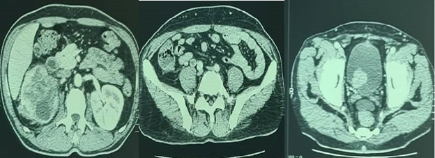
An abdominal ultrasound showed moderate dilation of the right pyelocaliceal cavities with echogenic tissue content. The right ureter is dilated ending in the bladder by a heterogeneous vegetative mass of 3 cm. A CT scan of the thoracic, abdominal and pelvic regions showed hydronephrosis of the right kidney as well as a tumor of the right upper excretory system (pyelon, pyelo-ureteral junction and iliac ureter) and the bladder. Multiple metastasis in the lung, spleen and liver as well as multiple metastatic lumbo-aortic lymph nodes were also noted (Figure 1).

Figure 1 CT scan showing bladder and iliac ureter tumor with dilation of the pyelo-calicial and ureteral cavities.
The patient received 3 blood transfusions after which the hemoglobin level was at 9.1g/dl. A trans-urethral resection of the bladder tumor was performed. The pathology report indicated that the tumor was a high grade papillary urothelial carcinoma infiltrating the lamina propria (pT1). The patient’s condition deteriorated rapidly. He developed skin lesions on the scalp and the back. They were round nodules with a diameter of around 0.5cm (Figure 2).
A biopsy was done which confirmed their metastatic nature (Figure 3). The patient was scheduled to receive palliative chemotherapy. However, he died 4 weeks after the initial diagnosis.
Cutaneous metastases from visceral malignancies are uncommon. In fact, their estimated incidence is at 5.3%. Breast cancer is the most common one to metastasize to the skin, with an incidence of about 24%, followed by renal, ovarian, bladder, lung, colorectal and prostate cancers with an incidence of 4, 3.8, 3.6, 3.4, 3.4, 0.7% respectively.1 In the case of our patient, who has both bladder and upper urinary tract carcinomas, the metastases probably originated from the upper urinary tract tumor. We have come to this conclusion due to the fact that the bladder tumor is non muscle invasive. The presence of lumbo-aortic lymph nodes with no iliac lymph nodes metastases also suggest that the upper urinary tract carcinoma was the one to metastasize. Cutaneous metastases from this type of tumor are very rare, and reports in literature are scarce.
The practitioner should be aware of this uncommon metastasis site and suspect cutaneous metastases when persistent, firm papulonodules develop acutely especially on the chest. However, they can take any form and they can be located anywhere on the body. Multiple sites can also be found,2 as is the case for our patient, who had lesions on his scalp and back. This can be explained by the lymphatic or hematological spread of the disease. In fact, there are 4 known mechanisms for cutaneous metastasis to develop: (1) Direct invasion; (2) implantation in an operation scar; (3) lymphatic spread ;( 4) blood-borne embolism.3
There is a wide range of differential diagnoses. Therefore, a biopsy is always necessary to confirm the metastatic nature of the lesions.
The standard treatment for metastatic bladder and upper excretory system is cisplatin-based chemotherapy.
Cutaneous metastases, having a universally bad prognosis, require mostly supportive care. They can cause considerable morbidity, as they can serve as a nidus for infection, bleeding, disfigurement, or pain. They are associated with a negative effect on quality of life. Therefore, symptom palliation is necessary. A recent meta-analysis evaluated five skin directed therapies: electrochemotherapy, photodynamic therapy, radiotherapy, intralesional therapy and topical therapy, all with promising results.4
The prognosis for metastatic urothelial cancer is poor. The presence of cutaneous metastases indicates an advanced disease and bears an even worse prognosis: most patients die within 6 months after the diagnosis and the 1-year survival rate is only at 2%. However, it has been reported that in unusual and rare cases, patients may live up to 42 months.5
None.

©2022 Kahla, et al. This is an open access article distributed under the terms of the, which permits unrestricted use, distribution, and build upon your work non-commercially.