eISSN: 2576-4497


Case Study Volume 6 Issue 4
Family Medicine Unit, Mexican Insurance Institute Social, Mexico
Correspondence: Daisy Alejandra Morales Lazcano, Family Medicine Unit No. 35, Mexican Insurance Institute Social, One of the Reform, Gentleman, Mexico
Received: December 15, 2023 | Published: December 22, 2023
Citation: Lazcano DAM. Breast cancer in men: a case of study. Hos Pal Med Int Jnl. 2023;6(4):98-100. DOI: 10.15406/hpmij.2023.06.00225
This work describes the case of a male patient with breast cancer, with histopathological report of malignant phyllodes tumor. Clinical case: 65-year-old man with clinical data in the right pectoral region, with pain in the right mammary gland, presence of a palpable, painful mass, with gradual increase in volume, movable, with a stony sensation, sudden weight loss in six months, approximately 15 kg, intolerance to oral intake, excretions unchanged. Evaluation by second level of care with report of mammography bi-rads 5 (Breast Imaging Reporting and Data System), suggestive of malignancy. Consulted by general surgery, oncologic surgery and medical oncology, fine needle biopsy was taken, histopathological report of high-grade sarcoma with necrosis, malignant phyllodes tumor, candidate for neoadjuvant therapy.
Keywords: mammography, breast neoplasms, male, phyllodes tumor
Breast cancer consists of the accelerated and uncontrolled proliferation of cells of the glandular epithelium, cells that have increased their reproductive capacity, which can spread via the blood or lymphatic vessels, adhere to tissues and form metastases.1,2 Breast cancer in men is an infrequent entity; in Mexico, the number of cases of confirmed male patients with malignant breast tumor represents 0.7% of the total number of reported cases; however, the incidence and prevalence of this pathology in male patients has increased due to the increase in life expectancy and unhealthy habits. Breast cancer occurs more frequently in women, in the age group 50 to 70 years, so there are screening programs aimed at this population, such as mastography from the age of 40, breast ultrasound, biopsy with fine or thick needle, among others; however, there are no screening programs for men, so those who present it may develop greater complications, coupled with the fact that this population, unlike women, do not attend preventive actions at the first level of care.1–4
Table 1 describes the main risk factors for breast cancer in men, some of which also play an important role in the development of this disease in women. In various settings, medical indicators for the first level of care are based on a specific methodology to assess the timely diagnosis of breast cancer, confirmatory diagnostic reporting, and the use of a specific methodology to assess the timely diagnosis of breast cancer, confirmatory diagnostic reporting, and confirmatory diagnostic reporting by histopathology, initiation of medical care at the second level of care and incapacity for work in women, which is why the evaluation of the breast cancer disease process in men should be included in some medical indicator.4,5
Family or personal history of cancer Hepatopathies |
Klinefelter's syndrome |
Administration of estrogens |
Ionizing radiation |
Gynecomastia (breast density) Obesity |
Alcoholism Smoking |
Table 1 Risk factors for the development of breast cancer in men
The diagnosis of breast cancer in men is more complicated than in women due to the lack of interest in preventive programs at the first level of care and to the lack of knowledge of this pathology; men come regularly when the nodule is palpable and painful, when they observe nipple retraction or when they present clinical data such as weight loss and general malaise.4–7 In the diagnosis, priority should be given to a complete clinical interrogation, identifying risk factors, clinical examination of the mammary gland and requesting a breast ultrasound; if the results are suggestive of malignancy, mastography should be requested and classified according to the BI-RADS radiological scale (Breast Imaging Reporting and Data System), which helps the radiologist to prepare a standardized report, reducing the possible confusion in the mammographic interpretation. If the breast ultrasound and mammography suggest a result
malignant or probably malignant biopsy should be performed as standard gold for the diagnosis of pathology; the most common tumor in male are ductal carcinomas or carcinoma lobular syndrome associated by Klinefelter.5–7 Table 2 shows the guidelines for the diagnosis of breast cancer in men.
Complete medical history, identification of risk factors |
History of radiation in the chest region, date of tumor appearance, size, symptoms of pain, nipple retraction, change of coloration, etc. |
Clinical examination of the breast |
If there is any palpable tumor, stony consistency, size of the tumor |
Mammography or mammography |
BI-RADS 4, 5 mastography with result suggestive of malignancy |
Ultrasound or breast ultrasound |
Breast ultrasound result BI-RADS 4, 5 |
Magnetic resonance imaging |
Target organ damage, evidence of metastasis |
Ultrasound-guided fine or core needle biopsy |
Diagnostic method to perform aspiration or core needle biopsy of the tumor. |
Histopathological report of the tumor Tumor size Histological type Histological grade Histological grade |
Gold standard for confirming the diagnosis of malignancy or ruling out malignancy of the tumor, works for the diagnosis, as well as to establish an adequate treatment for each case. Typical histopathologic features of phyllodes structures |
Hormone receptors: to estrogens or progesterone |
Important data to establish the tumor origin, as well as to provide specific treatment for each hormone receptor of the tumor. |
Chest X-ray |
Assessment of cardiothoracic index, assessment of lung fields by radiography. |
Abdominal ultrasound |
To evaluate the existence of metastases to abdominal organs. |
Bone scan |
Complete study of the bone skeleton to rule out bone metastasis. |
Computerized axial tomography |
Evidence of metastasis to target organs |
Table 2 Diagnosis of breast cancer in men
Treatment is related to the histological type of the tumor, degree of differentiation, axillary lymph node involvement, gene expression or amplification, and the type of tumor classification of the World Health Organization, breast phyllodes tumors are classified as benign, intermediate and malignant. These types of tumors occur most frequently in women and are very rare in men; they are biphasic tumors consisting of epithelial and stromal components that account for less than 1% of all breast tumors.5–9 Breast cancer is a complex entity, subject to epidemiological surveillance, which if detected early can have a survival rate of more than 90%. Treatment can last for years from the date of diagnosis confirmed by histopathological report, which can be emotionally draining for the patient, her family, as well as her work and social environment.10–13
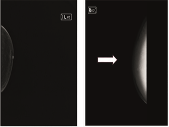
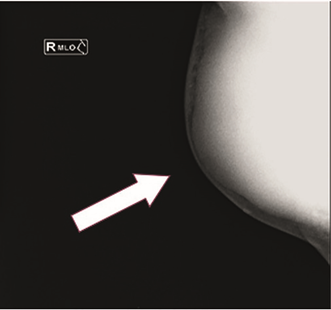
Male, 65 years old, originally from a rural community, farmer, incomplete primary school, married, Catholic. His mother had gastric cancer, the patient reported heavy smoking for 40 years, at a rate of ten to fifteen cigarettes per day, social alcoholism at a rate of two beers per month. Her condition began in April 2018 with pain in the right mammary gland, presence of a palpable, painful mass, with an increase in pau Latin volume, movable, with a stony sensation, sudden weight loss in six months, approximately 15 kg, intolerance to the oral route, unchanged excretions. In November 2018, she underwent an evaluation by a surgical oncologist, who indicated that she should undergo paraclinical and laboratory studies, with results of a BI-RADS 5 breast ultrasound and BI-RADS 5 mastography suggestive of malignancy (Figures 1 & 2).

Figure 1 Mammography with craniocaudal projection. Image of left breast without alterations, right breast with presence of radiolucent tumor, with architectural distortion, circumscribed margins, round shape.

Figure 2 Mammography with mediolateral-oblique projection and evidence tumor in the region of the right mammary gland.
Postero-anterior chest teleradiography showed data of tumor mass in the right hemithorax, as shown in Figure 3. The histopathological report showed a malignant phyllodes tumor, which can be treated with neoadjuvant treatment in January 2019. The patient began treatment with chemotherapy based on epirubicin, ifosfamide, without adequate tolerance to it, he presented upper respiratory tract infection after the application of the chemotherapy cycle, the hemoglobin report in the first days of February 2019 had a value of 9.4 mg/dl, an erythrocyte concentrate was transfused, treatment with clonazepam, tramadol and paracetamol. On February 10, 2019, he was admitted to the second level of care due to pain in the tumor area and generalized malaise, subsequently his relatives requested voluntary discharge to be transferred to his home, the patient died at home on February 28, 2019 due to complications of underlying pathology. A verbal autopsy was performed as part of the epidemiological surveillance of the reported case, the possible causes of death, direct or indirect, were analyzed, in addition to analyzing the failures of medical care. Of the possible causes in the critical links analyzed, a delay in diagnosis was found, since the patient did not go for medical examination in a timely manner, rapid progression of the disease, malignant phyllodes tumor with metastasis to abdominal organs, with the outcome of the case being death.
Phyllodes tumors of malignant origin are not rare among breast tumors. There is underreporting of cases such as presented in this study in the national and international literature.14–16 The idiosyncrasy, the lack of knowledge about certain diseases, as well as the prejudices and taboos in male patients make them take little care of their health, so early diagnosis of breast cancer in this sex is more difficult to obtain than in women, since the patient does not usually go to preventive appointments and there is no registration document for clinical breast examination in men, so the registration of such examination in the health record of these patients is required. Breast cancer in men is less prevalent and there are few cases in relation to the diagnosis in women. Phylloid tumors are rare tumors, which present epithelial and mesenchymal components, and have a particular recurrence or metastatic behavior. These types of tumors are diagnosed late when they occur in men, since the patient does not routinely go to the doctor and they are confused with other types of diseases.17 Early, timely and accurate diagnosis can provide a good prognosis for the patient, as well as quality medical care; it is important to raise awareness of the preventive model of health care in men in order to avoid the development and severity of diseases that can put their lives at risk.
To the patient's relatives for allowing this study to be carried out and thus contributing to the medical literature in Mexico on breast cancer in men. Special thanks to Dr Fernando González Figueroa for reviewing this work.
The author declare that there are no conflicts of interest.

©2023 Lazcano. This is an open access article distributed under the terms of the, which permits unrestricted use, distribution, and build upon your work non-commercially.