eISSN: 2469-2794


Case Report Volume 7 Issue 3
Department of Forensic Medicine, University of Sri, Jayawardenepure, Sri Lanka
Correspondence: Ariyarathna HTDW, Senior lecturer, Department of Forensic Medicine, Faculty of Medical Sciences, University of Sri Jayewardenepura, Gangodawila, Nugegoda, Sri Lanka, Tel +94 11 2802030
Received: April 20, 2019 | Published: May 24, 2019
Citation: Ariyarathna HTDW, Hulathduwa SR. A remote death following Whipple procedure with significant Forensic pathological dilemmas. Forensic Res Criminol Int J. 2019;7(3):129-133. DOI: 10.15406/frcij.2019.07.00276
Whipple procedure is a major surgical operation to remove the head of the pancreas, duodenum, gallbladder and the bile ducts most commonly performed for malignant tumors involving the head of the pancreas and distal bile ducts. It is also performed following pancreatic or duodenal trauma as well as chronic pancreatitis. The outcome depends on numerous factors. There are few recognized complications among which sepsis stands out prominently. The deceased in this case discussion had undergone Whipple procedure within three months after the onset of initial cluster of symptoms. Abdominal pain had been the most prominent symptom he presented with. The surgery was uneventful. The patient had been discharged on the 8th day following surgery. He had been advised to get the wound dressed every other day and visit the clinic in regular intervals. The deceased had not fully complied with the instructions and after observing a purulent discharge from the skin wound, a wound debridement had been done around the fourteenth day post op. The condition had gradually deteriorated since then and the deceased was re-admitted to the same ward on the eighteenth day following the surgery where he succumbed to death due to sepsis with metabolic acidosis. A judicial autopsy was performed following an inquest to exclude any possible allegations of medical negligence. Mild icterus was noted on external examination. Macroscopic features of sepsis with surgical site infection and generalized peritonitis were observed during dissection. Pancreaticojejunal, hepaticojejunal, gastrojejunal and jejunojejunal anastomosing sites were free of features suggestive of leakage. The cause of death was finalized according to the WHO guidelines.
1.a Septicemia,
1.b Whipple procedure and
1.c Distal cholangiocarcinoma.
The difficulty in diagnosis of sepsis at autopsy, identification of exact cause of sepsis in a post-surgical death, the significance of objective recording of the grade of surgical site infection and incorporation of surgical procedure in the wording of the cause of death are some of the forensic pathological issues to be addressed in this case. In addition to arriving at a diagnosis of sepsis, a forensic pathologist also owes an obligation to attempt to find out the root cause/causes for sepsis.
Keywords: sepsis, surgical site infection (classification), Whipple procedure, cholangiocarcinomaWhipple procedure is one of the most intricate and difficult surgeries in gastroenterology with several recognized complications.1 Peri-operative mortality and morbidity of this procedure had been over 20% and 50% respectively until 1970s.2 Due to recent advances in operative surgery, in most centers across the globe, the mortality rate has come down considerably and at present remains consistently below 4% though the morbidity still remains at a high figure.2 There are certain recognized predicting factors affecting the prognosis after surgery. The size of the tumour, degree of differentiation, lymph node involvement and the nature of the resected margins as well as the general co-morbid factors of the patient etc. do have a direct impact upon the outcome.3 Being a complex surgery, 43% of patients undergone this procedure still show post-operative complications in the form of cardiopulmonary complications, delayed gastric emptying and sepsis. Furthermore, an additional surgery was required in 3% of patients. Certain peri-ampullary complications, chronic pancreatitis, post-operative diabetes mellitus and other metabolic abnormalities are also recognized as rare but not-uncommon complications.4
A retired male school teacher of 69years of age had been apparently healthy until he developed severe abdominal pain as the first symptom. He sought treatment from a consultant physician. In the second visit a CT scan of the abdomen was done revealing a distal cholangiocarcinoma. For patients with the above condition, had the diagnosis been made early enough, pancreaticoduodenectomy (also called Whipple procedure) remains the best surgical option. Within a period of around three months of the diagnosis, Whipple surgery was performed on the patient in a specialized surgical unit in a tertiary care hospital in government sector. The operation was uneventful and he was discharged on the eighth day post-op. All necessary advice regarding postoperative care was given and he was referred to the surgical clinic to be seen at once-a-week intervals initially. He was well until two weeks after the surgery. Wound toileting was done every other day. At the end of the second week his wound on the surgical site appeared to be infected. He was admitted to the same surgical unit where debridement of the skin wound alone had been performed by a junior doctor. No ultrasound or any other form of imaging of the abdomen was performed. His abdomen was not examined by a consultant surgeon with a view of excluding any possibility of internal communication of the apparent cutaneous wound. He was discharged the same day. At home his general condition started to deteriorate further. The cutaneous surgical wound gradually got worsened despite strict adherence to instructions given by the ward upon discharge following wound debridement. More and more fowl-smelling purulent discharge was oozing from the wound. He developed high spiky fever with chills and rigors. His urine output was reduced and consciousness was frequently clouded. This deterioration took place rapidly within three days where he was re-admitted to the same unit on the eighteenth day post-op in a state of unconsciousness. The GCS was 3/15 and the blood pressure was unrecordable. The non-contrast CT brain was unremarkable. Haematological and biochemical investigations were in favour of sepsis with metabolic acidosis. The next day (day 20 post-op) he was pronounced dead. As the senior next of kin of the deceased had raised several medico-legal issues including the source of sepsis and its contribution to surgical procedure and the death being one with a potential for subsequent allegation of medical malpractice, an inquest into the death was requested to be followed by a judicial autopsy. At autopsy, the body was that of a thin built male with no external features of dehydration or emaciation. Mild icterus was noted. The sub-costal Kocher incision measured 28cmX2cm with necrotizing margins with visible underlying muscle. With the involvement of the distant organs in the form of peritonitis and meningitis etc. (discussed later), the surgical site infection could easily and safely be classified as “organ or space Surgical Site Infection (SSI)” (Figure 1).5
Routine dissection of all organs was done during internal examination. The brain weighed 1400g. It was dusky red in colour with marked congestion and features of meningitis. Cut sections revealed areas of necrosis with clear demarcation of grey and white matter (Figure 2). His neck structures were unremarkable. The lungs were congested and beefy red in colour, the right lung weighing 360g and the left lung 350g. No features of pulmonary embolism were evident to the naked eye. The pleural cavities contained blood-stained serous fluid, 160 ml and 200ml respectively on the left and the right sides. The pericardium contained 75ml of purulent fluid with dense adhesions to the outer surface of the heart (Figure 3). The heart weighed 360g. Major coronary arteries and their principal branches were not calcified and the maximum atherosclerotic narrowing was 30%. The heart valves appeared unremarkable.
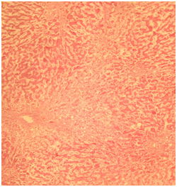
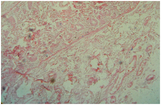
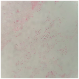
The endocardium appeared normal and the myocardium was flabby and pale. The epicardium was covered with yellowish, shaggy fibrous exudate. The undersurface of the diaphragm overlying the surface of the liver too was covered with a thick layer of frank pus. The peritoneal cavity contained approximately 350ml of sero-purulent fluid with offensive smell. Approximately a half of the left lobe of the liver was surgically resected. Pancreaticojejunal, hepaticojejunal, gastrojejunal and jejunojejunal anastomosing sites were free of features suggestive of leakage. A thin yellow purulent exudate was seen over entire peritoneal surfaces. Remaining segments of the small intestinal serosa was congested and flakes of pus were seen adhered onto the surface. No internal hemorrhages were noticed. The large intestinal serosa was also congested and was covered with a thick layer of pus. Both kidneys were normal in size and shape but pale in colour with no visible cortico-medullary demarcation. The bladder contained small amount of cloudy urine with an offensive smell. The bladder mucosa was not shiny and was covered with a thin layer of pus. The mucosa of the ureters too had the same appearance. The remaining portion of the pancreas did not show any overt macroscopic features of acute or chronic pancreatitis. The spleen was enlarged, soft and flabby with a thin layer of pus on the outer surface. It weighed 230g (Figure 4). The histopathology confirmed the gross changes seen and some micrographs are shown here Figures 5 A‒D.

5(A) Fatty liver.

5(B) Kidney.

5(C) Small intestine.

5(D) Cerebrum.
Figure 5 (A) Fatty changes shown in the liver. (B) The acute tubular necrosis with features of early renal failure. (C) Ischemic changes of small bowel. (D) Features of meningitis with acute inflammatory cells.
The cause of death was finalized according to the WHO guidelines.
1. a Septicemia
1. b Whipple procedure
1. c Distal cholangiocarcinoma
The above description gives an account of the general facts and findings of the case. The autopsy had been carried out by a consultant forensic pathologist in the presence of three post-graduate trainees in the form of a practical training session. During the external examination it was noted that none of the trainees has attempted to connect the description of the external surgical wound with the degree of surgical site infection. The naked eye autopsy features of sepsis were not very familiar to the trainees. Therefore, the authors wish to give a brief overview of some important medico-legal and forensic pathological issues encountered in this type of complicated cases for the benefit of the trainees in forensic pathology. Let us first have a glance on the criteria used in clinical practice to grade/classify Surgical Site Infections (SSI). Yet, this should not under any circumstances restrict a forensic pathologist in giving his opinion, which means that the clinical grading of SSI should only be used by a forensic pathologist to guide himself in expressing the autopsy findings as well as a tool for the clinicians to have a better glimpse of the naked eye autopsy finding as to the extent of surgical site infection and sepsis. A forensic pathologist may peruse surgical clinical notes, investigation results (such as CT or ultra sound imaging) and other ante-mortem data. Yet to his greatest advantage, he has the luxury of opening up of all body cavities, dissection of all organs and histopathoilogic examination of any relevant tissue with routine and special stains as well as sending postmortem samples such as heart blood and spleen cultures for microbiologic studies. Therefore, the forensic pathologist should be considered as the most reliable person to express an opinion regarding the degree and extent of SSI and its impact on the deceased. Forensic pathologists lack of knowledge on the exact surgical procedure performed on the deceased should be overcome by the clinician in charge of the deceased during life attending the autopsy to enlighten the pathologist on relevant aspects. This will build up a bi-directionally rewarding healthy clinico-pathologic correlation.
The objective data provided by the forensic pathologist regarding SSI especially in patients who die at home several days following operations or who die suddenly and unexpectedly after being discharged from the unit, would definitely be important to the clinicians to have a better understanding of the outcome of certain procedures not too often performed in their units. Retrospective analysis of postmortem reports should provide objective facts as to the degree of SSI. Three types of Surgical Site Infections (SSI) are described in operative surgery. Surgical site infection is defined as “an infection that occurs within 30 days after the operation and involves the skin and subcutaneous tissue of the skin incision and/or the deep soft tissues (Eg. fascia, muscle) of the incision (deep incisional) and/or any part of the anatomy (Eg. organs, cavities and spaces) other than the incision that was opened or manipulated during an operation (organ/space)”. (Source: European Centre for Disease Prevention and Control).6 The patient under discussion had shown a satisfactory recovery until up to the 14th day following the surgery. Thenceforth, the surgical site had shown swelling, oozing, redness and discomfort etc. A surgical wound toilet (debridement) had been performed once. Every other day wound toilet was done though day by day the wound got worsened along with general deterioration of the patient. So it is reasonable enough to assume that he had surgical site infection: “organ or space SSI” as per the above mentioned classification.
This patient has died shortly after the last admission. During that limited period of stay in hospital, few basic investigations were done and the diagnosis of ‘sepsis with metabolic acidosis’ was arrived at. The forensic pathologist was in agreement with this diagnosis. Therefore, the next duty of the forensic pathologist is to find out the contributory causes for sepsis. Any significant infection in a vulnerable individual could progress into Systemic Inflammatory Response Syndrome (SIRS), sepsis, severe sepsis and septic shock depending on the degree of contribution of the infection towards deranging the physiological mechanisms. All these entities are clinical diagnoses. Yet, it does not exclude the duty of the forensic pathologist in trying his best to find out the contribution of multiple factors in the causation of sepsis. Human sepsis is a spectrum of patho-physiological changes in the host system resulting from a generalized activation and systemic expression of the host's inflammatory pathways in response to infection. This is often characterized by end-organ dysfunction usually away from the primary site of infection.
Medical literature identifies few common sites of infection that could often act as sources of sepsis. They include pneumonia, haematogenous infections, intravascular catheter-related sepsis, intra-abdominal infections, urological sepsis and surgical wound infections etc.7,8 Surgical sepsis accounts for 30% of all patients with sepsis.9 In this particular patient, Whipple procedure itself, old age, presence of a malignant tumour, lack of nutrition etc. could have contributed in different proportions in the causation of sepsis.10 In addition, the more recognized sources of sepsis such as a focus of pneumonia, urinary tract infection, intra-abdominal infection etc. should also be considered. Along with other possibilities, the surgical site infection too could have led to sepsis in the background of his already mentioned risk factors. It is believed that most surgical site infections are due to contamination with the patient’s own flora as involvement of outside pathogens is relatively rare.10 As the senior next of kin was very keen in knowing the exact source of sepsis, the above possibilities were explained to him together with the inherent limitations in the diagnosis of exact source of sepsis at autopsy. Further, the authors wish to express the difficulties encountered by a forensic pathologist in arriving at a diagnosis of sepsis in relation to deaths occurring outside hospital as there are no specific, qualified and objective autopsy features to diagnose sepsis during post-mortem examination. Autopsy cultures (spleen and heart blood etc.) are most of the time unrewarding. Most of the other methods conventionally used in favour of the diagnosis of sepsis are similarly non-specific and unconvincing. As such, attempts have been made to utilize more innovative biochemical and immunohistochemical methods in better defining the criteria for autopsy diagnosis of sepsis.11–13 A reliable autopsy diagnosis of sepsis remains challenging.
Extensive sampling of blood and tissues for microbiology has been attempted but the validity is doubtful mostly because of postmortem contamination. For the macroscopic findings of sepsis to be of any value, the degree of sepsis should be severe. For example, in this case under discussion, pneumonic consolidations of lungs, features of meningitis, peritonitis and pyelonephritis etc. were evident macroscopically. Yet, in early cases of sepsis, such gross findings may be absent though the condition may well prove to be fatal. Thus, the diagnosis of sepsis remains challenging in forensic pathology in most occasions, despite improved investigative methods of collecting blood and tissue samples for postmortem bacteriology and extensive research in biochemical and immunohistochemical investigations. As explained earlier, the macroscopic findings are nonspecific and microscopic findings are elusive. CRP, procalcitonin and interleukin-6 are already is being used in certain centres for the diagnosis of sepsis though there is still no settled proposition as to the specificity of such markers. Identification of reliable markers of sepsis is quite difficult as the biochemical profile after death is subjected to considerable changes due to molecular leakage, molecular redistribution and molecular denaturation encountering restrictions in the application of the postmortem biochemistry.14 The existing challenge of diagnosing sepsis in autopsy work should be overcome by carrying out more research on this aspect. Forensic pathologists should pay due caution in wording the cause of death in the absence of solid evidence in favour of sepsis.
This case under discussion showed obvious features of sepsis both clinically and during the autopsy. Yet, in the absence of such glairing findings, due caution should be exercised before concluding the cause of death as sepsis. An autopsy report should be objective, factual and based on scientific evidence. It should contain material for an independent third party to arrive at the same or similar conclusions upon retrospective perusal on a later date; this perhaps being even several decades later. Accordingly, objective validity of the autopsy report may be improved if the forensic pathologists too use the same clinical classification for grading the Surgical Site Infections (SSI). After diagnosing sepsis, a forensic pathologist should also attempt to find out the root cause/causes for sepsis. This will help clinicians in better understanding the complete clinical picture of the deceased.
The author declares that there are no conflicts of interest.

©2019 Ariyarathna, et al. This is an open access article distributed under the terms of the, which permits unrestricted use, distribution, and build upon your work non-commercially.