Advances in
eISSN: 2377-4290


Case Report Volume 5 Issue 3
Department of Oculoplastic and Orbit, Centro Ocular Corredor Oftalmología Especializada, Venezuela
Correspondence: Rafael Corredor-Osorio, Av Bolívar, CC Las Acacias, local 31, Valera (Trujillo) Venezuela, Tel 58-0241-8255336
Received: October 20, 2016 | Published: November 25, 2016
Citation: Corredor-Osorio R. Unilateral congenital double lacrimal sac fistula. Adv Ophthalmol Vis Syst. 2016;5(3):262-263. DOI: 10.15406/aovs.2016.05.00157
A 10 years old girl presents to our eye clinic with a chief complaint of watering from right eye and for the evaluation of two small holes present inferonasal to the medial canthus since birth. On physical examination two small holes were present near the nose below the medial canthal of right eye that were associated with congenital double lacrimal sac fistula. Lacrimal sac regurgitation test was positive on both fistulas. Evaluation of the lacrimal system was performed under general anesthesia. Both fistulae were connected to the sac lacrimal by probing the orifice of the fistulae and finding that probe ran in the direction of the sac lacrimal, and she was treated successfully by primary fistulectomy. The postoperative course was free of symptom at 4 years follow-up. In review of the literature, only 1 case of congenital double lacrimal fistula has been previously reported.
Keywords: lacrimal fistula, lacrimal sac, congenital fistula, fistulectomy
A congenital lacrimal fistula is a rare developmental anomaly of the lacrimal system typically located in the inferomedial aspect of the medial canthus.1-3 The fistulae can originate from common canaliculus, lacrimal sac or nasolacrimal duct of skin.4-7 In 1675, the first description of congenital lacrimal sac was reported by Rasor.7-9 This paper is to report on atypical case of unilateral congenital double lacrimal sac fistula.
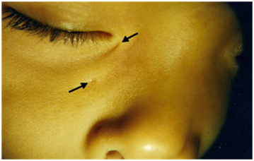
A 10 year-old girl presented to our clinic with chief complaint of epiphora from right eye and for her evaluation of two small holes that his parents noticed inferonasal to medial canthal of this right eye. His mother stated that the holes had been present from the birth. She had no complaints and was in good health. There was no past history of systemic disease, eyelid surgery, trauma or any relevant family history. Slit lamp examination of the anterior segment and indirect ophthalmoscopy of the posterior segment were unremarkable. On physical examination showed a 1mm hole approximately 5mm inferonasal to the medial canthal and a second 1 mm hole approximately 14 mm near the nose below the lower eyelid of the right eye (Figure 1). There was minimal discharge occasionally from both holes. Lacrimal irrigation of saline through the lower punctum demonstrated nasolacrimal competence and a communication with both holes. The fluorescein dye disappearance test was normal. Surgical removal of the fistulae was planned as the course of treatment.

Figure 1 The presence of congenital lacrimal fistulas (arrows) located just inferonasal to the medial canthal angle.
Under general anesthesia, standard surgical preparation were carried out, Lidocaine 2% with adrenaline was infiltrated as required along the incision for the purpose of haemostasis. The lacrimal fistula was probed with a No 0 Bowman lacrimal probe to the lacrimal sac. A second probe was inserted into lower canaliculus and sac lacrimal (Figure 2). Fine subcutaneous dissection under microscope is performed. A skin fusiform incision was made around conservatively the orifice of the fistula. Both the tracts were dissected completely down to their junction the lacrimal sac with Westcott scissors. The probe was removed and the remnant tract was ligated with 6/0 polyglactin 910 suture and no regurgitation of fluid was noted on syringing. The fistulous tract was then cut above the tie, and the subcutaneous tissue and skin was closed in layers (Figure 3). At follow-up after four years, the patient was asymptomatic.
Congenital lacrimal fistula are uncommon developmental anomalies of the nasolacrimal excretory system with an estimated incidence of one in, 2000 births3,10,11 and occasionally can be inherited in an autosomal dominant1,4,7,11 or recessive pattern.1,3,4,9 They are usually unilateral2,3,5,12 but familial cases are associated with higher incidence of bilaterally.4,9 There does not appear to be a sex or race predilection.2,6,8,12 In most of the cases they are asymptomatic3,6,11 however in some cases it may present with epiphora or discharge2,3,9 or mucoid secretion may be expressed by placing pressure on the sac, causing reflux.11,12 The nasolacrimal system is usually patent.2,8 The clinical presentation may delay for many years after birth due evaporation of small amounts of discharge.2 In this case, the patient had history of epiphora since birth and had no associated infectious complication. A review of the literature, only 1 case of congenital double lacrimal fistula has been previously reported with both fistulae connected to the common canaliculus.13 In our case, the fistulae were connected to the lacrimal sac.
During the sixth week of embryological development, neuroectodermal cells which are in the naso-optic groove between the lateral nasal and maxillary processes form a solid epithelial cord. After canalization the upper portion of the cord forms the canaliculus and the lower part forms the nasolacrimal canal. In the middle part forming the lacrimal sac, the epithelial cords are thicker, and therefore when the sac canalizes it has a larger diameter than the canaliculus or the nasolacrimal duct.12 An incomplete separation of the cord from the surface epithelium or an abnormal out-budding of the buried ectodermal cord can result in supernumerary puncta or canaliculus.2,5,9
The pathogenesis of lacrimal sac fistula is uncertain.7,8,10,12 Some authors suggest the following explanation: a failure of the surface ectoderm to fuse after invagination of the epithelial cord;10 an overdevelopment or out budding of the lacrimal duct ; an abnormality of amniotic bands ; a primary developmental arrest with secondary inflammation; an incomplete closure of the embryonic facial fissure;7,8,12 that the lacrimal fistula is really an extra canaliculus extending from the common canaliculus to the skin in the medial canthal area while the superior and inferior canaliculi are also being formed7 and that a defect interfering with the invagination, burial, and later tissue remodeling of the surface ectoderm cord was responsible.12 Most of these fistulae originate in the common canaliculus, in other cases they may arise from the lacrimal sac or nasolacrimal duct9 They can be seen externally as small orifices o pits characteristically located inferior and or medial to the medial canthi how to the present case.
Congenital lacrimal fistula can be associated with ocular pathologies including dacryocystitis, agenesis of the lacrimal punctum and canaliculus, lacrimal tract stenosis, strabismus, hypertelorism,3,9 double lower lid puncta, canalicular atresia , orbital meningocele, mucocele and nasolacrimal duct stenosis.4 Systemic associations with lacrimal fistula are so rare it requires genetic testing. Associated systemic disorders including CHARGE syndrome (coloboma, heart anomalies, choanal atresia, retardation of growth and development, genital and ear anomalies), Waardenburg-Klein syndrome, ectrodactyly-ectodermal-dysplasia-clefting syndrome,4 VACTERL syndrome (vertebral anomalies, anal atresia, cardiac malformations, trachea-esophageal fistula, renal and limb anomalies), preauricular fistulae, hypospadias, thalassemia and Down syndrome,3,4,6 uterus didelphys, renal agenesis ectopic pelvic kidney2 have been reported.
The treatment of choice for a symptomatic fistula is surgery,7,9,11 these options includes complete excision of the fistulous tract, excision with intubation1,5,6,11 or dacryocystorhinostomy.1,4,8,10 In the case of lacrimal fistula without symptoms the observation is the best choice.3,4,8 In this case, the girl had only lacrimation through the lacrimal fistulas, and there was no combined nasolacrimal duct obstruction. Fistulectomy alone caused the girl to be free of symptoms at 4 years post operatively a surgical procedure on a congenital lacrimal sac fistula, it is necessary to demonstrate adequate drainage from the nasolacrimal apparatus through a patent nasolacrimal duct. This can be done by dye disappearance and irrigation testing.
None.
Author declares that there is no conflict of interest.

©2016 Corredor-Osorio. This is an open access article distributed under the terms of the, which permits unrestricted use, distribution, and build upon your work non-commercially.