Advances in
eISSN: 2377-4290


Opinion Volume 12 Issue 1
Ophthalmologist- Retina specialist Marashi Eye Clinic Aleppo, Syria Consultant Ophthalmologist, Head of Retinal Imaging Centre, Syria
Correspondence: Ameen Marashi, Ophthalmologist- Retina specialist Marashi Eye Clinic Aleppo, Syria, Tel 0092 51 8439993
Received: January 21, 2022 | Published: February 11, 2022
Citation: Marashi A, Khan H. Subretinal hard exudates. Adv Ophthalmol Vis Syst. 2022;12(1):1. DOI: 10.15406/aovs.2022.12.00407
Hard exudates are an accumulation of lipid-laden macrophages which develop due to leakage from compromised inner retinal blood barrier.i On SD-OCT, hard exudates appear as hyperreflective intraretinal lesions casting a dense shadow on the deeper layers. They are usually seen in the mid to outer retina. We present a series of 3 diabetic patients with center involving chronic cystoid macular edema (CME) with an unusual OCT appearance of hard exudates, seen herniating and settling in the subretinal space.
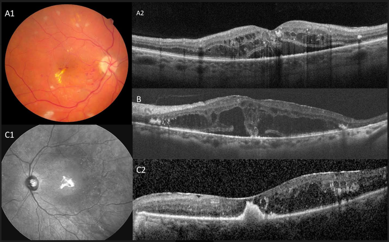
Case 1, a 64 yrs old diabetic female with best corrected visual acuity (BCVA) 20/40 OD and was treated with intravitreal bevacizumab injections (Figure A1) She had chronic CME and a break in the outer retinal layers is visible. Central hard exudates are seen herniating from intraretinal into subretinal space (Figure A2). Case 2 (Figure B), a 45 yrs old male with chronic CME, BCVA 20/80 OS shows hard exudates settling onto the RPE. Case 3, a 58 yrs old diabetic male treated with multiple intravitreal bevacizumab injections. The central hard exudates have condensed in the subretinal space (Figure C1) and the outer retinal break can no longer be seen (Figure C2). This gives a false appearance of a pigment epithelial detachment.

Figure 1 All cases are patients with non proliferative diabetic retinopathy with chronic exudative macular edema. Case 1 Fundus photograph shows right eye of 62 yrs old female with non proliferative diabetic maculopathy and chronic macular edema with central hard exudates (A1). SD-OCT shows a break in the outer retina and the hard exudates are seen herniating into the subretinal space (A2). Case no.2, SD OCT central macula of a 45 yrs old diabetic, a similar break and herniation is seen and the hard exudates are seen settling on the RPE (B). Case 3, SD OCT of 55 yrs old diabetic. With treatment and as the edema resolves these hard exudates condense (C1). On OCT, these give an appearance of a pigment epithelial detachment (C2).
Chronic cystoid macular edema is associated with loss of tissue and intervening septa between the cystic spaces.ii This may result in a break in the outer retina which has been postulated as the cause of subretinal fluid in diabetic maculopathy.iii Similarly, hard exudates are seen in these cases to migrate sub retinally. Since the level of hard exudates are difficult to determine on fundus examination alone, the level of these sub retinal exudates are more accurately seen on OCT. This subretinal location is unusual in diabetic maculopathy and may cause diagnostic confusion. Furthermore, similar to subretinal fluid, these subretinal hard exudates may indicate suboptimal treatment response.iv
None.
There are no financial conflicts of interest.
None.

©2022 Marashi, et al. This is an open access article distributed under the terms of the, which permits unrestricted use, distribution, and build upon your work non-commercially.