Advances in
eISSN: 2377-4290


Case Report Volume 8 Issue 6
1Director at Oftalmo Abad, Clínica de Ojos, Argentina
2Resident of Ophthalmology, Argentina
3Director HORA, Brazil
Correspondence: Dr. Luis Emilio Abad, Director at Oftalmo Abad, Clínica de Ojos, Río Gallegos, Santa Cruz, Patagonia Argentina, Argentina
Received: December 07, 2018 | Published: December 26, 2018
Citation: Abad LE, Mauro CRG, Carlos ECJ. Hamartoma simple congenital of retina, with OCT and ultrasonorography evaluation. Adv Ophthalmol Vis Syst. 2018;8(6):246-247. DOI: 10.15406/aovs.2018.08.00330
Purpose: To report in detail all the findings of optical coherence tomography (OCT) and ultrasound in a case of simple congenital hamartoma of the retinal pigment ephitelium (RPE).
Design: Observational case report.
Methods: Fundus examination, retinography, ultrasonography and OCT were done in one case of simple congenital hamartoma of the retinal pigment epithelium.
Results: a considerable, paramacular, solitary and highly pigmented lesion was observed in the right eye of an 11-year-old girl. Visual acuity was 20/60 in the eye with paramacular lesion. The ultrasound showed a hyperreflective nodule with echoes of medium and high reflectivity. The OCT revealed a high reflectivity of the surface of retinal mass with full increase thickening, with abrupt and total shadow of the optical transmission. It presented a solid vitreomacular adhesion on the border of tumor lesion. Macular thickening did not affect the surrounding RPE and choroid.
Conclusión: OCT is an important diagnostic method, useful and non-invasive to evaluate this type of tumor lesion. This diagnostic study may show in some cases (and this in particular), a vitreoretinal adhesion with thickening of borders of the tumor and adjacent retina.
This type of tumor (simple congenital hamartoma of the retinal pigment epithelium (RPE) is a fairly rare lesion of low prevalence. Diagnosis is usually made casually in healthy and asymptomatic children, as well as in young adults, during a routine fundoscopy in general ophthalmological consultation. Optical Coherence Tomography (OCT) has recently become an important tool for the evaluation of retinal anatomy and retinal abnormalities. Few reported cases have been written about the role of OCT in the evaluation of ocular tumors¹. In this report, we describe the OCT findings of this highly pigmented retinal tumor.
Keywords: eye, tumor, retina, congenital simple hamartoma, retinal pigment epithelium, optical coherence tomography, diagnosis
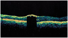
An 11-year-old girl consulted with her parents and reported that the girl had low vision with her right eye. Her parents reported that the girl did not have trauma to that eye nor had a history of eye inflammation. Snellen's visual acuity was 20/80 in her right eye and 20/20 in her left eye. In the biomicroscopy, there were no significant alterations, the ocular pressure remained within normal limits (14mmHg in both eyes) and, in the fundoscopy of the right eye, revealed a circumscribed lesion in the paramacular area, with irregular borders, nodular type and dark brown pigmentation affecting the RPE, which presented slight hyperplasia, with an increase in retinal thickness in the lower paramacular area with minimal invasion into the vitreous cavity (Figure 1-retinography). The lesion measured 586 microns of basal dimension and 656 microns of thickness in the OCT. The temporal arches were slightly tractioned to the lesion area with elevation of paramacular retina. There was no atrophy of the RPE at the level of lesion or macular edema or subretinal fluid. Ultrasound showed a nodular mass of high echogenicity in the paramacular area. The OCT revealed a prominent reflectivity of the retinal surface at the level of lesion, with an increase in the local thickness of the retina and slight disorganization of the paramacular RPE, causing an abrupt and complete shading of the optical transmission. The RPE outside the limits of lesion next to the choroid did not present alterations. The vitreous presented adhesion to the borders of lesion, by invasion of it to the vitreous cavity.1–3
The retinal Hamartoma was named by Dr. Donald M. Gassen in 1989 and described this pathology as raised, superficial and full thickness. The lesion presented in this case describes it as a tumor lesion with hyperplasia components of the RPE, without apparent vascularization.2,4 The hamartoma is supposedly congenital, with stable presentation and located in the lower paramacular area, favoring this diagnosis to maintain an acceptable visual acuity and a good prognosis. The lesion is not the cause of low vision, it is located in the paramacular area away from the fovea, but the hyperplasia of the EPR is what causes the visual decrease. There are not many valuable OCT data in cases of tumors in the RPE and retina (Figure 1-3).

Figure 1 (C) OCT revealed prominent surface reflectivity of the full thickness retinal mass with abrupt and complete shadowing of optical transmission.
The OCT in this case showed increased margins of the tumor as well as the adjacent retina. All the information that was obtained through the diagnostic methods together with the bibliography found confirms that it is a rare and infrequent tumor.
OCT is an important diagnostic method, useful and non-invasive to evaluate this type of tumor lesion. This diagnostic study may show in some cases (and this in particular), a vitreoretinal adhesion with thickening of borders of the tumor and adjacent retina.
This type of tumor (simple congenital hamartoma of the retinal pigment epithelium (RPE) is a fairly rare lesion of low prevalence. Diagnosis is usually made casually in healthy and asymptomatic children, as well as in young adults, during a routine fundoscopy in general ophthalmological consultation. Optical Coherence Tomography (OCT) has recently become an important tool for the evaluation of retinal anatomy and retinal abnormalities. Few reported cases have been written about the role of OCT in the evaluation of ocular tumors¹. In this report, we describe the OCT findings of this highly pigmented retinal tumor.
None.
Author declares there is no conflict of interest towards the manuscript.

©2018 Abad, et al. This is an open access article distributed under the terms of the, which permits unrestricted use, distribution, and build upon your work non-commercially.