Advances in
eISSN: 2377-4290


Case Report Volume 13 Issue 1
1Contract tutor of Optic& Optometry, University of Milano Bicocca, Italy
2Materials Science Department, University of Milano Bicocca, Italy
Correspondence: Gian Luca Verrenti, Contract tutor of Optic& Optometry, University of Milano Bicocca, Milan, Italy
Received: November 10, 2022 | Published: January 12, 2023
Citation: Verrenti GL, Biancofiore N, Mocciardini E, et al. Application of corneo-scleral contact lenses on a subject with aqueous deficient dry eye syndrome - case report. Adv Ophthalmol Vis Syst. 2023;13(1):5-6. DOI: 10.15406/aovs.2023.13.00429
In this study was customized a corneo-scleral contact lens for both eyes on a subject suffering from aqueous dry eye syndrome. Here below will be described the analysis of the various application attempts and changes made exclusively to the left eye, in order to identify the suitable lens for use, subsequently carrying out a follow-up of the case for a month.
Corneo-scleral contact lenses are made of rigid gas permeable material that allows to unload its weight both on the corneal portion and on the scleral portion of the eye. The corneo-scleral contact lenses are formed by a central portion shaped with the curvature of the corneal surface; a peripheral portion that allows to obtain a lifting of the lens to the limbal area of the cornea and a third portion that allows the rest of the lens to be alignined with the scleral portion. Each of these three portions is distinguished by a different curvature value. It is possible to modify the BOZR value, the value of the peripheral curve and the total diameter of the lenses separately. The lenses used are made of Ruflufocon D, a material formed by polymers of fluorine silicone acrylate which allows to make the lens more resistant to the formation of deposits on its surface and consequently improve tear stability and slow down its breakage.1,2
The subject of this study is a 25-year-old man with no previous eye conditions. In the preliminary phase were observed grade 2 of bulbar redness (with CCRLU grading scales), grade 1 of limbal redness and absence of corneal opacification and staining. In the conjunctival area there is micro-pointed and superficial grade 2 staining, widespread in the nasal and temporal areas. The subject obtained a score of 12.5 on the OSDI questionnaire, a parameter that can be considered as a "mild dry eye condition". After examining the tear dynamics, with the use of fluorescein, a linear rupture of the lacrimal fluid, post blinking, was observed (line break). This circumstance is associated with a mild condition of dry eye, caused by a deficiency of the aqueous component (ADDE).3 In the anamnestic phase, the subject complains of dissatisfaction, in terms of comfort, during the wearing of soft contact lenses.
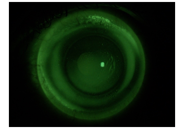
Following the execution of the corneal topography, the subject presents the parameters of curvature and size of the iris visible for the left eye, listed in the following Table 1. From the application protocol, the trial set lens applied is the one with 7.94 mm BOZR, PC unchanged compared to the BOZR and DT of 14.00 mm. The fluorescein pattern after 2 hours of wearing, shown in Figure 1, shows that there is excessive lifting in the central optical zone, so much so that the fluorescence, by gravity, settles downwards. In the pericheratic area, a too closed tear reservoir is generated, which assumes an inverse hourglass shape, due to the astigmatism against the rule of the subject's cornea.
Sim K1 |
8,07 mm |
41,82 D |
116 @ |
Sim K2 |
7,95 mm |
42,45 D |
26 @ |
Medium K |
8,01 mm |
42,13 D |
|
CYL |
-0,65 D |
116 Ax |
|
W-W |
12,06 mm |
|
|
Table 1 Execution of the corneal topography, the subject presents the parameters of curvature and size of the iris visible for the left eye

Figure 1 Fluorescein pattern of the lens with BOZR 7.94 PC 0.00 DT 14.00 in the left eye after 2 hours of wearing.
In theory, one could think of closing the vertical portion, in such a way as to allow greater compliance of the lens with the ocular asymmetry of the subject. This type of modification cannot be carried out by the manufacturer, therefore it was decided, as an alternative, to flatten the base curve by one step and increase the total diameter, in order to allow an improvement of the fluoroscopic picture. The new lens proposed will be one with BOZR 8.00 PC 0.00 DT 14.80. After 3 weeks of wearing this lens, the subject presented for the follow-up visit. After a 2-hour adaptation, the fluorscope picture was assessed (Figure 2). As soon as fluorescein was instilled, it was noted that the passage was limited only below the peripheral curve. Fearing possible adverse conditions, due to the excessive suction that this lens involves, the conformation of endothelial cells was observed by means of magnification increased to 40x and optical section, where it was precisely possible to observe the presence of small endothelial blebs (Figure 3). Therefore, it was decided to flatten the base curve by half a step, leaving the peripheral area unchanged. The subject was invited to wear this new lens with BOZR 8.05 PC 0.00 DT 14.80 for another 3 weeks.
By instilling fluorescein, unlike lenses applied during previous encounters, tear exchange occurs extremely quickly. The fluorescein pattern, which immediately formed, shows, compared to the application carried out during the first follow-up, an acceptable fluorescence in the center and, above all, a marked improvement in the lacrimal ring, due to the opening of the peripheral curve. The same peripheral aperture, however, causes excessive movement and instability of the lens (Figure 4).
None.
The authors declare that there are no conflicts of interest.

©2023 Verrenti, et al. This is an open access article distributed under the terms of the, which permits unrestricted use, distribution, and build upon your work non-commercially.