Advances in
eISSN: 2377-4290


Case Report Volume 7 Issue 2
Department of Ophthalmology, Rotherham NHS Foundation Trust, UK
Correspondence: Omar Rafiq, Consultant Ophthalmologist, Al Zahra Hospital, UAE, Rotherham, South Yorkshire, UK
Received: June 07, 2017 | Published: July 7, 2017
Citation: Lokovitis E, Rafiq O (2017) A Rare Case of Pigment Dispersion Syndrome in an 8-Year-Old Boy. Adv Ophthalmol Vis Syst 7(2): 00213 DOI: 10.15406/aovs.2017.07.00213
Pigment dispersion Syndrome (PDS) is a condition characterized by deposition of pigment throughout the anterior chamber and concave configuration of peripheral iris.1 Mid-peripheral iris transillumination defects and Krukenberg spindles constitute characteristic, but not universal findings of this clinical entity.2 PDS usually occurs in middle-aged myopic patients and it is uncommonly seen before 14 years of age.3
We herein describe a case of an 8-year-old boy with typical signs of pigment dispersion syndrome. The patient was referred to our department for further evaluation due to a “cobweb iris” seen by his optician. There was no family history of glaucoma and no history of trauma or surgery.
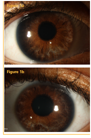
On examination the patient’s best corrected visual acuity was logMar 0.120 with -2.75/+1.75x111 in the right eye (OD) and logMar 0.20 with -2.25/+0.50 x 82 in the left eye (OS). Pachymetry showed the central corneal thickness was 679μm OD and 673μm OS. During slit lamp examination, the cornea and anterior chamberwere clear and quiet. Midperipheral radial iris transillumination defects were noted in both eyes (OU) (Figure 1a & 1b). The lens was clear and gonioscopy revealed open angles with 3+ pigmentation of the trabecular meshwork and concave configuration of the iris OU. The backbowing of the iris was confirmed with anterior segment optical coherence tomography (Figure 2a & 2b). The intraocular pressure was 15mmHg OD and 14mmHg OS. Fundoscopy identified optic disks with a cup-disk ratio of 0.7 OD and 0.6 OS (Figure 3a & 3b). Optical coherence tomography of the retinal nerve fiber layer indicated possible focal thinning of the temporal quadrant OS (Figure 4a & 4b). Humphrey visual field testing (24-2) was within normal limits OU.

Figure 1 Anterior segment colour photograph of the right eye (a) and left eye (b) demonstrating transillumination defects of the iris.
It is rare to find a child with typical signs of PDS in the age of 8 years old. To our knowledge this is the youngest case of typical PDS in the literature. A 14 year-old boy was the youngest patient in a clinical study which involved 407 patients with PDS.3 As our patient, this boy had an intraocular pressure within normal limits. In a series of 7 cases with PDS,4 the youngest patient was a 12-year-old boy, which was also normotensive. Although the patient had developed mild punctate transillumination defects at 7 years of age, there were no other signs of PDS until the age of 12 years.4 A12-year-old child was also reported with PDS, however, this case was associated with megalocornea.5 An atypical case of an 8-year-old child with PDS, elevated intra ocular pressure, miotic sluggish pupils, posterior subcapsular cataracts and peripheral anterior synechiae was presented.6 Even though our patient was the same age, the above mentioned clinical signs were not detected in our case, and although both cases presented increased central corneal thickness, our case was normotensive. Elevated intra ocular pressure was also a clinical feature reported in a case of an 11-years-old myopic girl with PDS.7 Although our patient was also myopic this feature was not universal for all the presented cases of PDS presented earlier.
The possible pathogenic mechanism responsible for the pigment dispersion in PDS, is the prolonged iridozonular contact between the concave peripheral iris and the anterior zonular packets.8 It is usually encountered in myopic patients, probably due to increased space within the Anterior Chamber and subsequent greater degree of iridolenticular contact. Moreover a recent study revealed an increased prevalence of iris concavity in young male population in UK without although indicating any increment in PDS prevalence.9 The authors stated that iris concavity and the associated iridocorneal contact must persist for a long period of time before the development of PDS occurs and that other features, intrinsic to the iris, must be present to induce pigment dispersion. It is possible that these features constitute the main factors of the early manifested PDS.
We have reported a rare case of early presentation of PDS without signs of glaucoma. There is no universal consensus on whether YAG laser peripheral iridotomy may prevent progression of PDS in pigmentary glaucoma.10,11 However, we decided not to perform YAG laser peripheral iridotomy, considering the possible adverse effects that could occur in a patient of such a young age, so we continued o monitor the patient.
None.
The authors declare no conflicts of interest.

©2017 Lokovitis, et al. This is an open access article distributed under the terms of the, which permits unrestricted use, distribution, and build upon your work non-commercially.