Advances in
eISSN: 2377-4290


Case Report Volume 8 Issue 3
Sri Sankaradeva Nethralaya, India
Correspondence: Dipankar Das, Department of Ocular Pathology, Uveitis & Neuro-Ophthalmology Services, Sri Sankaradeva Nethralaya, Beltola, Guwahati–781028, Assam, India, Tel 0361-2228879, 2305516, Fax #0361- 2228878
Received: January 01, 1971 | Published: June 15, 2018
Citation: Das D, Bhattacharjee H, Misra DK, et al. Evidence of zonular stress lines on human crystalline lenss. Adv Ophthalmol Vis Syst. 2018;8(3):186?187. DOI: 10.15406/aovs.2018.08.00299
Crystalline lens and zonules play an important role in accommodation. There are various clinical and theoretical explanations of tension zonules and its role in accommodative changes of the lens. We present the morphology of zonular stress lines (ZSLs) under bright field compound microscopy on the surfaces of transparent crystalline lenses in three human enucleated eyeballs removed for varied indications. ZSLs were evident on the periphery and paracentral part of crystalline lenses. Further study on these stress lines with advance imaging will give more important clue in the complex zonule-lens morphology.
Keywords: zonules, lens, enucleation, accomodation
The lens is an extremely planned system of focused cells which constitutes an important element of optical arrangement of the eye and fulfills the important functions of altering the refractive index of light entering the eye to focus on the retina.1,2 The transparency of lens is due to the shape, array, internal structure, and biochemistry of the lens cells or lens fibre.1 The lens is held in place by composite three dimensional systems of radially arranged zonules called zonules of Zinn or the suspensory ligament of the lens.1 These fragile fibres are attached to the lens capsule 2mm anterior and 1mm posterior to the equator and arise from the region of pars plana ciliary epithelium and pass forward closely related to the lateral surfaces of ciliary processes.1 The fibrous zonules blend with basal lamina of the lens capsule. The anterior and posterior zonules place in obliquely into superficial 1-2µm of pre- and post-equatorial lens capsule, while equatorial zonules insert at right angles.1–3 We present the documentation of zonular stress lines (ZSLs) for the first time in the scientific literature on the transparent crystalline lens from three enucleated eyeballs. These stress lines are the evidence of the zonules to take part in accommodation and also arbitrate accommodative movements. Although some believed that there are two types of zonules, first being ‘main zonules’ and other the ‘tension zonules’, the later being placed under tension during accommodation but there were no morphological different stress lines on the surface of lens in so called two varieties of zonules.1–3
This was a retrospective, observational and laboratory based study. Three crystalline lenses from three enucleated eyes for varied other indications were examined in ocular pathology laboratory in a tertiary institute of Northeast India. The informed consents were taken before the surgeries.
Case 1
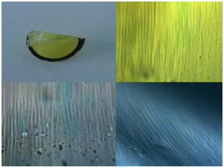
A 4 month old female child, clinically diagnosed as Group E retinoblastoma of the right eye. After enucleating the eye, gross examination of the eyeball was carried out and portion of transparent crystalline lens was brought out for direct examination under the bright-field compound microscope (Zeiss, AxioCam, MRc in Axioskop 40). An overview of stress lines of zonules on the surface of lens showed interesting observation. ZSLs were observed in the para-central portion and at the periphery of the lens. At the periphery, there were angulations of the stress lines which were documented (Figure 1).
Case 2
A 4 year old male child presented with white pupillary reflex of the right eye. A provisional diagnosis of retinoblastoma or Coat’s disease was made. Gross and microscopic examination of the specimen was consistent with Coat’s disease of the eyeball. Surface of the lens showed stress lines and were subsequently recognized (Figure 2).
Case 3
A 32 years old male patient presented to the institute with a history of penetrating trauma of the left eye and subsequently developed painful blind eye. Enucleation of that eye was done and after overnight fixation in 10% neutral buffered formalin, the eyeball was sectioned vertically. Lens was seen separately and ZSLs were observed on the anterior surface of the lens and brownish iris pigmentation was seen on the same surface with orientation of pigmentation along the stress lines (Figure 3). Microscopic examination of the eyeball showed there was evidence of endophthalmitis and special stains were carried out to know the causative organisms. We could clearly identify the presence of clefts by zonules on the surface of lenses particularly at the periphery of lenses with its extension to the para-central areas. They were multilayered and created linear impression on the surfaces of the lenses (Figures 1–4).

Figure 1 Specimen1 with gross photo of transparent crystalline lens. Adjacent microscopic picture showed the ZSLs visualized directly under compound microscope (Zeiss, AxioCam, MRc in Axioskop 40). Bending of the stress lines obliquely could also be seen in the inset (x 400).
The lens of the eye is a clear, biconvex, oval, semisolid, avascular body of crystalline structure located between the iris and the vitreous.4 The lens is unique among organs in that it contains cells exclusively of a single type, in various stages of cyto-differentiation and retains within it all the cells formed during its life time.4,5 Ciliary zonule consists basically of a series of fibers passing from the ciliary body to the lens.4,5 It holds the lens in place and helps ciliary muscle to act on it during accommodation.4 The zonules consist of thick, smooth bundles 5-30 µm in diameter. Each bunch consists of a series of fine fibres (0.35-1µm in diameter) and they composed of 8-12µm fibrils.1–4 One potentially applicable consequence of continued lens growth relates to altered zonulo-lenticular-ciliary body geometry and the resulting changes in degree and vector of the ciliary body and choroidal forces applied on the lens.5–8 Since the axial posterior surface of the lens remains permanent in position relative to the cornea and retina while lens grows, the central sulcus bisecting the lens equatorially is translated anteriorly, as is the actual lens equator.5–8 We had established the evidence of ZSLs on the anterior surface of the transparent crystalline lens in three specimens. These stress lines have clefts in a characteristic pattern on the surface of lenses which were prominent at the periphery and they could be traced towards the para-central areas. One can hypothesize that many optical consequences to these changes and even the anterior shift of the lenses could increase these ZSLs or zonular tension on the anterior capsule, making accommodation more difficult.9,10 We had seen that younger lenses in our series had less prominent ZSLs while lens seen in 32 year old man had more prominent ZSLs marking on its surface.
Sri Kanchi Sankara Health and Educational Foundation.
The Author declares that there is no conflict of interest.

©2018 Das, et al. This is an open access article distributed under the terms of the, which permits unrestricted use, distribution, and build upon your work non-commercially.