Advances in
eISSN: 2377-4290


Ichthyosis is a heterogenous group of dermatoses characterized by the presence of fish-like scales. The scaling is generally worse in winter. Ichthyotic disorders are usually inherited but may sometimes be acquired. Present case report describes a case of Lamellar Ichthyosis.
Ichthyosis is classified into congenital and acquired types which are further subdivided as:1
Lamellar Ichthyosis
Lamellar Ichthyosis is a rare congenital condition with equal distribution in both genders. At birth, child usually presents as a collodion (lacquered) baby ensheathed in a membrane; when the membrane sheds, typical scales manifest. Inheritance is Autosomal Recessive. It involves generalized body involvement which begins as collodion baby; after the membrane is shed, patient develops large thick, brown, pasted scales (fish- like) scales which persists for life. Flexures may show continuous linear rippling. Erythema is minimal; when present, it is maximum on the face. It is associated with generalized lesions, accentuated on lower extremities and flexural areas.1
Other associated features may include
Molecular defect involves abnormality of gene present on chromosome 14q11, which encodes for transglutaminase.2 Herein, we are presenting a case of Lamellar Ichthyosis associated with Bilateral Ectropion.
Lamellar Ichthyosis is extremely rare condition with autosomal recessive inheritance and is found equally in both sexes. It usually presents at birth and is associated with diffuse large, thick, brown pasted (Fish-like) scales with ectropion.
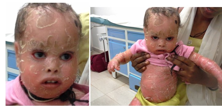
A 2-year-old female child was referred for the ophthalmology consultation with complaint of persistent watering in both the eyes since birth. As per narrated by patient’s mother, she was born out of full term normal vaginal delivery on 3rd of July 2012 at hospital with low birth weight of 2kgs for which she was kept in neonatal intensive care unit (NICU) after delivery. At birth, she was covered with a shiny membrane all over body (collodion membrane). She had history of scales all over the body like fish since birth (Figures 1–4). Three weeks after birth, ectropion of lower eyelids was observed by patient’s mother. The marriage of parents was consanguineous in nature as narrated by patient’s mother.

Figure 3&4 Generalize involvement of fish like scales all over body with Bilateral Ectropion in Lamellar Ichthyosis.
On general examination, the patient was malnourished with a weight of 5 kg, covered with fish-like dry scales with desquamation all over body. The child was fixing and following to light and responding to sound appropriately. Ocular examination revealed absence of eyebrows, presence of scales over lid skin and eye lashes and grade III ectropion in lower eyelids. Conjunctiva was normal in both the eye. Cornea was normal in both the eye and no evidence of xerosis or exposure keratopathy was present.
We reported the following clinical features in our case
On the basis of above mentioned clinical features, case was diagnosed as Lamellar Ichthyosis. It was due to the Bilateral Ectropion associated with Lamellar Ichthyosis, baby presented with watering from both the eye.
Topical Lubricant Eyedrops Carboxymethylcellulose 0.5% (CMC) 6 times a day was prescribed to the patient with lubricating eye ointment at night with proper eye patching while sleeping for proper lubrication of ocular surface and protecting cornea from exposure keratitis. Moxifloxacin 0.5% eyedrop 4 times a day was prescribed to prevent any infection. Surgical intervention for correction of ectropion in this case of Lamellar Ichthyosis is deferred as cornea is free from exposure keratopathy. Any surgical intervention at this stage can cause failure of surgery, due to scarring occurring in Lamellar Ichthyosis. Surgery is preserved for severe ectropion with exposure keratopathy.
At birth, child with Lamellar Ichthyosis usually presents as a collodion (lacquered) baby ensheathed in a membrane; when the membrane sheds, typical scales manifest. Generalized involvement; begins as collodion baby; after the membrane is shed, patient develops large thick, brown, pasted scales (plate- like) scales which persists for life. Flexures may show continuous linear rippling. Erythema is minimal or absent; when present, it is maximum on the face. Ectropion and eclabium may be associated with this condition.
In our case, history of collodion memberane at birth was present with generalized involvement of fish-like scales all over body, more on flexural regions and lower extremeties and face. Bilateral Ectropion was also an associated feature. Hence case was diagnosed as Lamellar Ichthyosis and hence it is congenital type of Ichthyosis. Mild cases are managed with hydration, lubrication and keratolytics, while severe cases are treated with Acitretin. Lamellar Ichthyosis is the rarest form of Ichthyosis with incidence of less than 1 in 3 lacs.3
None.
None.
The author declares there are no conflicts of interest.

© . This is an open access article distributed under the terms of the, which permits unrestricted use, distribution, and build upon your work non-commercially.