eISSN: 2378-3176


Case Report Volume 3 Issue 5
1Department of Pathology, NU Hospitals, India
2Department of Urology, NU Hospitals, India
Correspondence: Kiran Krishne Gowda, Consultant Pathologist, NU Hospitals, Bangalore, Karnataka, India, Tel 9902313888
Received: April 10, 2016 | Published: October 21, 2016
Citation: Gowda KK, Krishnamoorthy V, Pradeepa MG (2016) Carcinoma Cuniculatum of Penis: A Rare Variant of Squamous Cell Carcinoma with Excellent Prognosis. Urol Nephrol Open Access J 3(5): 00098. DOI: 10.15406/unoaj.2016.03.00098
Carcinoma cuniculatum of the penis is an extremely rare variant of squamous cell carcinoma characterized by an endophytic deeply branching and burrowing growth pattern. Seven cases of this rare SCC variant were originally reported in 2007, with the recent addition of 1 more case by Lau et al [1]. Despite its invasiveness, inguinal nodal metastases have not been reported and prognosis is excellent. We report the ninth case of carcinoma cuniculatum of penis in a 63 year old man, who is being managed by active surveillance post partial penectomy, rather than Ilioinguinal lymph node dissection.
Keywords: Squamous cell carcinoma; Carcinoma Cuniculatum; Active surveillance
SCC: Squamous Cell Carcinomas; CC: Carcinoma Cuniculatum; ILND: Ilioinguinal Lymph Node Dissection
Most primary malignant tumors of the penis are squamous cell carcinomas (SCC). Several histologic subtypes of SCC are recognized namely- basaloid, condylomatous, verrucous, papillary not, sarcomatoid, adenosquamous, mixed carcinomas, pseudohyperplastic carcinoma, carcinoma cuniculatum (CC), pseudoglandular and warty-basaloid carcinoma [2]. CC is a rare variant of SCC, with only eight cases reported in literature [3,4]. We present the ninth case of CC of penis in a 63 year old man.
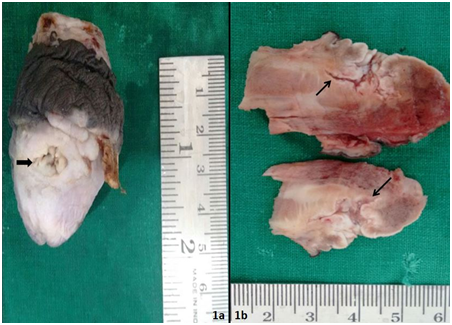
A 63 year old man, treated for nephrolithiasis five years back presented with penile growth since 11/2 years. Genitourinary examination showed ulcer on dorsum of penis, surrounded by verruciform lesion involving glans and corona sulcus. Biopsy of verrucous lesion done outside had revealed verrucous carcinoma, which on review was confirmed in our institute. Based on biopsy reports, partial penectomy was done. Histopathological examination revealed white lesion with nodular surface having both exophytic and endophytic components. Multiple sinuses were noted on surface of endophytic lesion which on cut surface displayed a branching and deeply burrowing pattern (Figure 1).


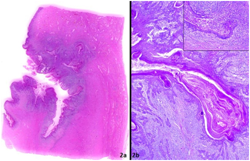
Microscopically, the deeply burrowing sinuses had hyperkeratotic and well-differentiated epithelium and were filled with layers of keratin. The interface between tumor and stroma revealed severe chronic inflammatory response (Figure 2). The tumor invaded corpora spongiosum and cavernosum. High-grade areas, vascular and perineural invasion, mitotic figures and koilocytic changes were not noted. Due to well differentiated morphology, prominent keratinization and unique burrowing endophytic growth pattern, the diagnosis of CC of penis was rendered.
CC is a verruciform tumor with characteristic exophytic/endophytic pattern of growth simulating rabbit’s burrow (cuniculus in Latin). CC tends to affect elderly patients with a mean age of 77 years and involves multiple anatomical compartments, mainly glans and coronal sulcus. Despite its invasiveness, inguinal nodal metastases have not been reported and prognosis is excellent [3,4]. The differential diagnoses include verruciform tumors like verrucous, warty and papillary carcinomas [3]. Verrucous carcinomas are cytologically and architecturally similar to CC, but they usually do not invade beyond lamina propria or superficial corpus spongiosum, nor do they exhibit a labyrinthine pattern of growth.
Although, warty carcinomas can sometimes depict a burrowing pattern, cutaneous fistulae and cyst-like structures are not common; this tumor is characterized by koilocytosis, higher degree of nuclear pleomorphism, papillae with prominent fibrovascular cores, and an irregular tumor-stroma interface. In papillary carcinomas, papillae are more irregular and complex, cyst-like and cutaneous fistulae are not present, the tumor front is jagged, and nuclear pleomorphism is marked. The correct diagnosis of CC of penis is important for post-resection management. Although locally destructive, absence of metastatic potential means that surgical resections can be performed with curative expectations and active surveillance seems to be more appropriate than llioinguinal lymph node dissection (ILND), a procedure associated with significant morbidity.
CC of the penis is an extremely rare variant of SCC. The main defining feature CC is the deeply burrowing growth pattern. Differentiating CC from other penile tumors is important for determining surveillance versus early ILND [5].
We would like to thank our institute NU hospitals, Bangalore, India for providing this opportunity to document this rare case.

©2016 Gowda, et al. This is an open access article distributed under the terms of the, which permits unrestricted use, distribution, and build upon your work non-commercially.