eISSN: 2378-3176


Case Report Volume 4 Issue 2
1Department of Urology, University Hospitals Coventry & Warwickshire, UK
2Department of Pathology, University Hospitals Coventry & Warwickshire, UK
Correspondence: Mr Aniket Deshpande, Post-CCT Fellow Urology, Department of Urology University Hospitals Coventry and Warwickshire, Clifford Bridge Road, CV2 2DX, Tel 024 7696 4000
Received: December 16, 2016 | Published: February 10, 2017
Citation: Pisavadia B, Mevcha A, Patel D, Sinha B, Blacker ARJ, et al. (2017) Case Report: An Unusual Presentation of Metastatic Renal Cell Carcinoma with Per-Vaginal Bleeding. Urol Nephrol Open Access J 4(2): 00118. DOI: 10.15406/unoaj.2017.04.00118
Renal cell carcinoma is an aggressive cancer, which could explain why as many as a third of cases are diagnosed as metastatic at presentation. With the incidence of renal cell carcinoma on the rise and a 5-year survival rate of just over 10% with metastatic disease, it is important to highlight the role of early detection of this cancer in order to optimize management and improve outcomes. Vulva and vagina represent rare sites for metastases from renal cell cancer, with clear cell renal cell cancer accounting for about a fifth of metastatic vulval cancer. We focus on this rare presentation of vulvo-vaginal metastases from renal clear cell carcinoma by presenting an unusual case of per-vaginal bleeding 3 months following detection of a renal lesion on CT imaging and highlight the importance of having a multidisciplinary approach to managing such complex cases to provide patients with the best possible outcomes.
Keywords: Vulvo-vaginal metastases; Renal cell carcinoma; Clear cell; Synchronous; Bartholin’s gland
RCC: Renal Cell Carcinoma; PV: Per-Vaginal; COPD: Chronic Obstructive Pulmonary Disease; CT: Computed Tomography; CTPA: Computed Tomography Pulmonary Angiogram
Vulvo-vaginal metastases from renal cell carcinoma (RCC) are rare presentation accounting for about 18% of metastatic vulval cancers [1]. There have been 11,873 new cases of renal cancer diagnosed in the UK in 2013, accounting for 3% of total cancer cases [2]. Approximately half of these cases were diagnosed in patients over the age of 70 years and 33% of cases were reported to be metastatic at presentation [2].
We present a case of per vaginal (PV) bleeding secondary to vulvo-vaginal metastasis from left renal clear cell carcinoma. The purpose of this case report is to highlight the relatively rare presentation of vulvo-vaginal metastases from RCC and the importance of early diagnosis of these cases due to its poor prognosis.
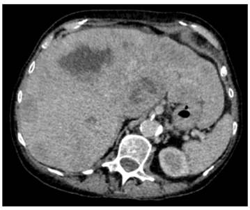
Our patient was a 79-year old female with severe progressive chronic obstructive pulmonary disease (COPD) on home oxygen, type 2 diabetes, hypertension and angina. She presented with type 1 respiratory failure and chest pain in December 2015. A computed tomography pulmonary angiogram (CTPA)4 months prior to this admission, showed no pulmonary embolism but did show a solitary liver lesion (Figure 1), measuring 2.5cm x 2.8cm that was suspicious for metastasis. A subsequent contrast-enhanced staging CT abdomen and pelvis demonstrated multiple liver metastases (Figure 2) with a 5.5cm solid-cystic lesion arising from her left kidney. After discussion with the Hepatobiliary multidisciplinary team (MDT), she underwent a percutaneous biopsy of the liver lesions, which showed metastatic carcinoma of indeterminate primary. She was seen by our Oncologists and was deemed suitable only for palliative treatment at this stage due to a presumed metastatic renal cancer and her multiple co-morbidities.
Three months later, she was presented to her general practitioner with vaginal bleeding and was evaluated in two weeks at the Gynecology clinic where on examination, a craggy 2-3cm mass, replacing the left Bartholin’s gland in the lower 1/3rd of vagina and vulva, was noted. This mass was biopsied which showed metastatic clear cell renal carcinoma. On a repeated contrast-enhanced staging CT chest, abdomen and pelvis, progression of her metastatic disease with an increase in the size of her left renal lesion to 7cm was observed (Figure 3&4). The patient was also noted to have become progressively thrombocytopenic. She was therefore, continued on the palliative care pathway with supportive treatment. At the time of submission of this article, patient was still alive under the care of the Oncologists who had discussed and commenced palliative Pazopanib at a reduced dose of 400mg daily. The patient’s main issue was bilateral lymphedema with not much symptoms from her vulvo-vaginal metastases.

Despite an increasing incidence of incidental finding of renal cancers on unplanned imaging, a third of them still have metastases at presentation [3]. This is most likely attributable to the relatively aggressive nature of RCC, with its tendency to metastasize [3]. Common sites of RCC metastases are lungs, bone, liver and lymph nodes [3]. However, there have been reports of metastases to rare sites including urogenital organs, particularly the vagina and the vulva, which we have presented in our case [3]. Approximately 70-80% of all RCC’s are of the clear cell histological subtype, which also happens to be the most common subtype associated with vaginal metastases [3,4]. Vaginal metastases are typically found in lower third and anterior vaginal wall, the former was the case with our patient [5]. These patients most commonly present with vaginal bleeding [5].
There have been a greater number of reported cases of RCC metastases to the vagina than to the vulva in the literature. Reports of clear cell carcinoma metastasizing to the vagina go back as early as 1936 [6]. Allard et al. [4] reported the first case of thrombocytopenia as a paraneoplastic phenomenon of metastatic RCC to the vagina. However, there has only ever been one reported case of synchronous vulvo-vaginal metastases from RCC and this was from the papillary subtype [1]. In our case, at initial presentation, CT abdomen and pelvis incidentally found the left renal lesion. When the patient presented 3 months later with PV bleeding, this led to an urgent biopsy, from which, clear cell metastatic RCC was confirmed. Before noting the vulvo-vaginal metastases she was already on palliative treatment.
Therefore, in post-menopausal women with clear cell carcinoma on histology following biopsy of a vaginal or vulval mass with features suggestive of a paraneoplastic syndrome, RCC must be a differential diagnosis. This is particularly relevant in light of the relatively poor prognosis of metastatic RCC, which has a 5-year survival rate of only 12.3%, as opposed to a localized RCC with a 5-year relative survival rate of 91.7% [7]. Even in the day of modern medicine with advances in management of metastatic renal cancer, such as immunotherapy, TKI inhibitors and mTOR kinase inhibitors, long-term survival of these patients remains poor, quite often only for months [3].
These patients are best served by a multidisciplinary approach involving Gynecologic oncologist, Uro-oncologist, dedicated histopathologists, radiologists and palliative care team [8].This case highlights the importance of bearing in mind the possibility of RCC in these patients, especially with proven histology (Figure 5&6), in order to optimize management to improve outcomes. If the patient is fit, a nephrectomy and excision of the metastases may be the best management options [8]. Due to multiple co-morbidities, our patient was not suitable for this and therefore, the aim of management was to achieve symptom control.

©2017 Pisavadia, et al. This is an open access article distributed under the terms of the, which permits unrestricted use, distribution, and build upon your work non-commercially.