eISSN: 2378-3176


Research Article Volume 1 Issue 3
1Consultant Surgeon, Port-Fouad General Hospital, Egypt
2Consultant surgeon, Services hospital, Pakistan
Correspondence: Aly Saber, Consultant Surgeon, Department of General Surgery, Port-Fouad General Hospital, Port-Fouad, Port-Said, Egypt, Tel 201223752032, Fax 20663400848
Received: October 22, 2014 | Published: December 13, 2014
Citation: Saber A, Bajwa TM. A simplified prognostic scoring system for Fournier’s gangrene. Urol Nephrol Open Access J. 2014;1(3):79-82. DOI: 10.15406/unoaj.2014.01.00018
Introduction: Fournier’s gangrene is an acute necrotizing fasciitis affecting the perineal, perianal regions and genitalia. The cornerstones of treatment Fournier’s gangrene are urgent necrotic tissue debridement, broad-spectrum antibiotics and resuscitation. Despite advanced management policies, mortality from Fournier’s gangrene is still high. The aim of this study was to present our experience in low-volume general hospital in the management of Fournier’s gangrene according to our advocated scoring system.
Patients and methods: A total of 68 patients were classified according to age, body mass index, early detection, area involved and comorbidity. The author advocated an eight- scale simplified prognostic scoring system with a maximum score of eighteen points denoting the highest risk of mortality and a minimum score of eight points carrying a relatively lower risk of mortality. The primary end point of the study was disease-related death and the secondary end point was length of hospital stay.
Results: There were three grades according out simplified prognostic scoring system; grade I from 8-10 points, grade II from 11-14 points and grade III from 15-18 points. Patients with grade I carried a lower mortality rate and less hospital stay than those with grade II and grade III.
Conclusion: We tried to develop a reliable tool to predict severity of the disease, not only to identify patients at highest risk of major complications or death but also to provide a target for medical teams and researchers aiming to improve outcome and to collect beneficial information for proper management of patients with Fournier's gangrene.
Keywords: Fournier’s gangrene, scoring system, simplified
sFG, Fournier’s gangrene; BMI, body mass index; LRINEC, laboratory risk indicator for necrotizing fasciitis; FGSI, Fournier’s gangrene severity index; UFGSI, uludag Fournier’s gangrene severity index
Fournier’s gangrene (FG) is an acute progressive infective necrotizing fasciitis affecting mainly the perineal, perianal regions and external genitalia of men, but can also occur in women and children.1 Fournier’s gangrene was considered as an idiopathic syndrome but in the majority of cases, urogenital and perineal traumas, including pelvic and perineal injury or pelvic interventions, are incriminated.2 Systemic comorbidities are also identified in patients with Fournier’s gangrene such as diabetes mellitus (DM), malignancy and malnutrition.3 The cornerstones of treating patients with Fournier’s gangrene are urgent necrotic tissue debridement, proper doses of broad-spectrum antibiotics and resuscitation with fluids.1 Despite advanced management policies, mortality from Fournier’s gangrene is still high.1,4 The aim of this study was to present our experience in low-volume general hospital in the management of Fournier’s gangrene according to our advocated scoring system.
A total of 68 patients presented with manifestations of Fournier’s gangrene were enrolled to the study from April 2000 to September 2013. Those patients were classified according to their age, body mass index (BMI), early detection, area involved whether single or multiple and comorbidity. According to this classification, the author adopted a simplified prognostic scoring system for prediction of mortality rate in his patients. Disease detection was considered early or delayed according to clinical and laboratory data. Medical comorbidity was traced as diabetes mellitus only or concomitant with other disease such as cardiovascular, renal or hepatic disease. Laboratory risk indicator for necrotizing fasciitis (LRINEC) was done for all patients upon admission. The parameters of this laboratory risk indicator are glucose level, C-reactive protein level, total leucocytic count, serum sodium, creatinine level, hemoglobin.
Early patient presentation was considered when there was localized pain, minor skin manifestations such as redness, hotness and crepitus but without apparent gangrene. Delayed presentation was noticed with body temperature above 38°C, rapid pulse, offensive wound discharge and cutaneous wound gangrene. Late presentation was considered with systemic manifestations of shock. The local ethics committee had approved all operative procedures. Ethical approval for this study was granted by the ethical review committee under supervision of the general director of Port- Fouad general hospital, Port-Fouad, Port-Said, Egypt.
The simplified prognostic scoring system
Here, the author advocated an eight-scale simplified prognostic scoring system with a maximum score of eighteen points denoting the highest risk of mortality and a minimum score of eight points carrying a relatively lower risk of mortality.
≤50years=I point. ≥50years=2 points.
BMI<25=I point. BMI <30=2 points BMI >30 =3 points.
<38°C= I point. >38°C= 2 points.
<100 beats/min=I point. >100 beats/min 2 points.
>90mm Hg= I point. <90mm Hg= 2 points.
Early=1 point. Delayed=2 points. Late=3 points.
Single=1 point. multiple=2 points.
DM=1 point. multiple=2 points.
Study sites and technique
Surgical interventions were performed in Port-Fouad general hospital, Port-Fouad, Port-Said, Egypt. Surgical interventions were of triple multimodal approach including hemodynamic stabilization, broad spectrum antibiotics, and surgical debridement. All necrotic and non-viable tissues were excised until the viable tissue was reached. Multiple sittings of surgical debridement were needed for the vast majority of cases. Very close patients’ observation and wound care were mandatory.
End points
The primary end point of the study was disease-related death and the secondary end point was length of hospital stay.
Patients were subdivided according to their ages and body mass indices as previously reported. The number of patients in each subgroup was traced as shown in (Table 1). Regarding the time of presentation, in vast majority of cases patients (82.6%) presented with delayed and late courses of the disease and only 12 patients (17.45%) presented with early disease manifestations according to (Table 2).
Item |
Subgroup |
Number |
||
|---|---|---|---|---|
Male |
Female |
Total |
||
Age |
≤50years |
14 |
6 |
20 |
≤50years |
28 |
20 |
48 |
|
BMI |
BMI<25 |
24 |
- |
24 |
BMI<30 |
12 |
10 |
22 |
|
BMI>30 |
6 |
16 |
22 |
|
Table 1 Showed patients distribution regarding their ages and BMIs
Item |
Age subgroup |
BMI subgroup |
|||
|---|---|---|---|---|---|
< 50 years |
> 50 years |
BMI<25 |
BMI<30 |
BMI>30 |
|
Early |
8 |
4 |
7 |
3 |
2 |
Delayed |
6 |
20 |
7 |
9 |
10 |
Late |
6 |
24 |
10 |
10 |
10 |
Total |
20 |
48 |
24 |
22 |
22 |
Table 2 Showed patients distribution regarding time of their presentations
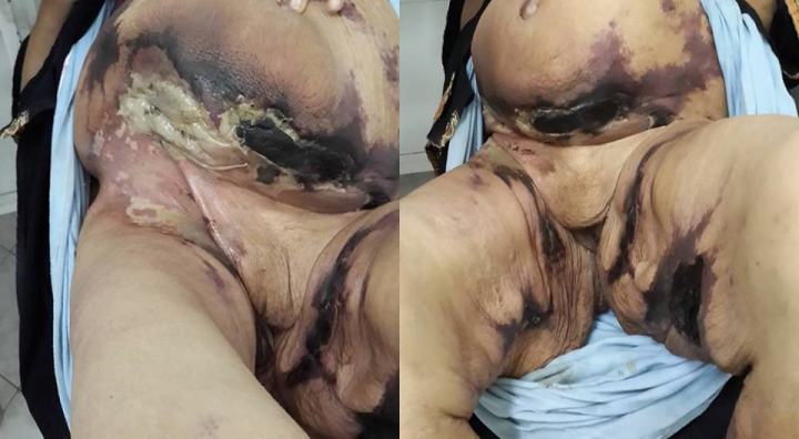
The extent of the disease was determined as single primary site (Figure 1), two or multiple sites including the primary sites and disease extension sites (Figure 2). Genitalia were the most common primary site of the disease in men (Figure 3), where gluteal region was the most common in women where there was no gluteal region involvement as a primary site in male patients of our series. Table 3 showed the number of patients regarding the primary site of the disease.

Site |
Male |
Female |
Total |
Genitalia |
20 |
8 |
28 |
Perianal |
9 |
4 |
13 |
Perineal |
13 |
4 |
17 |
Gluteal |
- |
10 |
10 |
Total |
42 |
26 |
68 |
Table 3 Showed patients regarding the primary site of the disease
In the present study, 28 patients (41.1%) died as a direct result of the disease. Age above 50years, higher BMI and delayed presentation were associated with much more incidence of mortality as shown in Table 4. The mortality rate was higher in females (69%) as we noticed that 18 of 26 female patients died as a result of the disease. In case of male patients, the mortality rate was 28.6% as a total of 12 of 42 patients died as a result of the disease. Regarding the adopted simplified prognostic scoring system, there were three grades; grade I from 8-10 points, grade II from 11-14 points and grade III from 15-18 points. Patients with score of 8-10 points [grade I] carried a lower mortality rate and less hospital stay than those with score of 11-14 points [grade II] and score of 15-18 points [grade III]. The twelve patients who presented at the early stage were belonged to grade I score while delayed and late presentations were evident with grade II and grade III scores. The simplified prognostic scoring system was directly proportional to the mortality rate where patients with higher grades [grades II and III] showed more mortality compared with those having grade I score.
Factor |
Male [ N=12] |
Female N=18 |
|
Age |
≤50 years |
2 |
- |
≤50 years |
10 |
18 |
|
BMI |
BMI<25 |
1 |
2 |
BMI<30 |
6 |
8 |
|
BMI>30 |
5 |
8 |
|
Presentation |
Early |
- |
- |
Delayed |
5 |
8 |
|
Late |
7 |
10 |
|
Table 4 Showed mortality in relation to patient’s age, BMI and presentation
The hospital stay was calculated as the time of admission, resuscitation, surgical interference and time needed for wound care until patient was discharged. The time of hospital stay ranged from 7 to 30days which was 7-10days in case of early presented patients with grade I score while patients presented with delayed or late course of the disease and required both grades II and III, showed much more hospital stay as 15- 30days. As regard to hospital stay, our simplified prognostic scoring system was directly proportional to the hospital stay where patients with higher grades [grades II and III] showed more hospital stay compared with those having grade I score.
Fournier’s gangrene is a progressive and fulminant necrotizing fasciitis of the genital, perianal and perineal regions that may extend to the abdominal wall between the fascial planes.5 There are two important validated scoring systems for outcome prediction of Fournier's gangrene. These systems are Fournier’s Gangrene Severity Index (FGSI) and Uludag Fournier's Gangrene Severity Index (UFGSI).6 FGSI score uses nine parameters including the body temperature, heart rate, respiratory rate, hematocrit, white blood cell count, and serum levels of sodium, potassium, creatinine and bicarbonate.7 Yilmazlar et al.,8 suggested a new scoring system, the Uludag FGSI (UFGSI), adding the age and the extent of the disease scores to the FGSI.
Although FGSI score is higher in non survivors, the difference was found statistically insignificant and some researchers concluded that the FGSI had no prognostic value4,9 and some other studies have shown no relationship between high FGSI scores and mortality.10 Conversely, some studies revealed that FGSI scores were sensitive and specific for predicting mortality rate11 and on comparing UFGSI against FGSI, it was concluded that despite including more variables, the UFGSI does not seem to be more powerful than the FGSI.12 Both scoring systems lack the timing of patient presentation, body mass index (BMI) and patient’s comorbidity.6–12
The present study advocated an eight- scale simplified prognostic scoring system with a maximum score of eighteen points denoting the highest risk of mortality and a minimum score of eight points carrying a relatively lower risk of mortality. The proposed system contains patient’s age, BMI, temperature, pulse, systolic blood pressure, timing of presentation, area involved and comorbidity. Many studies found obesity,13,14 the timing of patient presentation and patient’s comorbidity as important parameters for outcome prediction of Fournier's gangrene and is considered as a major risk factor for mortality.1,3,5,15,16
Mortality rate in patients with Fournier’s gangrene is usually high and depends on many related risk factors according the already validated scoring systems or our proposed simplified system. In the present study the overall mortality rate was 41.1% with 69% in females and 28.6% in males. In studies of same interest, the overall mortality rate was 25-30%16,17 and may reach up to 88%18 while was higher in females than males in series of Czymek et al.19 Higher mortality rate is usually detected with increasing age >50years and the time interval between the first symptom and surgical intervention.1,3,5,18 Our results came in agreement with these reports and others of same interest.
The extension of the disease beyond the primary site and the mortality rate are controversial themes in the literature. Some studies have reported that the spread of the disease is related to a higher death rate, while others reported that the extension of the gangrene does not relate to a poorer prognosis.5 It was found that the extension of the infection to the abdominal wall5 and thighs20 is a predictor of mortality.
The associated medical illnesses included diabetes, chronic renal failure, chronic liver disease; hypertension, alcoholism, and peripheral vascular disorder usually carry the higher rates of mortality.16 Other researchers reported that various co-morbidities are known to be associated with FG, of which diabetes is the most common but its association with increased mortality is controversial.21,22 In most series, the majority of the patients had DM4,23 and the percentage of their occurrence varied from 54%15 to 70%.1 Existence of one or more of such co-morbidities as DM, alcoholism, neurological diseases, malignancy, immunosuppression, and, liver and kidney diseases can affect morbidity and mortality rates in cases with FG. DM is the most common predisposing factor with an incidence of 46-76.9% and mortality has been reported in 36-50% of these patients.22
Fournier's gangrene, although rare, is usually a potentially lethal disease. Co-morbidities, especially diabetes mellitus increase the mortality rate. Repeated surgical excisions and debridement with broad spectrum antibiotic provide good results. We tried to develop a reliable tool to predict severity of the disease, not only to identify patients at highest risk of major complications or death but also to provide a target for medical teams and researchers aiming to improve outcome and to collect beneficial information for proper management of patients with Fournier's gangrene.
The corresponding author declares he has neither competing interest nor funding sources.
None.
The author declares no conflict of interest.

©2014 Saber, et al. This is an open access article distributed under the terms of the, which permits unrestricted use, distribution, and build upon your work non-commercially.