MOJ
eISSN: 2475-5494


Case Report Volume 7 Issue 1
1Department of Radiology, Women?s Yas Hospital, Tehran University of Medical Sciences, Iran
2Department of Radiology, Imam Khomeini Hospital Complex, Tehran University of Medical Sciences, Iran|
3Department of Radiology, Amiralam Hospital, Tehran University of Medical Sciences, Iran
4Maternal, Fetal and Neonatal Research Center, Women?s Yas Hospital, Tehran University of Medical Sciences, Iran
Correspondence: Behnaz Moradi, Department of Radiology, Womens Yas Hospital, North Nejatollahi Street, Tehran, Iran
Received: February 06, 2018 | Published: February 15, 2018
Citation: Moradi B, Asadi M, Kazemi MA, et al. Prenatal sonographic features of Fraser syndrome with multiple craniofacial abnormalities: a case report . MOJ Womens Health. 2018;7(1):26-29. DOI: 10.15406/mojwh.2018.07.00162
Fraser syndrome is a rare autosomal recessive disorder characterized by a combination of different anomalies including major and minor manifestations. Here we refer to a 17weeks fetus because of severe oligohydramnios. Along oligohydramnios, the first ultrsonographic obvious findings were echogenic and edematous lungs in favor of high airway obstruction. Craniofacial evaluation showed bilateral cleft lip/palate, depressed nasal bridge, hypoplastic nasal bone, unilateral cataract, hypertelorism, cryptophthalmos, and turricephaly. Based on these findings, diagnosis of Fraser syndrome was made which was confirmed by the autopsy and post mortem evaluation. So, the parents chose termination of the pregnancy. The present case revealed that oligohydramnios and high airway obstruction are two good clues for other associated criteria such as craniofacial abnormalities to be searched by prenatal ultrasonography.
Keywords: cryptophthalmos, fraser syndrome, facial abnormalities, prenatal diagnosis
Fraser syndrome (also called Cryptophthalmos-Syndactyly Syndrome) is an extremely rare congenital syndromic anomaly with auotosomal recessive inheritance.1 The incidence rate of this syndrome is estimated below 0.04 per 10.000 live born infants and 1.1 in 10.000 stillbirths.2 It is mainly characterized by variable expressions of cryptophthalmos, syndactyly, abnormal genitalia, laryngeal malformations and urogenital defects.2–4 Antenatally diagnosed cases had more manifestations result in a pathological amount of amniotic fluid.3 Prenatal diagnosis is getting more difficult in the case of inconstant and variable sonographic findings especially when oligohydramnios and a healthy previous sib are presented.2,5 Oligohydramnios can occur at any time during pregnancy, it is most common during the last trimester but its occurrence in earlier in pregnancy has poorer prognosis with about 80-90% mortality rates. It could result from placental insufficiency or congenital abnormalities in the fetus such as agenesis of the kidneys or atresia of the urethra.6 Here we described a prenatal case with Fraser syndrome indicated severe oligohydramnios, high airway obstruction, bilateral renal agenesis and multiple associated craniofacial anomalies in a nonconsanguineous couple.
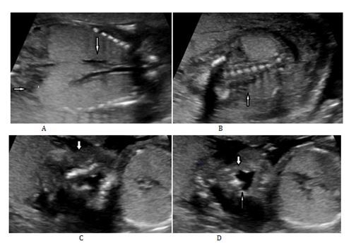
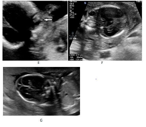
A 34–year-old G2 P1 pregnant lady was referred to our hospital because of severe oligohydramnios with no complain of risk factors. There was no positive history of similar anomalies in the previous pregnancy and her only child was healthy. The parents were not consanguineous. Ultrasound study was performed and a 17weeks alive male fetus with multiple anomalies was identified. The biometry was consistent with the expected gestational age. The first obvious finding was lungs enlargement with increased echogenicity and downward displacement of the diaphragm. The trachea and bronchi were distended (Figure 1). The heart was squeezed centrally but was anatomically normal. Findings were in favor of Congenital High Airway Obstruction Syndrome (CHAOS). Adrenal glands were enlarged and the kidneys and bladder were not identified which showed the bilateral renal agenesis (Figure 1). Mild ascites was found. In the 2D sonographic evaluation of craniofacial structures many abnormalities were detected such as no identified bilateral palpebral fissures (Figure 1); bilateral cleft lip/palate (Figure 1); depressed nasal bridge and hypoplastic nasal bone, unilateral cataract (Figure 1); hypertelorism (Figure 1) and turricephaly (Figure 1). Low set ears were suspected too. One hand and one foot were apparently normal but the other side could not be visualized well. No abnormality was detected in external genitalia and other structures.


Figure 1 A) Ultrasonography of a 17weeks fetus: A)Thorax and abdomen coronal cross section showing expanded and hyperechogenic lungs with everted diaphragm (horizontal arrow) and grossly dilated trachea and bronchi (vertical arrow); B) Bilateral renal agenesis with enlarged adrenal glands (arrow) and severe oligohydramnios; C) In coronal cross section of fetal face, there is no evidence of palpebral fissure infavor of Cryptophthalmos (arrow), D) Bilateral cleft lips (wider in one side) e: echogenic lens (cataract); F) Axial cross section of fetal head at the level of orbits. Binoccular distance is about 20weeks and inter ocular distance is increased (Hypertelorism): G) Turricephaly.
The fetus karyotype was compatible with normal male. The parents elected termination of the pregnancy and misoprostol was used for induction of pregnancy. The abortus was dead at birth with weight of 185gram. Post mortem evaluation confirmed the cryptophthalmos, low set ears and other mentioned abnormalities. No syndactyly was identified in hands and feet and external genitalia were normal. Autopsy revealed bilateral hyperplastic lungs with laryngeal atresia and bilateral renal agenesis. The liver was enlarged and edematous. One globe was normal and the other one was slightly hypoplastic with opaque lens (cataract). No other internal abnormality was found.
The current findings were considered diagnostic of Fraser syndrome. Such syndrome is a very rare autosomal recessive disorder; and its genetic background has been linked to FRAS1, FREM2 and GRIP1 genes.1 For the first time in 1872, Zehender described a child with cryptophthalmos and associated complex malformations including syndactyly, genital abnormalities and other minor abnormalities. In 1962 (FS) George Fraser recognized it as a clinical entity and named it as Fraser syndrome.3–5,7 At first, Thomas et al.,4 proposed diagnostic criteria for Fraser syndrome in 1986. They reviewed 124 cases of cryptophthalmos and associated syndromes and proposed diagnosis based on either at least two major criteria and one minor criterion or one major criterion and at least four minor criteria. After that in 2007, van Haelst et al.,2 provided a revision of the diagnostic criteria through the addition of airway tract and urinary tract anomalies as a major criteria and removal of mental retardation and cleft lip and palate. Based on this new diagnostic criteria three major of them, two major and two minor criteria or one major and 3 minor criteria are needed.2,4,8 The present case fulfilled both diagnostic criteria of Fraser syndrome (Table I).
|
Diagnostic criteria by Thomas et al.4 |
Case |
Revised diagnostic criteria by van Haelst et al.2 |
Case |
|
Major Criteria |
Major Criteria |
||
|
Syndactyly |
Syndactyly |
- |
|
|
Cryptophthalmos |
Bilateral |
Cryptophthalmos spectrum |
Bilateral |
|
Ambiguous genitalia |
- |
Urinary tract abnormalities |
Bilateral renal agenesis |
|
Sib with cryptophthalmos syndrome |
- |
Ambiguous genitalia |
- |
|
- |
Laryngeal and tracheal anomalies |
High airway obstruction and Laryngeal atresia |
|
|
- |
Positive family history |
- |
|
|
Minor Criteria |
Minor Criteria |
||
|
Congenital malformation of nose |
Depressed nasal bridge and hypoplastic nasal bone |
Anorectal defects |
- |
|
Congenital malformation of ears |
Bilateral low set ears |
Dysplastic ears |
Bilateral low set ears |
|
Congenital malformation of larynx |
Laryngeal atresia |
Skull ossification defects |
- |
|
Cleft lip and/or palate |
Bilateral left lip and palate |
Nasal anomalies |
Depressed nasal bridge and hypoplastic nasal bone |
|
Skeletal defects |
- |
Umbilical abnormalities |
|
|
Umbilical hernia |
- |
||
|
Renal agenesis |
- |
||
|
Mental retardation |
Bilateral renal agenesis |
||
|
- |
Table I Diagnostic criteria for Fraser syndrome based on two qualities and expressions in the presenting case
Prenatal detection of Fraser syndrome has been reported in several cases but it is worth noting as the most criteria are not easily diagnosed by 2D ultrasound in young fetuses (early second trimester as our case with 17 weeks of GA) especially when a previous sib was not affected.5 In literature review by Rousseau et al.,5 in 2002 prenatal diagnosis was made usually based on a history of a previous child born with Fraser syndrome (in 9/21 cases), and the detection of multiple malformations (in 12/21cases). Various sonographic features were observed, and oligohydramnios was the most common isolated finding which was detected before the (13/21cases) voluminous hyperechogenic lungs (9/21) and renal abnormalities (7/21). Their results were nearly similar to Berg et al.,3 literature review in 2001.5 Breg et al.,3 proposed that the association of bilateral hyperechogenic lungs and oligohydramnios with renal agenesis might be a marker of Fraser syndrome in prenatal ultrasound exam. They found 7 out of 16 prenatally detected cases with these association.3 The detection of some criteria as syndactyly and facial clefting is difficult in the background of oligohydramnios. Also in the case of enlarged adrenal gland an exact diagnosis of renal agenesis in prenatal ultrasound assessment will be a problem.
Van Haelst et al.,2 had found that cryptophthalmos was slightly more frequent in postnatally diagnosed cases. However abnormalities result in increase (laryngeal atresia) or decrease (bilateral renal agenesis) in amniotic fluid were more common and more detected in prenatal period. No difference was identified about other abnormalities such as syndactyly in both groups.2 The prenatal and postnatal fetal phenotype in 38 cases with Fraser syndrome was analyzed in 2016 by Aude Tessier. Among the 26 prenatally evaluated cases detectable anomalies consisted of oligohydramnios (in 22cases), ascites/hydrops (in 9cases), renal abnormalities (in 20cases), high airways obstruction (in 11 cases), ophthalmologic anomalies (4cases), ear dysplasia (2cases) and syndactyly (2cases). Contrary to Van Haelst et al.,2 study nearly all postnatally detected cases had abnormalities in nose, ears and syndactyly. Similar to Berg et al.,2 study, they concluded that a combination of oligohydramnios, renal agenesis and high air way obstruction could lead to this diagnosis in antenatal sonographic evaluation.3,9,10 The same as our case that firstly oligohydramnios, bilateral renal agenesis and evidence for high airway obstruction were detected and then craniofacial abnormalities were noticed. Antenatal detection of cryptophthalmos might be possible by sonographic evaluation of the fetal palpebral fissure but concomitant oligohydramnios makes its diagnosis more difficult.3,11 As it was mentioned specially based on the prenatally evaluated cases, in previous multiple literature review and case studies, nose, ear, ophthalmologic and facial cleft are among the less common abnormalities. Here we present a case with multiple craniofacial abnormalities detected by prenatal ultrasound.
Depending on the severity of their symptoms, Fraser Syndrome can be fatal before or shortly after birth (for example due to bilateral renal agenesis or severely malformed or missing larynx); less affected cases can live into childhood or even adulthood. Treatment options may include surgery in order to correct some of the relevant malformations (such as cryptophthalmos or ambiguous genitalia). Eye abnormalities usually lead to vision impairment or loss in post natal life. The recurrence risk of Fraser syndrome among siblings will be 25%. Therefore, counselling affected families is an important task for prenatal diagnosis.3
In conclusion due to the wide spectrum of possible anomalies the diagnosis will stand questionable in most prenatally evaluated cases. Oligohydramnios and high airway obstruction are two good clues in searching for other associated criteria of the Fraser Syndrome in prenatal ultrasound exam. Our case has multiple craniofacial abnormalities that complete the diagnostic criteria.
None.
The author declares no conflict of interest.

©2018 Moradi, et al. This is an open access article distributed under the terms of the, which permits unrestricted use, distribution, and build upon your work non-commercially.