MOJ
eISSN: 2475-5494


Review Article Volume 4 Issue 3
1Department of Gynecology and Obstetrics, Princess Grace Hospital, Monaco
2Department of Nuclear Medicine, Princess Grace Hospital, Monaco
Correspondence: Moussa Diallo, Department of Gynecology and Obstetrics, Princess Grace Hospital, 1 Avenue Pasteur, 98012, Monaco
Received: February 13, 2017 | Published: February 16, 2017
Citation: Diallo M, Raiga J, Boujenah J, et al. A rare cause of antigen cancer elevation 125 (CA 125): a case of uterine adenomyosis. MOJ Womens Health. 2017;4(3):53-55. DOI: 10.15406/mojwh.2017.04.00085
Until the recent past, the delays entered the first sign and the diagnosis of adenomyosis was several years. In the absence of a specific sign, its frequency is hardly appreciable because of the great heterogeneity of the studied populations (all on parts of hysterectomy). All research is converging towards reducing the time of diagnosis of adenomyosis and endometriosis. The dosage of CA 125 could be promising. The physiopathology of its synthesis and its secretion shows us that it can be elevated during endometriosis in general, sometimes in the absence of clinical signs. We believe that even in the absence of functional signs, its dosage should be systematic in the presence of uterine pathology even in the absence of endometriosis. We report here a case of isolated adenomyosis (in the absence of any uterine fibroid) associated with an elevation of Ca 125 and whose kinetics of values were influenced by hormonal treatment.
Adenomyosis is an ectopic localization of endometrial tissue (glands and stroma) and autonomous development in the thickness of the uterine muscle. The prevalence of adenomyosis ranges from 5 and 70%1 after hysterectomy according to the surgical indication,2 whatever them indications. Transvaginal ultrasound and magnetic resonance imaging exam (MRI) have currently an important place in the diagnosis before surgery. The antigen cancer 125 (CA 125) is currently, apart the ovarian’s epithelium cancer, used in the diagnosis of deep endometriosis, and does not appear as a determining element in the diagnosis of denomyosis. We report here, a case of isolated adenomyosis (in the absence of any uterine fibroid) associated with an elevation of CA 125 and whose kinetics of values were influenced by hormonal treatment.
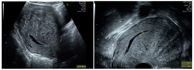
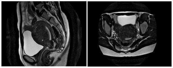
None.A 35-year-old women presented for an incidental findings of an increased level of antigen cancer 125 performed in systematic blood test check up. She reported pelvic chronic pain and abnormal menstrual bleeding for several months. The transvaginal ultrasonography showed a large antero-posterior asymmetry of the uterine walls, a striated appearance of the myometrium, and a neoformation rounded with fuzzy limits without mass effect and calcifications. No adnexal mass was observed. These signs were strongly suggestive of adenomyosis lesion (Figure 1). The magnetic resonance imaging (MRI) confirmed a diffuse adenomyosis without uterine fibroids (Figure 2). Neither the ultrasound nor the MRI revealed deep infltrating endometriosis (DIE). A Positron Emission Tomography (PET) scan was performed to rule out other differential diagnosis of an increased blood level of CA125. Focal hypertense hypermetabolism was only observed in the myometrium (Figure 3). Standard pipelle endometrial sampling did not reveal atypic cells. The other tumor’s markers including ACE, CA153 were normal. Conservative management was proposed as first line therapy. Therefore successive hormonal suppression was given (gonadotropin-releasing hormone agonist-GnrHa, and then Ulipristal Acetate). The Figure 4 summarizes the changes of CA 125 level and menorrhagia with treatment. However, symptoms did not release and a radical management was performed. At the laparoscopy, a globally enlarged uterus was found. No other macroscopic focus suggestive of pelvic endometriosis was highlighted. No complications occurred after hysterectomy. The histological examination of the specimen showed numerous foci and diffuse adenomyosis consisting of heterotopic glands subtended by a cytogenic chorion and hyperplasia muscle fibers (Figure 5). Also the endometrium was hypotrophic and the Fallopian tubes was normals.


The prevalence of adenomyosis ranges from 5 and 70%1 after hysterectomy according to the surgical indication.2 While the diagnosis of adenomyosis is histopathologic, transvaginal ultrasound and MRI can be strongly suggestive. The antigen cancer 125 (CA 125) is currently, apart the ovarian’s epithelium cancer, used in the diagnosis of deep endometriosis, and does not appear as a determining element in the diagnosis of adenomyosis. The finding of an incidental increased level of CA 125 challenges the diagnosis. Ovarian epithelial tumors, severe endometriosis, cardiac and chronic renal failure should be ruled out. Our case demonstrates that adenomyosis could be another differential diagnosis.
This case raises further questions
Increase blood level of CA125 can be found in adenomyosis and should be considered in the differential diagnosis. The effect of hormonal therapy on the CA125 level remains unclear.
None.
The author declares no conflict of interest.

©2017 Diallo, et al. This is an open access article distributed under the terms of the, which permits unrestricted use, distribution, and build upon your work non-commercially.