MOJ
eISSN: 2574-9935


Case Report Volume 2 Issue 1
Member of the Shoulder and Elbow Group of the Orthoservice Hospital, Sao Jose dos Campos, Brazil
Correspondence: Alexandre Tadeu do NascimentoI, Member of the Shoulder and Elbow Group of the Orthoservice Hospital, Sao Jose dos Campos, Brazil
Received: December 15, 2017 | Published: January 23, 2018
Citation: Nascimento AT, ClaudioI GK, RochaI PB. Arthroscopic treatment for a chronic isolated avulsion fracture of the lesser tuberosity of the humerus: a case report. MOJ Sports Med. 2018;2(1):24-26. DOI: 10.15406/mojsm.2018.02.00040
Fractures of the lesser tuberosity of the humerus usually occur in association with three or four part fractures associated with fracture-luxation of the proximal humerus (e.g., the Neer classification).1 They are also commonly reported as isolated avulsion injuries of the subscapularis insertion4 or as injuries associated with posterior shoulder dislocation5. Moreover, such fractures occur in only 0.46 per 100,000 persons per year, compared with an annual incidence of 110 per 100,000 persons per year for other fractures of the proximal humerus.2,3 Isolated avulsion fractures of the lesser tuberosity mostly occur in young patients3-6 with the primary mechanism of injury being shoulder trauma in abduction and external rotation. With the arm in this position, there is a strong eccentric contraction of the subscapularis muscle that causes avulsion of the lesser tuberosity.7 Given that many of these injuries are only diagnosed at a chronic stage, it appears that many are overlooked at the time of the initial trauma.8
Acute displaced fractures of the lesser tuberosity should be treated by surgical reduction and internal fixation.9 This is because failure to achieve anatomical reduction may cause functional impairment due to weakness of the subscapularis or due to the secondary effects of malunion.10 Indeed, the unpredictable results of treatment for chronic injury7 and the potential for progressive displacement of the lesser tuberosity fragment due to contraction of the subscapularis, all patients who have a radiographically visible fracture with any degree of displacement should be treated surgically.11 In general, open reduction and fixation with screws is the preferred surgical option. In this report, we present the management of a patient who had a chronic isolated fracture of the lesser tuberosity that we treated via arthroscopy, using three suture anchors.
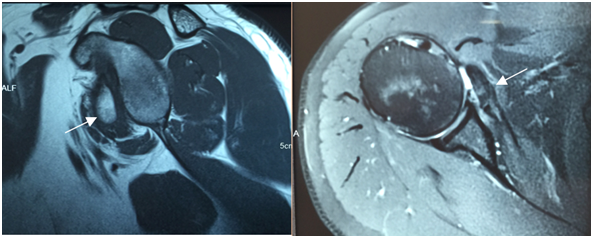
The patient was a 47-year-old man who worked as a sales representative. He presented with a history of falling down stairs two years previously, at which time he sustained trauma to the right shoulder. Since then, he had experienced pain and functional impairment, which he described as being unable to perform intimate hygiene or grab a wallet from his back trouser pocket and difficulty in using his arm when above shoulder height. The patient did not seek medical assistance at the time of the trauma because he did not have medical insurance and did not want to be treated in the public healthcare sector. Our initial physical examination showed a normal but painful range of movement; with positive tests for evaluate subscapularis tendon injury (e.g., bear-hug, Gerber, abdominal compression and lift-off tests). His DASH (Disabilities of the Arm, Shoulder and Hand) score was 60, his UCLA (University of California at Los Angeles) Shoulder Score was 16, his SF-36 (Short Form Health Survey) Score was 32.7 and his visual analog scale (VAS) score was 10. Shoulder x-rays revealed an avulsion fracture of the lesser tuberosity of the proximal humerus (Figure 1). Magnetic resonance imaging was done to support this, which confirmed the diagnosis of an isolated displaced fracture of the lesser tuberosity (Figure 2).

Figure 1 Preoperative radiographs showing a deviated fracture of the lesser tuberosity, which was at the level of the glenoid (white arrow).

Figure 2 Magnetic resonance imaging showing lesser tuberosity fragment at the glenoid level (white arrow).
The patient underwent surgery in a beach chair position under general anesthesia and a brachial plexus block. Diagnostic arthroscopy was performed via a posterior port. The area of subscapularis insertion was examined in the superior-to-inferior direction at the anterolateral port. This showed that the medial biceps pulley system was injured and the long head of the biceps was medially dislocated, but that there were no other intra-articular injuries. After creating the anterior and anterolateral ports, the fragment adherent to the adjacent soft tissues was observed as a single large fragment that included the subscapular insertion. The axillary nerve was identified at the exterior face of the subscapular tendon, deep and medial to the conjoined tendon. Caution was taken not to damage the nerve during the release procedure. The fragment was then released circumferentially. After provisionally tying an Ethibond® thread on the subscapular tendon to aid mobilization and with the arthroscope in the anterolateral port, tendon traction was facilitated by radiofrequency-based debridement and a soft tissue shaver to debride the external and internal surfaces of the tendon, respectively. This resulted in sufficiently mobilizing the fragment so that it could reach its anatomical location. Next, with the arthroscope in the posterior port, we performed cruentation of both the internal surface of the free fragment and of the area of reinsertion of the lesser tuberosity, taking care to remove the fibrotic tissue without removing any bone. Fragment reattachment was done in the same manner as a subscapular tendon repair (Figure 3), except for the need to use a third anchor at the lateral margin of fracture site to settle the bone fragment.
From left to right, initial aspect of the lesser tuberosity adhered to soft tissue, the aspect after debridement and mobilization, passage of sutures using "Bird Beak", partial reduction of the fragment and final appearance after final fixation with 3 anchors. The first suture anchor was placed in the inferior region of the medial margin at the fracture site, with the subscapularis tendon penetrated immediately adjacent to the bone-tendon interface at the most inferior aspect, using a bird-beak passer. The second anchor was placed in the medial margin of the fracture area in a more superior position, with the suture passing through the most superior aspect of the subscapularis tendon. The third anchor was placed in the most lateral aspect of the fracture area and its suture threads were passed through the bony fragment of the lesser tuberosity to ensure its fit in the fracture area. In addition, the threads of this third anchor were used to perform tenodesis of the long head of the biceps. Tenotomy was only performed after the bone repair and tenodesis. Checking was done through the anterolateral port. In the immediate postoperative period, the shoulder was immobilized with a sling for four weeks before the patient started physiotherapy. It has now been two and a half years since the operation and the patient has shown significant improvement. Of note, the DASH score decreased to 8.3, the UCLA Shoulder Score increased to 34, the SF-36 Score increased to 83.8 and the VAS score decreased to 2. All clinical tests for subscapularis lesions were negative at the most recent evaluation (Figures 4 & 5).
Isolated fractures of the lesser tuberosity are rare and can often be overlooked. Consequently, it is not uncommon for treatment to be delayed2 and for treatment of chronic fractures to be complicated by the need to release the avulsed fragment and subscapularis tendon from local adherences. The treatment of these fractures is typically by open surgery, either because of the technical difficulty in repairing them arthroscopically or because the surgeon is more familiar with the open procedure.8 However, we have shown that it is possible to perform successful arthroscopic surgery, which allows reduced postoperative pain and that facilitates earlier rehabilitation.12 We believe that these benefits of arthroscopic surgery may justify its preferential use over the open procedure.
The typical signs of acute fracture of the lesser tuberosity are pain on passive external rotation and limited internal rotation. Because the subscapularis tendon is linked directly to the fractured fragment, avulsion causes insufficiency of the muscle-tendon unit, which can be detected by various tests and clinical signs.12 In cases where fragments are dislocated, the injury can usually be detected by simple x-rays, with an axillary view often required to detect small fragments that have a small degree of dislocation.13 Computed tomography or magnetic resonance imaging can be useful for precise diagnoses and can determine the size and extent of the dislocation and any additional injuries. Most acute cases reported in the literature have been treated by open reduction and internal fixation and excellent clinical results have been obtained. Ogawa and Takahashi analyzed the long-term results of isolated fractures of the lesser tuberosity of the humerus and found that the results obtained by open reduction and internal fixation were superior to those obtained by conservative treatment.8
There is a possibility that this injury is associated with subluxation of the biceps or luxation of the long head of the biceps because of the proximity and interdigitation of the fibers of the subscapularis insertions in the humerus and superior complex of the glenohumeral and coracohumeral ligaments (i.e., the biceps pulley system), especially if the most superior part of the lesser tuberosity is also fractured .12 In our case, the fragment was dislocated by approximately 2 cm and preoperative magnetic resonance imaging showed that the fracture involved the whole tuberosity, including the most superior region. Therefore, we anticipated the reflection pulley of the biceps would also be injured, with associated dislocation of the long head of the biceps. Finally, two and a half years after the surgery, the patient in this report only had sporadic low-intensity pain, with a complete range of active and passive movements. The diagnostic tests and clinical signs for subscapularis lesions were also negative, suggesting a functional muscle-tendon unit of the subscapularis. This satisfactory outcome is consistent with existing case reports and small case series published on this topic.6-9,12
Arthroscopic surgery for isolated avulsion fracture of the lesser tuberosity can produce excellent clinical and radiological outcomes. In this cases report, we showed that this was possible even with chronic injury and concomitant impairment of the long head of the biceps.
None.
Author declares that there is no conflict of interest.

©2018 Nascimento, et al. This is an open access article distributed under the terms of the, which permits unrestricted use, distribution, and build upon your work non-commercially.