MOJ
eISSN: 2574-9935


Research Article Volume 1 Issue 1
1Graduate Program in Science of Human Movement, Methodist University of Piracicaba, Brazil
2Physical Education Faculty, Brazil
3Physical Education Faculty, Nove de Julho University (UNINOVE), Brazil
4Center for Sport Performance, Department of Kinesiology, California State University, USA
Correspondence: Paulo H Marchetti, Methodist University of Piracicaba (UNIMEP), Graduate Program in Science of Human Movement, Brazil, Tel 13400-911
Received: April 19, 2017 | Published: May 9, 2017
Citation: Marchetti PH, Fioravante GZ, da Silva JJ, et al. Effects of squat amplitude on pelvic tilt and tibial inclination. MOJ Sports Med. 2017;1(1):6-7. DOI: 10.15406/mojsm.2017.01.00002
Strength training is commonly performed at two different knee flexion amplitudes: partial (to 90 degrees) or total (to 140 degrees). During these amplitudes, both the pelvis and the tibia are moved to ensure control of the center of gravity and displacement of the external overload. Forward or backward movement of the pelvic tilt may indirectly influence the internal load on the spine. Objective: To measure the effect of squat amplitude on pelvictilt and tibia inclination. Eighteen male subjects (age: 26 ± 6 years, height: 178 ± 7 cm, total body mass: 81.3 ± 11 kg, resistance training experience: 5 ± 4 years) were evaluated. Pelvic tilt and tibial inclination were measured by a digital inclinometer (Max Measure, USA, accuracy: ± 0.02°, resolution: 0.05°) during isometric squatting at partial and full amplitudes. The digital inclinometer was fixed on the sacrum and on the tibia, with aneutral spine position. A paired student t-test and a significance of 5% were used. There were significant differences in pelvic tilt between partial and full amplitudes (+ 32.4° ± 10.9 and -21.7° ± 12.3, respectively, P<0.001). Maximum tibial inclination values were not significantly different between partial and total amplitudes (19.1 ± 6.6 and 20.1 ± 7.4, respectively, P = 0.225). It was concluded that the partial squat position produces anterior pelvic tilt while the full squat produces backward pelvic tilt. Inclination of the tibia is similar in both amplitudes of the squat.
Keywords: Exercise; Posture; Amplitude
The squatexerciseis a multi-joint task, and canbeconsidereda fundamental exercise for lower body strength, general fitness, and rehabilitation. Several studies have shown that manipulating the amplitude ofthe squat exercise results in altered muscle activity1-3 however, research on pelvic movements in the squat are limited.4
Some research methodologies suggesta correct way to perform the squat,5 but the correct technique is still controversial, with suggestions that thelumbar curve should be maintained throughout the squat,6 where as others suggest avoiding a rounded lumbar spine.7 For heavy squats8,9 suggest the squat should be performed to full depth as long asthe lordotic curve is maintained. The alignment of the pelvis is correlated with spine curvature and it has also been found to influence lifting function, withan anterior tilt of thepelvis providing increased trunk muscle activity.10 The majority of research on squat technique provide no quantified measure or description of the pelvic tilt. Therefore, the purpose of the present study was to measure the effect of squat amplitude on pelvictilt and tibia inclination.
Participants
Eighteen male subjects (age: 26 ± 6 years, height: 178 ± 7 cm, total body mass: 81.3 ± 11 kg, resistance training experience: 5 ± 2 years) were evaluated. Subjects had no previous lower back injury, surgery in the lower extremities, and no history of injury with residual symptoms (pain, “giving-away” sensations) in the lower limbs within the last year. This study was approved by the University research ethics committee and all subjects read and signed an informed consent document (#68/2016).
Procedures
Subjects were instructed in properisometric back squat technique for both conditions (partial: at 90° knee flexion, and full: at 140° knee flexion). Knee angle was measured by a goniometer. Their feet were positioned at hip width and vertically aligned with the barbell. The barbell was positioned on the shoulders (high-bar position) and all subjects performed each isometric squat condition three times for 3-s (rest between reps?). During each squat, the degree of pelvic tilt and tibial inclination were measured, and the highest value was used. Pelvictilt and tibial inclination were measured by a digital inclinometer (Max Measure, USA, accuracy: ± 0.02°, resolution: 0.05°) fixed on the sacrum and on the tibia, at an orthostatic position with a neutral spine. For pelvic tilt, positive values refer to anterior/forward and negative to posterior/backward positions. A rest period of 5-min was provided between conditions. All measures were performed at the same hour of the day, between 5 and 7 PM, and by the same researcher. A paired student t-test and a significance of 5% was used.Cohen’s formula for effect size (d) was calculated, and the results were based on the following criteria: <0.35 trivial effects; 0.35-0.80 small effect; 0.80-1.50 moderate effect; and >1.5 large effect, for recreationally trained subjects.11
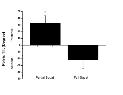
There were significant differences in pelvic tilt between partial and full amplitudes (+ 32.4° ± 10.9 and -21.7° ± 12.3, respectively, P<0.001, d = 0.95, Δ% = 33.8%) (Figure 1). Maximum tibial inclination values did not show significant differences between partial and full amplitudes (19.1° ± 6.6 and 20.1° ± 7.4, respectively, P = 0.225, d = 0.14, Δ% = 4, 9%).

The present results demonstrate important differences between partial and full squats based on pelvic tilt. During the partial squat, the pelvis hadan an terior tilt, increasing the lordotic position, while the full squat moved the pelvis backward creating lumbar retification.
The back musculature supports the spine in a neutral position. Increased and potentially harmful compressive and shear forces of the lumbar spine may result during intense squat conditions.12 Therisk of disc herniation is increased during heavy resistance squatting, with both the flexed spine position, and the backward pelvis tilt as a resultof excessive stress placed on intervertebral discs.13
Spinal flexion and extension have been shown to significantly impact joint kinetics during squat performance. Squattin gwith a flexed lumbar spine decreases the moment arm for the lumbar erector spinae, reduces tolerance to compressive load, and results in a transfer of the load from muscles to passive tissues, heightening the risk of disc herniation. Moreover, shear forces during squatting have been found to be significantly greater as lumbar flexion increases from the neutral position.12
Previous studies have shown that compressive forces increase during excessive lumbar extension.14-17 Therefore, it is advisable to maintain a neutral spine throughout performance of the squat, avoiding any excessive flexion or extension. Furthermore, the lack of tibial inclination differences demonstrates that it does not represent a major influence on control of the center of mass during both squat amplitudes
The partial squat produces anterior pelvic tilt, while the full squat produces backward pelvic tilt. Inclination of the tibia is similar in both amplitudes of the squat.
None.
Author declares there is no conflict of interest in publishing the article.

©2017 Marchetti, et al. This is an open access article distributed under the terms of the, which permits unrestricted use, distribution, and build upon your work non-commercially.