MOJ
eISSN: 2374-6939


Case Report Volume 10 Issue 1
1King Saud Bin Abdulaziz University for Health Science (KSAU-HS), King Abdullah Specialized Children Hospital (KASCH), Saudi Arabia
2Department of Orthopedics, King Saud Bin Abdulaziz University for Health Science (KSAU-HS), King Abdulaziz Medical City, Saudi Arabia
Correspondence: Saleh Alfaisali, King Saud Bin Abdulaziz University for Health Science (KSAUHS), King Abdullah Specialized Children Hospital (KASCH), Ministry of National Guard -Health Affairs, P.O. Box: 22490, Riyadh 11426, Saudi Arabia
Received: December 27, 2017 | Published: January 19, 2018
Citation: Fannouch G, Alfaisali S, Bobseit A, Bayounis A (2018) Masquelet Technique for Reconstruction of Extensive Bone Defect of Tibia after Gunshot Injury in a Child: Case Report. MOJ Orthop Rheumatol 10(1): 00382. DOI: 10.15406/mojor.2018.10.00382
Reconstruction of extensive traumatic bone loss in children especially after gunshot injury represents a complex and challenging clinical entity with significant long-term morbidity. In 1986, A.C. Masquelet conceived and developed an original reconstruction technique for large diaphyseal bone defects, based on the notion of the induced a pseudo-membrane, This technique will be described in our case report.
Different techniques have been proposed to reconstruct extensive bone defects, and restoring original bone anatomy and strength to regain its functions. IIlizarov technique, vascularized fibular grafts, and acute limb shortening have been used previously to address defects of various lengths. In 1986, A.C. Masquelet conceived and developed an original reconstruction technique for large diaphyseal bone defects, based on the notion of induced membrane.1 The principle of the induced membrane technique involves provoking a foreign body reacts by placing a cement spacer in the bone defect. That leads to formation of a membrane which is in fact a biological chamber. This prevents graft resorption by inducing vascularization and provides growth factors, it is a two-step procedure. The first step includes an extensive excision of infected and non-viable tissue, it is followed by careful stabilization of the bone and placement of cement spacer in the bone defect .and covered with a flap if required .The second step is performed at least 6 to 8 weeks after the first step, it includes removing the spacer and filling the biological space which has been created with cancellous graft or bone substitutes.1-4
A13 y old boy previously healthy, sustained to gunshot injury, with segmental bone defect in the middle third of the right tibia and lose of soft tissue, Initial irrigation and debridement with fixation of the tibia by external fixator were done in the peripheral hospital, Five weeks later the patient was transferred to our institute (level I trauma center) for definitive management. On examination we found a 4x6 cm wound at the anteromedial aspect of the right tibia (Figurs1a & 2b), with foot drop. X-ray revealed mid-shaft fracture of the tibia with bone loss of about 6 cm (Figure 2a & 2b).
Nerve conduction study showed evidence of axonal motor nerve injury of the right peroneal nerve. In addition, the right tibial nerve also showed mild motor axonal injury. We decided to do Masqulete procedure, in association with our plastic team to do a muscle flap and nerve grafting. Careful debridement and irrigation with refreshment of the fracture ends were done. The defect filled with polymethylmethacrylate (PMMA) cement spacer rotation al flap and skin graft were performed at the same operation.
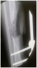
After seven weeks, second stage was performed. The bone was approached through an incision on the edge of the muscle flap, the membrane was well formed, it incised, the cement spacer removed and the space was filled with autograft and synthetic bone graft (Figure 4). Peroneal nerve was explored and tagged with aim to do nerve grafting after 3 months.

Figure 4 AP x-ray of the leg post removal of the cement spacer and filling the membrane with bone graft.
Three month later, the external fixator was removed; back slab was applied for 10 day to decrease the possibility of pin site infection. Final augmentation of the grafting was done by fibular allograft and fixed by 12 whole LCP. The nerve grafting performed (Figures 5a-5c). Two months later, the patient was seen in the clinic, he was able to move his ankle, radiography of the tibia demonstrated a remarkable filling of the gap by bone (Figures 6a-6d).
Treatment of large segmental bone defects in children can be challenging for orthopedic surgeons. Alain C. Masquelet described a bone reconstruction technique combining induced membranes and cancellous grafting. This induced membrane technique consists of two surgical steps. The first step comprises soft tissue and bone debridement with implantation of a cement spacer that induces a pseudosynovial membrane, stabilization of the bony segment with a stable fixation, and soft tissue coverage or free tissue transfer. The second step is performed approximately 2 months later and comprises removal of the cement spacer and filling of the cavity with cancellous bone graft and bone substitutes.1-3
David Jean Biau et al.5 described a case report of 16 cm defected treating by the Masquelet technique. Their patient was 12-year-old with right femoral diaphyseal Ewing’s sarcoma. Seven months after the initial procedure during which adjuvant chemotherapy was given, the second- stage procedure was performed. Touchdown weight bearing was allowed immediately, partial weight bearing was resumed 6 weeks after the operation, and full weight bearing was allowed 4 months later. Successive plain radiographs showed rapid integration of the autograft to the host bone with bone union and cortical reconstitution.5
R gouron et al.6 published a retrospective study in 2013 of 14 children who underwent bone reconstruction using Masqulete technique in the context of trauma, tumor resection or congenital pseudo-arthrosis, bone union was achieved in 9.5 months (range: 2 to 25 months), non-union was reported in about 35% of cases. Technical errors were identified in all the non-union case, insufficient cement overlap of the bone ends or fixation not stiff enough.6
J sales de Gauzy et al.7 conducted retrospective multicenter study. They evaluated diaphyseal bone defects in cases in which bone reconstruction was performed. Bone defect was either associated with trauma or secondary to infected non-union. The aim of this series was to determine the epidemiological characteristics and to evaluate the results of different treatments in this entity. Traumatic bone defects were found to have a better prognosis in children than in adults. The thicker, more active and richly vascularized periosteum in children was considered an important prognostic factor. They concluded that the treatment of bone defect requires good initial bone stabilization, reconstruction depends on the integrity of the periosteum, and in case of an intact periosteum, bone reconstruction does not seem necessary in young children. If one part of the periosteum is intact, a simple autograft seems sufficient even with extensive bone defects. In the absence of the periosteum or especially in case of infection, the induced membrane technique seems preferable, with bone transport or a vascularized bone transfer.
Karger et al.8 have reported the largest case series that used this technique. This was a retrospective study which included 84 posttraumatic diaphyseal long bone reconstructions (61 tibias) of which 50% were infected. Bone defect size ranged between <20 mm to >100 mm. At a mean duration of 14.4 months after the first surgery, the authors observed union in 90% of the cases. Eight patients (10%) were reported as failures and six patients required amputation. The authors concluded that reconstruction of large bone defects can be achieved with this procedure and its indications include non-traumatic bone defects.
The induced membrane technique makes the reconstruction of big bone defects possible, with high rate of successful. The two-step surgical procedure has an advantage in case of primary infection and where repeated debridement is necessary. The notion of a biological chamber which is described in this method provides a vast experimental field that needs further exploration.8
None.
None.

©2018 Fannouch, et al. This is an open access article distributed under the terms of the, which permits unrestricted use, distribution, and build upon your work non-commercially.