MOJ
eISSN: 2374-6939


Case Report Volume 10 Issue 1
Department of Orthopedic Surgery and Traumatology, Hospital Universitario Doctor Peset, Spain
Correspondence: Sergio Perez Ortiz, Orthopedic Surgery and Traumatology Department, Hospital Universitario Doctor Peset, Av. Gaspar Aguilar, 90. 46017, Valencia, Spain
Received: November 28, 2017 | Published: January 9, 2018
Citation: Pérez OS, Borrás CJC, Blas DJA (2018) Pure Isolated Medial Peritalar Dislocation: A Case Report. MOJ Orthop Rheumatol 10(1): 00374. DOI: 10.15406/mojor.2018.10.00374
Introduction: Peritalar dislocations are a rare lesion caused by the dislocation of the talocalcaneal and talonavicular joint with a variable prognosis depending on the associated structures that are injuried. It is a very rare when they occur without associating bone fractures.
Case report: We report the case of a 32-year-old Caucasian male patient brought to our hospital after suffering a fall with ankle inversion while practicing sports. Physical examination revealed swelling and deformity of the hindfoot with medial plantar flexion and no neurovascular alterations. In conventional X-ray a peritalar dislocation was observed. The dislocation was reduced and the limb was immobilized with a non-weight bearing below-the-knee splint. The reduction was assessed with two X-ray projections, and computed tomography (CT) ruled out associated bone fractures. The splint was maintained for four weeks.
Results: The patient started with with passive ankle dorsiflexion and plantar flexion exercises after the removal of the splint and partial weight bearing followed by full weight bearing in the 10th week after reduction. At the 6-month follow up, the patient achieved a score of 99 points in the questionnaire of the AOFAS and had resumed sport activity without clinical repercussion.
Conclusion: Isolated and closed peritalar dislocations are very rare lesions that have a good prognosis if an early reduction is carried out, no associated injuries are present, which can be ruled out with more precise imaging studies, and immobilization is maintained during the appropriate period of time.
Keywords: Peritalar, Subtalar, Dislocation, Talus, Hindfoot, Traumatology
CT, Computed Tomography; AOFAS, American Orthopedic Foot and Ankle Society; MRI, Magnetic Resonance Imaging
Peritalar dislocations are caused by a simultaneous dislocation of the talonavicular and talocalcaneal joints. They are infrequent injuries due to the inherent stability of the talus, representing 1% of total dislocations.1,2 They occur more frequently in middle-aged adult male patients and, in most cases, they are associated with fractures of the malleoli or other bones of the foot, being exceptional their presentation as an isolated injury.3 We report a case of a 32-year-old Caucasian male patient with an isolated medial peritalar dislocation without associating foot or ankle fractures.
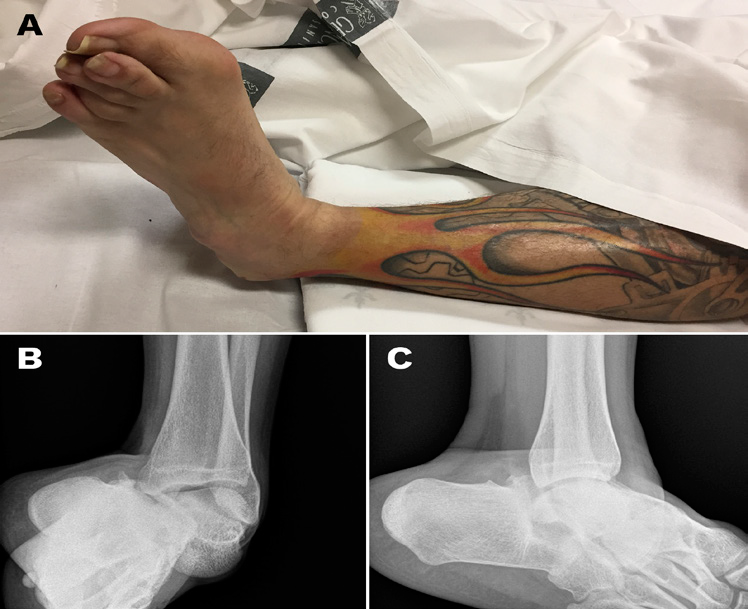
We report the case of a 32-year-old Caucasian male patient who was brought to the Emergency Room of our hospital after suffering a fall with ankle inversion mechanism while practicing sports with a BMX-type bicycle. Physical examination revealed intense pain located in the left ankle, swelling and deformity of the hindfoot with medial plantar flexion, prominence of bony structures in the lateral region of the joint, abrasions and ecchymosis without deep wounds (Figure 1A). The patient was hemodynamically stable and did not associate other lesions. The neurovascular status of the limb was evaluated by checking the presence of the dorsalis pedis and posterior tibial pulse, correct nail capillary refill and preservation of sensitivity by two-point discrimination test.
Conventional X-ray revealed a peritalar dislocation with displacement of the calcaneus and rest of the bones of the foot towards the medial side without observing associated fractures (Figure 1B & 1C).

Figure 1A ankle appearance on arrival to the Emergency Room; B: conventional X-Ray (B, anteroposterior view; C, lateral view) showing pure peritalar dislocation.
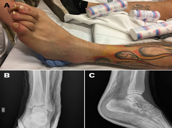
Closed reduction of the dislocation was carried out under sedation by traction with the knee flexed, followed by eversion of the foot with plantar flexion of the ankle and, finally, dorsal flexion of the ankle (Figure 2A). After the reduction, the presence of dorsalis pedis and posterior tibial pulses, capillary nail refill and distal sensitivity, as well as the correct oxygenation by digital pulse oximetry were clinically verified. The affected joint was immobilized with a below-the-knee splint. The correct reduction of the dislocation was confirmed by conventional X-ray in two projections (Figure 2B & 2C).

Figure 2A ankle appearance after close reduction; B: conventional X-Ray (B, anteroposterior view; C, lateral view) showing correct reduction of the dislocation.
In computed tomography (CT), associated fractures in other bones of the foot, osteochondral lesions and intra-articular free bodies were ruled out (Figure 3).
The below-the-knee splint was maintained for 4 weeks, beginning with passive ankle dorsiflexion and plantar flexion exercises without doing eversion or inversion movements after the removal of the cast and later with partial weight bearing followed by full weight bearing in the 10th week after reduction.
At the 6-month follow up, the patient achieved a score of 99 points in the questionnaire of the American Orthopedic Foot and Ankle Society (AOFAS).4 and had resumed sport activity without clinical repercussion (Figure 4).
Peritalar dislocation, also called subtalar or luxatio pedis subtalo.2 was first described by Dufarest and Judey in 1811.5 and is defined as the dislocation of the talocalcaneal and talonavicular joint without associated fractures or involvement of the tibiotalar or calcaneocuboid joint.6 It is a very rare lesion when it occurs without associating fractures in other bones and a very complex injury when involving joint surfaces of load bones.2 with a variable prognosis depending on factors related to the patient and the structures involved.7 and a complication rate close to 20%, among which include osteoarthritis, avascular necrosis or instability of the subtalar joint.8
The subtalar or talocalcaneal articulation is a complex joint that can be divided into an anterior part, where the medial and anterior concave calcaneal facets articulate with the head of the talus; and a posterior convex facet, separated from the other facets by the interosseous talocalcaneal ligament, that articulates with the talus facet.9 This precise articulation of stabilizing ligamentous structures, which can be divided into extrinsic (calcaneofibular and tibiocalcaneal portion of the deltoid ligament) and intrinsic (cervical and interosseous talocalcaneal ligament).10
The talonavicular joint is formed by the ellipsoidal convex surface of the head of the talus and the concave facet of the navicular, whose longitudinal axis is oriented as the one of the head of the talus.11 but which does not cover it in its entirety.12
The head of the talus fits into a structure delimited medially and inferiorly by the plantar calcaneonavicular ligament or "spring ligament", a portion of the bifurcated ligament laterally, the anterior and middle articular facets of the subtalar joint proximally and the articular facet of the navicular distally.10 which some authors call acetabulum pedis because of its similarity to the hip joint.13 Therefore, it could be considered that the subtalar joint is formed by the posterior talocalcaneal joint and acetabulum pedis.
The peritalar dislocation is classified according to the modification that Malgaigne and Buerger.14 made of Broca's classification (1853).15 in medial, lateral, posterior or anterior dislocations depending on the direction in which the distal part of the foot moves with respect to the talus.
The medial dislocation, the most common with a frequency close to 80%.2 is produced by an external rotation of the talus with the foot in inversion and flexion, with the sequential rupture of the dorsal talonavicular ligament, anterior and posterior interosseous ligaments.16 It is the one that presents less associated bone or soft tissue injuries.17,18
The lateral dislocation, second in frequency with a percentage close to 15%, is produced by an eversion of the foot that leads to rupture of the deltoid ligament, interosseous ligaments and dorsal talonavicular ligament.19 It is most frequently associated with bone and soft tissue lesions, making it difficult to reduce it in many cases by interposition of the adjacent structures.18,20
The posterior and anterior dislocation correspond to 2.5% and 1% of the total respectively and have a great tendency to instability, which can easily become medial and lateral, which would explain the low incidence of this type of dislocation.3 Some authors consider that these two types are, in fact, subcategories of the previous ones.21 They are produced by a forced plantar flexion or translation in the anterior direction, leading to a rupture of the interosseous ligament and medial and lateral ligaments.22,23
They are clinically presented with ankle deformity with the foot in plantar flexion and swelling, with more or less soft tissue involvement, protruding the head of the talus dorsolaterally with the foot in supination or dorsomedially with the foot in pronation according to the medial or lateral dislocation respectively.19 This deformity is lower in the case of anterior or posterior dislocations when there is less alteration of the axis and less misalignment.24 The incidence of open lesions in subtalar dislocations is close to 20-25%, being more frequent in lateral dislocations when they are produced by high energy trauma.25,26
It is important to perform an exploration of the possible soft tissue and neurovascular involvement of the limb to rule out concomitant pathology, since neurovascular alterations have been demonstrated in up to 70% of cases.19
A radiographic study should be made for diagnosis with an anteroposterior and lateral projection of the hindfoot to assess the diagnosis and rule out the possible association of bone fractures in the foot or ankle because, if the occur, they will condition the definitive treatment.2,6,19,25
A closed reduction under sedation should be attempted as soon as possible.8 to avoid additional complications such as avascular necrosis of the talus, neurovascular or soft tissue lesions such as skin necrosis.6,25 To reduce the dislocation, the knee and ankle are flexed towards plantar, in order to relax the triceps surae.6,7 longitudinal traction is performed and force is applied in the opposite direction to the dislocation in order to reduce the head of the talus on the navicular joint facet.19 Occasionally, direct soft pressure on the head of the talus can make reduction more easy.25 In the event that the reduction is not achieved by interposition of bone or soft parts.8,19,20,27,28 multiple attempts with brute force should not be undertaken under any circumstances, and urgent open reduction should be considered.1,18 since, otherwise, we can cause iatrogenic injuries such as fractures of the talus or neurovascular injuries.6
After reduction, the correct neurovascular state of the limb is checked by palpation of the dorsalis pedis and posterior tibial pulses and distal oxygenation by pulse oximetry. If signs of ischemia are detected, additional imaging tests such as Eco-Doppler or angio-CT will be performed.3
Once the dislocation has been reduced, the joint is immobilized with a non-weight bearing posterior short-leg cast or below-the-knee splint, since it prevents a high pressure in the compartments of the lower limb and allows a better control of the condition of the skin and soft tissues.19 Joint congruence will be checked by conventional X-ray with at least two projections.20
In 10% of the medial dislocations and 40% in the lateral dislocations, approximately.25 closed reduction may be impossible due to the interposition of soft parts: in the case of medial dislocations, reduction can be impeded by the superior extensor retinaculum, the bellies of the extensor digitorum brevis, the capsule of the talonavicular joint, the posterior neurovascular bundle or the peroneal tendons.25,29 on the lateral dislocations, the tibialis posterior tendon or the flexor digitorum longus can obstruct it.27,29 In these cases, an open reduction will be made in the shortest time possible to avoid soft tissue injuries. A longitudinal approach will be made on the head of the talus in the medial dislocations and between the medial malleolus and the head of the talus on the lateral dislocations.19
CT or magnetic resonance imaging (MRI) are useful to rule out or confirm associated bone lesions, that are present in up to 60% of cases, since we must not allow osteochondral lesions of the joint surfaces or fractures of the lateral talar process and sustentaculum tali to go unnoticed, which could lead to the development of posttraumatic arthritis of the subtalar joint.6,25 However, some authors indicate that MRI would not be very useful in this pathology, since ligamentous injury is assumed as a constant and it is CT that will provide more information about bone lesions.19
The time of immobilization is controversial, with diversity of opinion among authors, being variable between four and six weeks, and avoiding weight bearing.30 Some authors consider that it may be less time in the case of isolated dislocations without signs of instability, maintaining the splint for three weeks and then beginning with an early mobilization.28,31 while others consider that a period of less than four weeks is insufficient for the healing of the lesions resulting in chronic ligamentous instability or recurrent subluxation.3,24,25,28 In our case, we maintained the immobilization with a below-the-knee splint and non-weight bearing for four weeks and, after removing it, and checking the stability of the subtalar joint and the absence of soft tissue injuries, progressive partial weight bearing and the initiation of rehabilitation with active mobilization exercises were authorized with full weight bearing at six weeks after the removal of the immobilization.
Among the notable complications we can find stiffness, being the most frequent complication with rates of up to 70%.32 especially in cases of long periods of time immobilization.19 arthritis, the main cause of pain.33 avascular necrosis of the talus, mainly in dislocations caused by high energy trauma resulting in open dislocations and/or associated fractures, regardless of whether it is medial or lateral.8 skin necrosis or deep tissue infections, only described in open dislocations.34 and instability.35
The prognosis of these injuries is usually good in cases in which the injury mechanism has been low energy, there are no associated fractures and the lesion has been exclusively ligamentous. Closed lesions, those that have been treated early, and immobilizations that have not been maintained for an excessive period of time have a better prognosis.3,19,25,28,30,36 Regarding the type of dislocation, there are different opinions in the published literature regarding the prognosis of each: while some authors have observed that medial dislocations have a worse prognosis.37 others indicate that lateral dislocations present a worse recovery and prognosis due to frequent association with fractures or open dislocations.25,37 Finally, a third group of authors indicates that there is no difference between the type of dislocation if it has not associated injuries.35
Isolated and closed peritalar dislocations have a good prognosis regardless of their classification when an early reduction is carried out and immobilization is maintained during the appropriate period of time. Therefore, a quick action on the lesion and the performance of more precise imaging studies such as CT are necessary to rule out associated fractures that require an additional treatment, and which could condition a worse prognosis.
None.
All the Authors state they do not have any conflict of interest related to this article.

©2018 Pérez, et al. This is an open access article distributed under the terms of the, which permits unrestricted use, distribution, and build upon your work non-commercially.