MOJ
eISSN: 2374-6939


Case Report Volume 6 Issue 5
Orthopedic Department, Greece
Correspondence: Panagiotis G Tsailas, Orthopedic Department, Larissa General Hospital, 6 Mitika str, Larissa, 41447, Greece, Tel 307000000000
Received: September 14, 2016 | Published: November 29, 2016
Citation: Tsailas PG, Hartonas G (2016) Bicon Threaded Acetabular Cup Fracture, Case Report, Mode of Failure and Review of the Literature. MOJ Orthop Rheumatol 6(5): 00235. DOI: 10.15406/mojor.2016.06.00235
Threaded acetabular cups have been equally successful as hemispheric cups. However, based on our case, as well as the reported literature, these thin shelled cups may be at risk of fracture in cases with posterocranial or anterocranial bone defects, even if a reconstruction of the defect is planned. We believe press-fit cups with their thicker walls are preferable in cases with osseous defects.
Threaded acetabular cups have been equally successful as hemispheric cups. Despite this success there is always the chance of failure. The commonest mode of failure being polyethylene wear, osteolysis, and loosening. A more implant specific mode of failure of threaded acetabular shells has been cup migration.1-3 Another rare mode of failure seems to be stress fracture of the acetabular shell. After a review of the literature we found two reports of fractured threaded Bicon acetabular shells.4,5 numbering three cases each. As well as a brief mention in a series of 153 cases, where there were 4 cases of fatigue fracture of the titanium shell.6 This mode of failure is very rare in hemispheric cups with only one published case of cup fracture.7
The purpose of this case report is to present our experience with this rare, however, implant specific complication, as an addition to the existing literature, in an effort to shed some more light in the involved mechanism of fracture.
Keywords:Threaded acetabular, Anterocranial, Posterocranial, Acetabular, Polyethylene, Osteolysis, Bicon
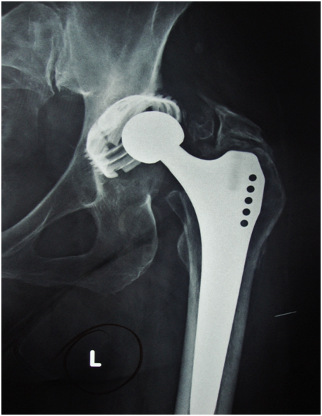
The patient was a 77 years old female (160cm and 70Kg, BMI 27.3) who was operated for the first time in 2001 due to dysplasia (Figure 1). A Zweymüller stem combined with a second generation Bicon cup were used (Sz. 2 stem, 46-mm Bicon® titanium threaded cup, metal/PE combination; Plus Endoprothetik, Rotkreuz, Switzerland) (Figure 2). The patient had an uneventful post-operative period. During the subsequent follow-ups there was good osteointegration of the cup with no signs of loosening, cup migration or polyethylene wear, and the hip was completely pain free. Thirteen years postoperatively, (2014) the patient suddenly presented acute pain in the operated hip, difficulty walking and was forced to use crutches. During these thirteen years the patient did not have regular follow-ups and on admission she did not present any previous radiographs, however, the primary surgeon (G.H.) could not recall of any radiological signs that could have predicted the problem. On radiographic examination a fracture of the acetabular shell was evident (Figure 3), and she was admitted for revision. During surgery it was observed that the PE was intact, without significant wear. There were some signs of impingement of the neck of the stem at the antiluxation lip of the insert, but without any signs of impingement against the metal shell (Figure 4A). The fractured part was anterior superior and it was completely loose (Figure 4A-4D). Upon removal there was no bone support from 8 o’clock to 1 o’clock (left hip). The rest of the cup was well fixed and it was removed with curved osteotomes, with care to preserve as much host bone as possible. On gross examination there was osteointegration at the level of the threads of three quarters of the perimeter of the well-fixed part and no sign of osteointegration of the rest of the implant or the fractured part (Figure 4D). The anterior-superior bone defect was reconstructed with a femoral head allograft contoured to a figure of 7, so as to achieve positive interlock at the rim and at the same time have the stronger femoral neck bone over the ilium for screw fixation. Following fixation of the graft with 2 screws, the head and acetabulum were rimed until the graft and the host bone were flash. The host bone was about 55-60% of the acetabular area to receive the cup. A 48mm hemispheric cup (Mallory-Head Acetabular Shell, porous coated, Biomet, Bridgend, UK) was impacted in place and further augmented with 2 screws. On the post-operative radiographs there was complete coverage of the new cup (Figure 5). The patient at eighteen months postoperatively is doing well, although she has some pain over the greater trochanter due to the trochanteric hook plate. A written informed consent of the patient, for the print and electronic publication of the text and any additional components, such as photographs, as well as, an approval by the institution review board were obtained.

Figure 2 Immediate postoperative X-Ray showing a somewhat high hip center of rotation, and uncoverage of the implant superiorly.

Figure 3 13 years postoperatively, X-Ray shows an anterior-superior fracture of the insert, without any sings of loosening or migration.

Figure 4
Figure 4A Reconstituted retrieved implants showing the direction of the fracture, which was anterior-superior from 8 o’clock to 1 o’clock (Left hip). There was no significant wear of the PE, and some signs of impingement and delamination of the elevated lip.
Figure 4B Backside of the Polyethylene insert showing the lines where the fracture occurred.
Figure 4C Metal insert showing that the fracture line was through diametrically opposed notches.
Figure 4D On gross examination there was osteointegration at the level of the threads of the well-fixed part and no sign of osteointegration of the rest of the implant or the fractured part.
Using the Zweymüller cementless threaded cups has had satisfactory long-term results.3,8-10 The Bicon is a double-cone cup made of hot forged pure titanium of a mean wall thickness of 0.9mm. The aim for the reduced wall thickness of the implant was to match the elasticity of the cup to that of natural bone.5 The self-cutting thread design provides sufficient primary stability that prevents tilting of the cup. Along the evolution of the Zweymüller threaded cup there were three generations. The first-generation was the Alloclassic cup, which was developed in 1985. The second-generation Zweymüller Bicon cup was introduced in 1993, which initially had eight notches along the outer circumference intended to accommodate the seating instrument. In 1995, the eight notches of the Bicon cup were reduced to four notches and then eliminated in 2003 with the development of the third-generation Bicon cup to protect against titanium shell fatigue fractures.6 There are no holes for screw fixation, for added stability. Compared with press-fit hemispheric cups, most threaded cups have considerably thinner walls, increasing the probability of implant fracture. Hemispheric cups are at least 3 times thicker. Despite the wall thinness, shell fractures are rare complications that have been recorded only in individual cases so far.4-6
In the literature we found three articles reporting fractures of the Zweymüller Bicon threaded cup.4-6 Two case reports.4,5 with three patients each (in one of them only one patient is described in detail.5 and a case series mentioning briefly that they had four implants with fatigue fracture.6 which were well fixed and well oriented except one which was implanted in extreme valgus. Altogether there were ten cases reported.
From the available information we can deduct that fatigue fractures occur mostly in overweight patients (mean BMI of 26.15) with absent or inadequate bone supporting the acetabulum 4 or failed craniolateral osteointegration.5 In the report by Hoburg et al.4 the position of the defect was uniformly in the dorsocranial area of the acetabulum, and it was only with defects in this area that fractures of the threaded cups were found. This region has been shown to constitute the main direction of the transmission of force into the pelvis.11 In one patient.4 although the wall defect was reconstructed by an allograft at the same time with the implantation of the threaded cup, there was no osteointegration in this area, because of the limited biologic potential, which led to the fracture. All three cups presented in this report were firmly integrated to the existing bone. All these patients presented with symptoms between 3.5 and 7 years postoperatively. For this reason it was suggested that defects in the anterosuperior area, which are typical of dysplasia, can be treated with a threaded cup, since there have been no cup fractures when such implant were used in the treatment of patients with congenital hip dysplasia .4
This is contrary to our findings, in which the defect was anterior-superior (Figure 4A). Our patient has been neglecting her follow-ups, since she had no clinical problems with her hip, up until the day the cup failed. The fact that almost two third of the circumference of the cup were well osteointegrated and thus were rendering the cup stable, may have been the reason why the patient had a well-functioning hip replacement without any complaints. The anterior-superior part of the cap fractured due to repeated loading of a non-osteointegrated area, secondary to the lack of bony support. We believe that the mechanism of fracture is superior stress loading of a partially well fixed cup, on its inferior side, transforming compressive loads from the femur to shearing stresses, for a longer period of time. All the other cases reported are early to mid-term failures compared to our patient, which was a late case of fracture.
By looking at our cup and at the images of the fractured cups provided by the two other reports, the arc of the fractured piece or pieces passes through the notches for the seating instrument, which makes them a weak point of the cup (Figure 4C). This coincides with the supposition in the case series by Korovessis et al.6 that the notches at the peripheral rim of Bicon shell represent points of stress concentration that create fatigue fracture, resulting from acetabular remodeling, independently from the biologic fixation of the cup.
Threaded cups have been recommended for implantation in hip dysplasia.12-14 and in revision situations.15 In case of dysplasia either a high hip center technique has been used, which compensates for the bone defect.12 or reaming was carried out by removing as much of the medial wall as necessary to engage at least one thread without using any structural bone graft for reconstruction of the deficient anterolateral acetabular margin.13 The aim always being to have sufficient acetabular cup coverage (mean cup coverage of 96%).14 While the general consensus for the threshold coverage required for long-term cup stability is supposed to be over 70% [16-18], this percentage refers to the thicker hemispheric cups and may not apply to the thinner Bicon threaded cup. In our case a somewhat high hip center was used as well to achieve as much coverage as possible (Figure 2). In case of revisions the use of this cup was not advised if peripheral segmental acetabular margin defects are present because all the implants have showed signs of migration.15
Despite the fact that a case report of a fractured hemispheric cup has been presented in the literature.7 there has not been any other occurrences of such a mode of failure for this particular type of cups. Furthermore the fracture was secondary to failure of the polyethylene liner and, subsequently, fracture of the acetabular component from biomechanical wear between the head and the metal shell.
In conclusion, based on our case report, as well as the observations of the two other publications, these thin shelled cups may be at risk of fracture in cases with posterocranial or anterocranial bone defects, even if a reconstruction of the defect is planned. We believe press-fit cups with their thicker walls are preferable in cases with osseous defects. One case of failure of an otherwise successful cup can certainly not be generalized. It is possible that there may have been a manufacturing problem with the specific implant which led to its failure after years of loading. However, the fact that this specific generation of implants has been substituted by the manufacturer, in addition with the other cases reported in the literature, suggests that leaving them with a large part of their circumference uncovered by bone may lead to problems in the long run.
None.
None.

©2016 Tsailas, et al. This is an open access article distributed under the terms of the, which permits unrestricted use, distribution, and build upon your work non-commercially.