MOJ
eISSN: 2374-6939


Case Report Volume 4 Issue 2
1Trainee Resident, Orthopedics Department, King Fahad Specialist Hospital, Saudi Arabia
2Consultant of Foot and Ankle Surgery, Orthopedics Department, King Fahad Specialist Hospital, Saudi Arabia
Correspondence: Ramadan Al-Tamimi, Department of Orthopedics, King Fahad Specialist Hospital, Building 7, 1st Floor, Room#111, PO Box 15215, Dammam 31444, Saudi Arabia, Tel 967000000000, Fax 0096613-844-2680
Received: January 07, 2016 | Published: January 28, 2016
Citation: Tamimi RH, Ali SM (2016) Traumatic Multi Ligament Ankle Injury, Associated with a Displaced Articular Calcaneus Fracture, in the Absence of Ankle Fractures. MOJ Orthop Rheumatol 4(2): 00134. DOI: 10.15406/mojor.2016.04.00134
Traumatic, Ankle instability, Anterior talofibular ligament, Deltoid ligament, Calcaneus fractures
EUA, Examination Under Anesthesia; ATFL, Anterior Talo Fibular Ligament; CFL, Calcaneo Fibular Ligament; PTFL, Posterior Talo Fibular Ligament
We are reporting a rare case of fracture calcaneus associated with traumatic lateral and medial ankle ligamentous injury, in the absence of ankle fracture or syndesmotic disruption.1,2
A 31 years old female, presented to our institution as a case of acute trauma, with history of fall from approximately six meters of height landing on her feet. Her chief complaint was generalized back, and right foot and ankle pain.
Initial assessment
Lower back tenderness with lower limb neurological deficit below L3, A closed injury to the right foot and ankle with global instability.
Radiological assessment shows
L3 unstable spine fracture, Right intara-articular calcaneal fracture of joint depression type, Saunder’s III AC3,4(Figure 1 & 2).
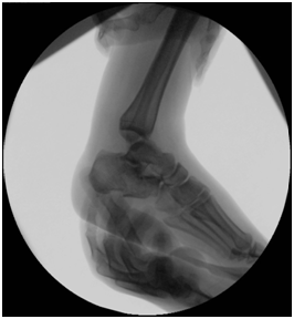
Treatment Management began with stabilization of the patient’s general condition, and later definite treatment on 2nd stages. 1st stage: Patient went for decompression spine surgery and examination under anesthesia (EUA) of the right ankle which demonstrated medial and lateral ligament complex instability as evident by a positive anterior drawer test (Figure 3), a posterior drawer test (Figure 4) and a positive varus and valgus stress tests with intact syndesmosis clinically (Figure 5 & 6). Because of the local swelling, the patient was put in a cast. 2nd stage: 5 days later, when the swelling subsided, patient underwent open reduction internal fixation of the right calcaneus fracture and repair of the medial and lateral ligament complex tears.

Figure 3 Anterior drawer test: an anteriorly directed force is applied to the heel while supporting the leg.

Figure 4 Posterior drawer test: a posteriorly directed force is applied to the foot while supporting the leg.
Intraoperative findings
Complete tear of superficial and deep components of the deltoid ligaments. Complete tear and attenuation of the anterior talofibular ligament (ATFL), calcaneofibular ligament (CFL), and the posterior talofibular ligament (PTFL).
An Open reduction internal fixation of right calcaneal fracture using extensile lateral calcaneal approach and the anatomic repair of the medial deltoid ligaments and the reconstruction of the lateral ankle ligaments (ATFL, CFL) using a bone tunnel and a tendon allograft.
Patient was kept non weight bearing in a below knee cast for 6 weeks. Later she was put in an Air Cast Boot and started on active ankle range of motion exercises at 2 months of assessment.
Clinically
She maintained a power of 3/5 of her lower extremities heel varus was corrected, no tenderness at the fracture site ankle was stable in Varus, Valgus and Anterior drawer tests.
Radiologically
The calcaneus fracture has healed with no Talar shift or tilt (Figure 7-9).
High energy trauma such as, fall from heights and motor vehicle collisions are the usual cause of intra-articular calcaneal fractures. During axial loading, depending on the foot position, different fracture line and varying degrees of communition and articular involvement develop.1,3,5
Calcaneal fractures are associated with spine and pelvic fractures and lower extremity injuries,1,2 however their association with lateral or medial ankle instability has not been reported so far in English literature, which makes this case unique.
We are using the classification system of Sanders et al.6 for classification of Calcaneal fractures,6 which is based on CT coronal plane images, and takes into account the number and location of primary fracture lines. Another useful classification is the one described by Essex Lopresti,3 which demonstrates the impact of the mechanism of injury on calcaneal fracture geometry.
Three major configurations contribute to stability of the ankle joint; which are, the congruity of the articular surfaces, the static ligamentous restraints, and the musculo tendinous units, which allow for dynamic stabilization of the joint.7
These static restraints fail in extremes of position, which might be the case in acute trauma. Studies done by Rasmussen8 have shown that during forced ankle dorsiflexion the PTFL ruptures, and in forced internal rotation the ATFL ruptures followed by injury to the PTFL. Extreme external rotation causes the deep deltoid ligament to tear. Adduction in neutral foot position injures the CFL, while in plantar flexion the ATFL is injured.8
Stress radiographs are better performed under sedation or anesthesia for evaluation of ankle stability. A positive anterior drawer stress test indicates an ATFL rupture.4,9 A positive talar tilt on varus ankle stress tests indicates an injury to the ATFL and CFL.7,9,10 Injury to the deltoid ligament is manifested by a widening of the medial clear space and a positive valgus stress test.11-13 The syndesomsis can be assessed by the tibio fibular overlap and the tibio fibular clear space on AP and mortise views. An external rotation stress tests is useful for assessment of the deep deltpoid ligament and the syndesmosis.14
Controversies exist in management of displaced intra-articular calcaneal fractures.15 Operative intervention with the use of plate and screws through a lateral calcaneal approach was superior to conservative measures in various studies.2,16,17 We used an AO locking calcaneal plate for fracture stability and to maintain height.
Several surgical techniques have been advocated for management of lateral and medial ankle ligament tears, these include direct anatomic repair and reconstruction.18
The creation of boney tunnels and the use of auto- or allo- grafts for ligament reconstruction have been described. Reattachment can be done with the use of anchor or trans-osseous sutures.17,19 We used a direct anatomical repair of the deltoid ligament, because good approximation of the tissue was achieved. On the lateral side; because of tissue attenuation of the ligaments, we used bone tunnel techniques through the talus and fibula, and used a tendon allograft for reconstruction.
In summary, we have achieved good results with this approach in this patient, and we advise for careful examination and timely management in such complex rare injuries.
None.
None.

©2016 Tamimi, et al. This is an open access article distributed under the terms of the, which permits unrestricted use, distribution, and build upon your work non-commercially.