MOJ
eISSN: 2373-4442


Research Article Volume 2 Issue 2
1Maisonneuve-Rosemont Hospital Research Centre, Canada
2Department of Microbiology, University of Montreal, Canada
3Canadian Space Agency, Canada
Correspondence: Laurent Sabbagh, Maisonneuve-Rosemont Hospital Research Centre, 5415 l’Assomption boulevard, Montreal, QC, H1T 2M4, Canada,, Tel 450-680-4606
Received: August 29, 2014 | Published: June 5, 2015
Citation: Geneviève DL, Luchino YC, Sabbagh L (2015) Corticosterone Signaling in Central Memory CD8+ T Cells Triggers Apoptosis that is Inhibited by Interleukin-15. MOJ Immunol 2(2): 00044. DOI: 10.15406/moji.2015.02.00044
Immunological memory plays a vital role in monitoring the body against specific pathogens. CD8+ memory lymphocytes allow a rapid response and elimination of any threat previously encountered in addition to keeping latent some chronic viruses in the host. However, certain situations such as chronic stress decrease immune function and could thereby promote viral reactivation. We demonstrate here that the stress hormone corticosterone, a cortisol analog in mice, induces apoptosis in CD8+ memory T cells. Interleukin-15 (IL-15), a cytokine essential for the homeostasis of memory T cells rescues corticosterone-induced apoptosis in a dose-dependent manner. IL-15 and glucocorticoid signaling pathways were investigated to explain the inhibitory effect of this cytokine. NF-kB, IkBα and Bim protein expression did not vary significantly when memory lymphocytes were cultured with corticosterone. However, expression of the glucocorticoid-induced TNFR-related protein (GITR), a member of the TNF receptor family, did significantly increase in the presence of IL-15 in a dose-dependent manner. Since GITR is required for the survival of memory CD8 T cells in vivo, this suggests that it may be involved in concert with IL-15 to counter glucocorticoid-induced apoptosis of CD8 memory T cells.
Keywords: corticosterone, IL-15, TNF-receptors, apoptosis, CD8 memory T cells
IL-15, interleukin-15; GITR, glucocorticoid-induced TNFR, related protein; GCs, Glucocorticoids; GCR, GC receptor; CST, corticosterone
Small populations of effector memory CD8 T cells have been identified in the vicinity of neurons infected by the HSV-1, an alpha-herpes virus.1 The same type of immune surveillance has been observed in cases of latent infection with CMV and EBV, beta- and gamma-herpes viruses respectively.2,3 In each case there are a small proportion of memory CD8 T cells surrounding the reservoir of latent viruses, even if there is no active production of virions. These effector memory T cells also present a certain cytotoxic capacity confirmed by their production of IFNg and non-lytic granules. Among the physical, psychological and environmental stimuli which trigger the reactivation of latent viruses, most involve a stress imposed on the organism. For example, UV exposure and hyperthermia induce the proliferation of HSV-1.4,5 Verbal and mental examinations, shift working as well as space missions have all been identified as conditions leading to the reactivation of EBV, detectable by high viral titers in the blood.6–8
Immunological memory is essential to protect the organism against repeated exposure to the same infectious pathogen or chronic viral reactivation. Recent studies have shed light on the factors governing the survival and homeostasis of antigen-specific memory T cells, emphasizing the role of specific cytokines and receptors of the TNFR family.9,10 Interleukin-15 (IL-15) is known to regulate cell proliferation, activation and differentiation of CD8+ lymphocytes and NK cells.11 This member of the gc receptor cytokine family binds to a specific cell surface receptor (IL-15-R) composed of three subunits: IL-2Rβ, IL-2Rα (gc) and IL-15Rα.12 Its role in promoting CD8+ memory T cell homeostatic proliferation and survival in the absence of antigen has been demonstrated using IL-15Rα-/- and IL-15-/- mice. These genetic models displayed a significant reduction in memory phenotype CD8+ CD44high T cell survival and proliferation. In addition, mice deficient in IL-15 produce a small number of CD8 memory cells in response to infection and this population has a proliferative defect in the absence of antigen.
Stress leads to the secretion of steroids hormones, mainly glucocorticoids (GCs), by the adrenal glands. Binding of GCs to the GC receptor (GCR) represses the transcription of proinflammatory cytokines and chemokines genes such as IL-2, TNFα, IFNg and RANTES.13 These effects are caused by the interaction of the GC-GCR complex with the NF-κB signaling pathway through direct interaction with NF-κB or upregulation of IκBa, an inhibitor of NF-κB activation14,15 GCs have also an important immunosuppressive effect as they trigger apoptosis in multiple cell types such as CD4+CD8+ thymocytes, osteoclasts, dendritic cells, and T cells.16,17 The mechanisms involved in GCs-induced apoptosis are poorly understood; however Bcl-2 and Bcl-xL inhibition and increased Puma and Bim expression are potentially involved.18–20 GCs also inhibit IL-2 secretion and Jak-Stat signaling in T cells by interfering with Stat5 DNA binding upon IL-2 stimulation.21 These results suggest that GCs could affect the survival of memory CD8 T cells which could explain the relation between elevated stress levels and viral reactivation. This study was undertaken to determine the impact of a stress hormone (corticosterone) on central memory CD8+ T cells in vitro. The results show that corticosterone induces apoptosis in resting memory T cells and that the addition of IL-15 has a potent dose-dependent anti-apoptotic effect.
Mice
OT-I mice22 were obtained from Jackson Laboratory (Bar Harbor, ME, USA). Mice were maintained under specific pathogen-free conditions in sterile micro isolator caging. All mice experiment protocols were performed following the Canada Council on Animal Care (CCAC) guidelines and were approved by the Maisonneuve-Rosemont animal care committee.
Lymphocyte isolation, stimulation and survival assay
Splenocytes were purified after red blood cells lysis of total spleen from OT-I mice using ammonium-chloride-potassium lysing buffer (ACK). They were cultured (5x10⁶cells/ml) in complete RPMI medium (RPMI 1640 with L-glutamine (Life Technologies, Ontario, Canada) complemented with 10% fetal bovine serum (FBS), 1% penicillin 10000 units/mL, 1% streptomycin 10000 ug/mL, 0.1% β-2-mercaptoethanol, 1% non essential amino acides (NEAA) 100X, 1% sodium pyruvate 200mM, and 1% Hepes 1M) and stimulated with SIINFEKL peptide of ovalbumine (0.1µg/ml) (provided by Dr. N Labrecque) for 18h in a 6-well plate at 37°C/5% CO2. After washing excess peptide, the cells were cultured for 48h at 1.5x10⁶cells/ml in complete medium. On the 3rd day after activation, density gradient centrifugation (Lympholyte-M, Cedarlane, Ontario, Canada) was used to isolate live CD8+ T cells. Flow cytometry analysis confirmed that the purity of live CD8+ T cells was >90%. The percentage of live cells was assessed using Trypan blue exclusion.
Central memory CD8+ T cells generation and stress assay
Activated CD8+ T cells were cultured in 6-well plates (106cells/ml) in the presence of 20ng/ml of human recombinant IL-15 (R&D Systems, Ontario, Canada) for 6 days as described previously by Pulle (2006). Analysis of T cell survival, activation and memory differentiation was performed by flow cytometry as described below. Once the phenotype of resting memory T cells was confirmed, they were placed in culture (106cells/ml) with increasing concentrations of IL-15 as indicated, and in the presence of 10-6M of corticosterone (Sigma-Aldrich, Ontario, Canada) or the same concentration of carrier (DMSO) before assessment of cell viability by flow cytometry.
Flow cytometry
Surface staining for flow cytometry assays was done using anti-CD8α-PerCP, anti-CD62L-FITC, anti-CD44-APC, anti-CD69-PE (BD Pharmigen, Ontario, Canada). Apoptosis levels were assessed by staining with Annexin-V-APC (BD Pharmigen) and propidium iodide (PI, company). Flow cytometry data was acquired on a FACSCalibur (BD Biosciences, CA, USA) and analysis was performed with the FlowJo software (TreeStar, OR, USA).
Western blot analysis
Aliquots of 106 CD8+ T cells were harvested from cell cultures, centrifuged and stored at -20°C until analysis. After thawing, cell pellets were re suspended in lysis buffer for nuclear extraction (1% NP-40, 0.05M Tris HCl 50mM, 0.15M NaCl 150mM, 0.25% Na-deoxycholate) for 30 minutes on ice. After centrifugation, the cell lysates were subjected to SDS/PAGE and then transferred to PVDF membranes. The membranes were blocked in TBS-T (Tris-buffered saline supplemented with 0.05% Tween20) with 5% skim milk and probed overnight with gentle shaking using specific antibodies for phospho-STAT5, STAT5, IκBα or Bim (Cell Signaling Technology, Ontario, Canada) or β-actin (Sigma). Membranes were washed 3 times in TBS-T and then incubated with HRP-conjugated anti-rabbit (Sigma, St-Louis, MO) or anti-mouse Ig antibody (Cell Signaling). HRP signals were revealed using the Amersham ECL detection system (GE Healthcare Life Sciences) and the Image Quant LAS-4000 imager (GE Healthcare Life Sciences) was used for the digital acquisition. Quantification of western blot signals was performed using the Multi Gauge Software (Fujifilm, Tokyo, Japan) and pSTAT5/total STAT5, BimEL/β-actin or IκBα/β-actin ratios were calculated. The statistical analysis for significance was done using a paired Student t-test (n=3 unless otherwise specified).
Corticosterone (CST) induces apoptosis of CD8 memory T cells
GCs have been shown to stimulate apoptosis in different lymphocyte subsets. Since stress factors produced during space missions trigger viral reactivation, the impact of the stress hormone CST on survival of central memory CD8+ T cells was assessed. These cells were generated by exposing splenocytes from OT-I mice to the SIINFEKL peptide followed by culturing with IL-15.23 The phenotype of central memory CD8 T cells was verified 9 days after stimulation, where these cells expressed high levels of CD62L and CD44, and most of the cells had lost the expression of CD69 (Figure 1A). Addition of CST at physiological concentrations observed in condition of stress24 triggered high levels of cell death in vitro, as shown by changes in cell morphology and the appearance of Annexin-V positive cells (Figure 1B). This response was observed despite the presence of 20 ng/ml of IL-15 in the cultures, a survival factor for central memory CD8 T cells.10 Kinetic experiments showed that cell death occurred between 6 and 24h following the addition of CST Figure 1C.
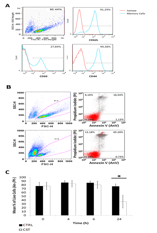
Figure 1 Corticosterone induces apoptosis in CD8 memory T cells
T cells were isolated from OT-I mice, stimulated with the SIINFEKL peptide for 18h and differentiated into CD8 memory T cells with 20ng/ml of IL-15 (n=4). (A) Representative FACS plot of the resting central memory CD8 T cells phenotype (CD62Lhi, CD69low, CD44hi). (B) CD8 memory T cells (106) were incubated with 10-6 M of corticosterone or DMSO (CTRL) and 20ng/mL of IL-15 for 24h. Representative cells were surface staining using Annexin V (AnV) and propidium iodide (PI) viability markers analyzed by flow cytometry. (C) Mean percentage of live cells were calculated using AnV-/PI- gated cells. The same viability analysis was performed at 0, 4, 6 and 24hrs. Error bars represent the SEM *p<0.01
IL-15 inhibits apoptosis triggered by CST in CD8 memory cells
Central memory T cells are dependent on survival signals for homeostatic persistence after a successful immune response. Since IL-15 is a major survival factor for this cell subset, the hypothesis that it could antagonize the pro-apoptotic effect of CST was verified. Figure 2 shows that addition of IL-15 to the cultures inhibited CD8 central memory cell death in vitro in the presence of CST in a dose-dependent manner. The protective effect of IL-15 correlated with activation of STAT5 phosphorylation, as soon as 1 h after addition of IL-15 Figure 3A. Although STAT5 phosphorylation levels were slightly higher in the presence of IL-15 and CST, the stress hormone did not seem to have a significant impact on this early event of IL-15 signaling Figure 3B.
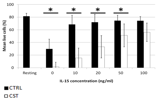
Figure 2 IL-15 inhibits CST-induced apoptosis in CD8 memory T cells in a dose-dependent manner T cells were isolated from OT-I mice, stimulated with the SIINFEKL peptide for 18h and differentiated into CD8 memory T cells with 20ng/ml of IL-15. Resting CD8 memory T cells (106) were incubated with 10-6 M of corticosterone or DMSO (CTRL) and increasing doses of IL-15 ranging from 0 to 100ng/mL of IL-15. Error bars represent SEM. *p<0.005, n=5.
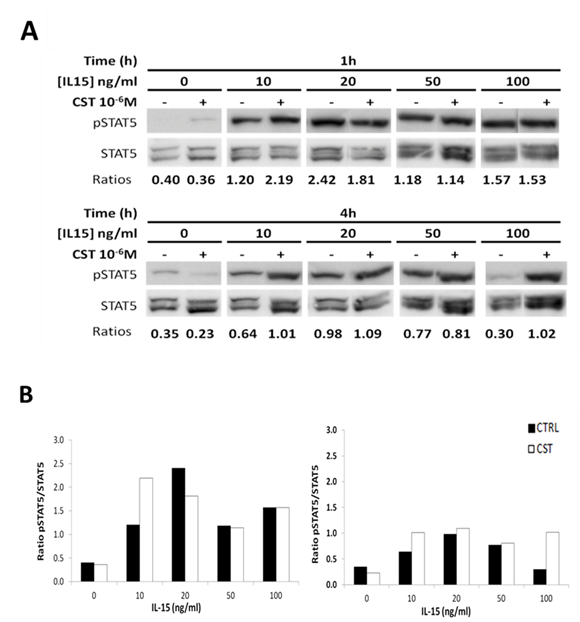
Figure 3 Activation of STAT5 pathway in presence of corticosterone and increasing doses of IL-15
T cells were isolated from OT-I mice, stimulated with the SIINFEKL peptide for 18h and differentiated into CD8 memory T cells with 20ng/ml of IL-15.
Regulation of NF-kB expression is not involved in the response of memory cells to CST or IL-15
Glucocorticoids have been shown to trigger apoptosis in T lymphocytes by inhibiting NF-kB activity.14 The GCR can affect the function of this transcription factor either by direct interaction or through upregulation of IkBa expression. To investigate the role of this factor in CST-induced apoptosis of memory CD8 cells, the level of IkBa was determined in the presence of CST, IL-15 or both. Results of a preliminary experiment are presented in Figure 4 and show that in this model, IkBa levels expressed as an IkBa/actin ratio were not affected by CST or IL-15 after 1 h or 4 h of stimulation. More experiments are required to confirm this result and to investigate the role of NF-kB in this response.
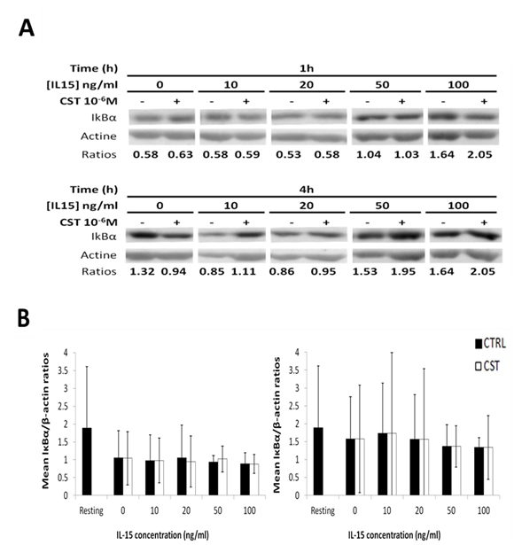
Figure 4 IκBα expression is not affected by the presence of corticosterone or increasing doses of IL-15
T cells were isolated from OT-I mice, stimulated with the SIINFEKL peptide for 18h and differentiated into CD8 memory T cells with 20ng/ml of IL-15.
Role of Bim in central memory CD8 cell survival in the presence of corticosterone
The GCR induces the expression of the pro-apoptotic Bcl-2 family member Bim in pre-B ALL cell lines.25 Bim inactivation also reduces double positive thymocytes mortality induced in vivo by dexamethasone injection.19 Therefore the role of Bim in this model of stress-induced apoptosis of memory CD8 cells was assessed through determination of Bim levels in these conditions. Following addition of increasing concentrations of IL-15, in the presence or absence of CST, BimEL levels were analyzed by western blot. Results presented in Figure 5 suggest that there is no significant change in BimEL levels in the presence of CST and that addition of IL-15 does not decrease the level of this pro-apoptotic factor.
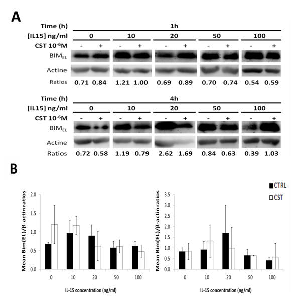
Figure 5 Expression of BimEL in central memory CD8 T cells in presence of corticosterone and increasing doses of IL-15
T cells were isolated from OT-I mice, stimulated with the SIINFEKL peptide for 18h and differentiated into CD8 memory T cells with 20ng/ml of IL-15.
Role of GITR in IL-15 protective effect
IL-15 sends a survival signal in central CD8 memory T cells26 and a recent study demonstrated that the glucocorticoid-induced TNFR-related protein (GITR) plays an important role in this protective effect. IL-15 can up-regulate the expression of GITR in memory CD8 T cells27 and the presence of GITR on memory CD8 T cells is required for their persistence in vivo.28 Confirming previous results, we found that IL-15 increased GITR expression in a dose-dependent fashion in our model Figure 6.
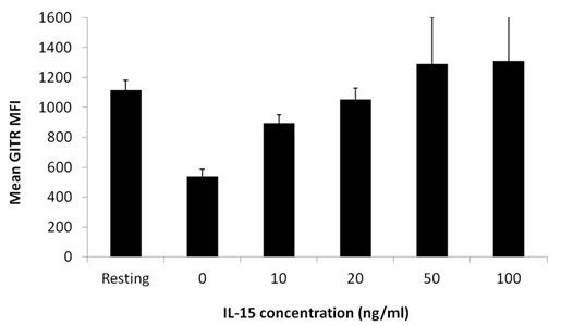
Figure 6 GITR surface expression on CD8 memory T cells is upregulated by IL-15 in a dose-dependent manner.
T cells were isolated from OT-I mice, stimulated with the SIINFEKL peptide for 18h and differentiated into CD8 memory T cells with 20ng/ml of IL-15. Resting central memory T cells were surface stained with anti-GITR antibody and analyzed by flow cytometry. Mean fluorescence intensity (MFI) of three independent experiments in presence or absence of increasing dose of IL-15 ranging from 0 to 100ng/mL is illustrated. Error bars represent SEM.
Resting memory T cells constantly monitor the entire system to ensure that no foreign threat to the host can be found. This is especially critical in the case of latent viruses such as EBV, CMV or VZV. Some significant events have been shown to trigger viral reactivation. Stressful events including UV exposition, mental examination or intense physical activities have been shown to result in an elevation of viral titers in the blood. In this study, we demonstrated that treatment of resting CD8+ memory T cells with physiological amount of corticosterone corresponding to chronic stress triggers the apoptotic process. This response seems to require transcriptional regulation since cell death appears is not detectable before 6 h of culture with CST. Addition of IL-15 concurrently with the stress hormone inhibited in a dose-dependent manner the pro-apoptotic effect of CST.
Association of IL-15 to its receptor triggers the recruitment and phosphorylation of molecules of the Janus kinase-signal transducer and activator of transcription 5 (Jak-STAT5) pathway.29,30 Jak kinase phosphorylates tyrosine residues of the gc and IL-15Rα subunits of the IL-15 receptor, leading to the recruitment and subsequent phosphorylation of the Stat5a/b complex followed by its translocation to the nucleus.31 In Stat5a/Stat5b double knockout mice, there is a significant decrease in hematopoietic stem cells repopulation capacity and a clear lymphopenia .32,33 Although thymic development is normal in these mice, Stat5a/b-/- T cells lack the ability to proliferate and migrate to sites of inflammation while NK cells are totally absent.34 Inversely, constitutive activation of Stat5 significantly enhances the survival of terminally differentiated effector T cells and the consequent formation of memory T cells.35 Another mechanism by which IL-15 ensures the survival of memory T cells is the regulation of the antiapoptotic proteins Bcl-2 and Bcl-XL.36,37 Indeed, the addition of IL-15 to resting CD44high CD8+ T cells induced the expression of Bcl-2 family proteins in vitro preventing spontaneous apoptosis. Glucocorticoid signaling inhibits the NF-κB transcription factor by preventing its translocation to the nucleus or increasing the expression of its cytoplasmic inhibitor, the IκBα protein. Our results suggest that CST does not induce apoptosis through this mechanism in CD8 memory T cells. Glucocorticoids have also been shown to trigger apoptosis of leukemia B cell lines by inducing the expression of the pro-apoptotic protein Bim, however CST did not affect Bim levels in CD8+ memory T cell cultures. We found that the presence of corticosterone and increasing doses of IL-15 had no impact on the protein levels of Bim. These results suggest that glucocorticoid-induced apoptosis of memory CD8 T cells does not involve this Bcl-2 family member.
The glucocorticoid-induced TNF-related (GITR) protein is required for the survival of CD8 memory T cells in vivo.28 GITR signaling results in NF-κB activation and a consequent upregulation of the Bcl-xL expression which is responsible for the increased survival of antigen-specific CD8 T cells.38 The expression of this TNFR family member on memory T cells increases in the presence of IL-15.27 In our in vitro model, GITR expression by CD8 memory T cells was also enhanced in the presence of IL-15 in a dose-dependent manner. These results suggest that GITR may be involved in the protection from glucocorticoid-induced apoptosis by IL-15. Ligation of IL-15 with its receptor activates Stat5, which triggers the expression of BCL10, an upstream regulator of NF-kB activity.39 Since IL-15 has been shown to induce NF-kB activation in neutrophils and cerebral endothelial cells, this suggests that IL-15 could also use another anti-apoptotic pathway to promote survival in resting memory T cells.40,41
Our results demonstrate that IL-15 protects against glucocorticoid-induced cell death. Our analysis of the mechanisms by which the survival advantage is induced by IL-15does not implicates IκBα or the proapoptotic protein Bim. However, significantly higher level of GITR in the presence of increasing doses of IL-15 shows that GITR could be involved in the anti-apoptotic effect of IL-15. This TNFR family member is essential for the 4-1BB receptor expression on memory CD8 T cells as well as their survival in vivo.28 GITR is also known to interact with the TNF associated factor (TRAF1, TRAF2 and TRAF3), which downstream signaling leads to enhanced survival of the CD8 memory T cells.38 Hence, our results suggest that IL-15 up regulates GITR expression at the T cell surface which, as in TCR-mediated activation, could inhibit corticosterone-induced apoptosis. Results presented in this study are relevant to chronic viral infection therapy and vaccine development. The anti-apoptotic capacity of IL-15 suggests potential new therapeutic interventions against latent infections that are influenced by stress hormones, such as Herpes infection. Since GITR also enhances the stimulation of effector and regulatory T cells, there is a need to better understand the interaction between the signaling of this TNFR family member and IL-15.
We thank Rocksan Dubé for technical assistance with the mouse colony. This work was supported by a Maisonneuve-Rosemont Hospital Foundation grant (to L.S.) and the Canadian Space Agency (to L.Y.C). G.D.L. was the recipient of a departmental scholarship from Université de Montréal.
The authors declare that there have no conflicts of interest.
None.

©2015 Geneviève, et al. This is an open access article distributed under the terms of the, which permits unrestricted use, distribution, and build upon your work non-commercially.