MOJ
eISSN: 2373-4442


Research Article Volume 5 Issue 1
College of Science, Al-Muthana University, Iraq
Correspondence: Weam Saad Al-Hamadany, College of Science, Al-Muthanna University, Samawah Governorate, Iraq, Tel 964791000000
Received: January 01, 1971 | Published: January 12, 2017
Citation: Al-Hamadany WS (2017) Estimation of Some Immunoglobulins Classes and Innate Immunity Parameters in BOM Cases. MOJ Immunol 5(1): 00143. DOI: 10.15406/moji.2017.05.00143
Immune response against OM infections depends mainly on Humoral mediated Immunity (HMI). From this scientific fact, the present study took care with the estimation of some immunological parameters related to these infections immunological response. Total W.B.C.s count was estimated. Furthermore Radial Immune Diffusion (RID) method was used in determination of Immunoglubulins IgG, IgM and IgA; also complement components C3 and C4 levels were estimated using Endoplate kits too. Results: OM Infections resultant from gram positive cocci increased total W.B.Cs counts, IgG, IgM, IgA, C3 and C4 complement components levels; in non–immunologicaly compromised patients comparing with the values of the peoples of control groups for all the parameters that estimated. Conclusions: Otitis media is a common infection in both children and adults. Gram positive suppurative OM caused; significant elevations in Total W.B.C.s counts, Immunoglubulins IgG, IgM and IgA and the complement components C3 and C4. Since HMI is the immunological response against these infections.
Keywords: om infections, rid, hmi, igg, humoral mediated immunity, igm, iga, total wbcs count, gram positive cocci
CG(s): Controls Group(s); CMO: Chronic Otitis Media; ENT Ear: Nose and Throat; HMI: Humoral Mediated Immunity; OM: Otitis Media; PG(s): Patients Group(s); PMNs: Polymorphnucleous cells; RID: Radial Immune Diffusion; RTI(s): Respiratory Tract Infection(s); W.B.C.s: White Blood Cells
Otitis media (OM) infections represent a serious problem for many peoples, especially children.1 Recurrent suppurative OM with discharge, pain, and hearing impairment are the most common symptoms of this disease. Complicated cases may develop deafness.2 Gram positive cocci bacteria are the predominant pathogens responsible for such infections; pointing to Staphylococcus aureus and Streptococcus pneumoniae.3–5 The complications upon OM infections made this disease very important, since when left untreated; the infection may spread to the surrounding tissues causing Mastoiditis, Brain abscess and Meningitis mainly in children and patients with impaired immune system. Prolonged chronic otitis media (COM) in children lead to problems in speech and social skills.6
Immune response against OM infections depends mainly on humoral mediated immune response (HMI). It starts with local inflammation at the site of pathogen invasion. Polymorphnucleous cells (PMNs) arrive combined with cytokines production and release; e.g. IL–1. These events end with specific antibodies production. Especially IgA; since sIgA plays a major role in the protection of body mucosal surfaces against bacterial colonization and infections.7
Pyogenic cocci bacteria like Staphylococcus aureus and encapsulated Streptococcus pneumoniae are able to resist body defenses by escaping phagocytosis, releasing enzymes and toxins. That will lead to W.B.Cs death and pus forming due to colonization of pathogen. Any defect in body immune system can cause recurrent suppurative OM infections that are usually called COM infections.4,8
Patients: Otitis media cases were collected from the outpatients of (Ear, Nose and Throat) ENT Dep. Al–Numan Hospital in Baghdad (2000–2006). The patients were clinically diagnosed by ENT physicians. A total of (60) OM cases were collected. They were infected with gram positive bacteria as causative pathogens as single or mixed infections. Bacteriological identification for them was done by the microbiological laboratories in the same hospital mentioned.2,5
Controls: Healthy volunteer people represented controls; they were (30) individuals taken randomly with no clinical signs for any disease. Whereas, ages were taking in account during controls collection as shown in Table 1. Smokers were avoided and no woman during menstruation period was involved as recommended by.9
Clinical specimens collection: A total of (5) ml blood sample was taken from all patients and controls by venipuncture.
Clinical specimens' treatment: Each blood sample was divided into two parts. One ml was put in EDTA tubes (whole blood) and kept in the refrigerator (4 °C). The second part (4 ml) was used to separate serum after clotting; obtained serum was kept in freezer (–20 °C) until used.9
Total W.B.C.s counts: whole blood was used in total W.B.C.s count estimation using Hemocytometer chamber as in.9
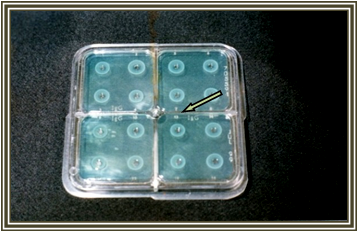
Immunoglubulins and complement components estimation: Antibodies IgG, IgM and IgA and complements C3 and C4. These parameters were estimated using Endoplate Kits. The method of Radial Immunodiffusion (RID) was the Immunological Assay depended by the kit manufacturing company (Sanofi/ Italy). After serum samples application on the specific wells in plates, the antibodies and complement components diffused in the agarose gel. The gel contains a monospecific antiserum against the Ab class or complement component under test. Then precipitation zoon will form and able to be read after incubation (the leaflet of kit and 7).
Statistical analysis of data: All results that obtained for cases and controls were statistically analyzed using mean calculations (M) and standard Error (SE) using Statistical Analysis System SAS (2000). Each group results were compared with the other group’s results using student t–test to find the significance of probability level (P) of increase or decrease for all the studied parameters, the level (p≤ 0.05) was the level of significance.10
The (60) OM patients included in this study were infected with gram positive cocci; Bacteriological culturing and identification was accomplished by microbiology laboratories in the same hospital. There were (49) patients (81.6%) among them infected with Staphylococcus aureus. And 11 patients (18.3%) were infected with Streptococcus pneumonia.
The total (60) Patients were divided into 6 groups according to age; also the (30) controls had the similar distribution; (the patients group (PG1) involved the youngest patient with three months age only). Each group results were compared with the similar age group of controls as shown in Table 1.
|
No. |
Age Range |
Patients Group |
No. of Cases |
Control Groups |
No. of Controls |
|
1 |
≤ 10 |
PG1 |
3 |
CG1 |
5 |
|
2 |
11-20 |
PG2 |
6 |
CG2 |
5 |
|
3 |
21-30 |
PG3 |
16 |
CG3 |
5 |
|
4 |
31-40 |
PG4 |
18 |
CG4 |
5 |
|
5 |
41-50 |
PG5 |
13 |
CG5 |
5 |
|
6 |
51-60 |
PG6 |
4 |
CG6 |
5 |
Table 1 Otitis Media Patients and Control Groups with Age Ranges distributed according to Ages and Cases number.
Generally; all patients' ages ranged (4–60) years while controls ranged (10–58) years. There were 26 (43.3%) males and 34 (56.7%) females recorded among patients, while controls represented 13 females and 17 males. Patients recorded 86.6% with chronic Otitis Media (COM), while Acute Otitis Media (AOM) recorded (13.4%) from total (60) cases.
Total W.B.C.s results are shown in Table 2 whereas PG5 recorded a significant increase (P ≤ 0.5) comparing with CG5, other groups results increased but in significantly. The depended range of normal values was as in.11
|
No. |
Age Range |
Total W.B.C.s count ×103 |
Complement C3 |
Complement C4 |
|||
|
PG |
CG |
PG |
CG |
PG |
CG |
||
|
1 |
≤ 10 |
6.3±0.1 |
10±0.01 |
213.0±19.3* |
117±0.02 |
36.9±6.6 |
26.8±1.8 |
|
2 |
20-Nov |
7.2±0.6 |
6.9±0.5 |
188.0±23.0* |
137±14.1 |
35.2±7.9 |
31.8±1.8 |
|
3 |
21- 30 |
8.7±1.5 |
8.6±1.1 |
178.0±36.2* |
142±7.7 |
45.9±11.1* |
33.3±5.6 |
|
4 |
31- 40 |
8.4±2.5 |
8.9±0.5 |
194.9±44.4* |
169±20.1 |
45.1±12.8* |
38.6±1.9 |
|
5 |
41- 50 |
9.0±2.1* |
8.3±0.1 |
175.8±19.8 |
166±4.2 |
39.7±9.7 |
31.9±3.7 |
|
6 |
51- 60 |
8.8±0.7 |
9.5±0.3 |
189.0±28.8 |
161±11.3 |
37.2±12.3 |
32.0±7.4 |
|
Normal Range |
(4.3-11) ×103 cell/ml |
(101-186)mg/dl |
(16-47)mg/dl |
||||
Table 2 Total WBCs, Complement Components C3 and C4 values obtained for all groups, (M ± SE).
The Endoplates (RID) results are shown in Table 3, Figure 1, IgG values increased significantly (P ≤ 0.5) in all groups except PG4 which increased insignificantly comparing with CG5 mean value. The values of IgM increased significantly in all PG5 except PG2 which increased insignificantly, comparing with CG5, IgA values increased in PG4 and PG6 significantly (P ≤ 0.5) and insignificantly in PG2, 3 and 5. While the mean of PG1 values decreased insignificantly.
|
No. |
Age Range |
IgG |
IgM |
IgA |
|||
|
PG |
CG |
PG |
CG |
PG |
CG |
||
|
1 |
≤ 10 |
1450.0±137.8* |
1004.0±102.0 |
190.3±43.8* |
94.0±28.0 |
183.3±56.9 |
187.2±2.34 |
|
2 |
11-20 |
1687.2±210.0* |
1399.5±220.0 |
205.2±139.0 |
108.5±10.6 |
250.0±27.3 |
276.5±17.8 |
|
3 |
21- 30 |
1832.0±33.7* |
1346.0±144.2 |
224.3±61.7* |
113.0±26.9 |
305.5±95.4 |
165.5±23.0 |
|
4 |
31- 40 |
1942.9±662.5 |
1666.0±156.9 |
209.0±129.8* |
120.5±28.0 |
360.3±115.6* |
201.5±13.5 |
|
5 |
41- 50 |
1576.9±287.0* |
1226.0±313.9 |
204.0±66.5* |
116.5±10.6 |
256.7±82.0 |
244.5±21.9 |
|
6 |
51- 60 |
1968.8±458.6* |
1450.0±148.5 |
244.3±131.9* |
136.0±5.7 |
400.0±104.3* |
214.0±35.4 |
|
Normal Range |
(844-1912) mg/dl |
(50-196) mg/dl |
(68-423) mg/dl |
||||
Table 3 AThe Immunoglubulins IgG, IgM, and IgA values obtained for all groups, (M ± SE).

Figure 1 The Endoplate Kit (Radial Immune Diffusion, RID), showing precipitation zones obtained. Arrow shows a control well.
Complement components C3and C4 results are shown in Table 2. The complement C3 levels increased in all PGs outcomes. But, in PGs 1, 2, 3 and 4 the elevation was significant (P ≤ 0.5) comparing with CGs. The complement C4 values increased significantly (P ≤ 0.5) in PGs 3, 4 and 5 while other groups increased insignificantly comparing with CGs.
Chronic Otitis Media (COM) cases are more common than (AOM), and that can be attributed to the fact that OM infections are the most common consequences after Respiratory Tract Infections (RTI). This opinion is in agreement with.1 They found in a study done in Pittsburgh hospital that the chronic infections with effusion are more common than the AOM especially in winter outbreaks, which was also supported by.12
Concerning total W.B.C.s results, OM infection caused elevation in total W.B.C.s count; these outcomes can due to OM infection after RTIs. Whereas RTI lead to elevation of total Leukocytes count as stated by.13 That was also in documented by.11
The outcomes of Immunoglobulin's estimation showed that OM caused increase IgG and IgM values and that can be explained by the induction of HMI, OM bacterial infections lead to rise Immunoglobulin levels due to specific Abs production especially when the infections were caused by the pathogens Staphylococcus aureus and Streptococcus pneumoniae, that was in consistent with the opinions of both.14,15
Also.16 pointed to the fact that bacterial OM caused by encapsulated Streptococcus pneumoniae usually causes elevation in IgM value because this type of antibodies is effective during immune response against bacterial capsules and there were 11 (18.3%) patients in the present research whom had the same causative pathogen. The type IgA immunoglobulins represents the first line of defense against bacterial colonization in mucosal surfaces in the Upper Respiratory Tract. That because the subclass secretary sIgA antibodies are able to be secreted with mucus and share with other factors in local immune response against pathogens colonization in the middle ear.7,17
The results of IgA levels were as expected all PGs levels increased except PG1. This group IgA levels decreased insignificantly that can be attributed to that this group represents children under the age 10 years. The members of this group are more prone to bronchiolitis and other RTI during winter outbreaks.18 Moreover Eustachian tube mucosa in children is less differentiated and differs from adults as mentioned by.19,20
Complement components C3 and C4 increased in OM patient's blood because of the important role of these complement components in the inflammatory process during infections, these results are also documented by.21 they fully described complement role in the middle ear infections and they attributed that increase to the increase of the complement and inflammatory mediators production during inflammation of middle ear after activation of phagocytic cells. The authors.7,22 stated that many complement proteins and Cytokines levels usually rise during inflammatory diseases as a part of innate immunity and as a start of specific Humoral response and that is supporting to the findings of our study.
Otitis media is a common infection in both children and adults epically after winter season. Gram positive suppurative OM caused; in imunologically uncompromised patients; significant elevations in Total W.B.C.s counts, Immunoglubulins IgG, IgM and IgA and the complement components C3 and C4 due to induction of HMI response against these infections.
Special thanks to Dr. Rajwa H. Al–Rubaei (Dep. Bio. Coll. Sci. Al–Mustansiriya Univ.) And to Dr. Hamid M. Ghani (let the mercy of God be upon him). I am grateful to Al–Nuaman Hospital ENT Dep, and the inner and outer Laboratories staff. I am much obliged to patients and healthy volunteers.
None.

©2017 Al-Hamadany. This is an open access article distributed under the terms of the, which permits unrestricted use, distribution, and build upon your work non-commercially.