MOJ
eISSN: 2576-4519


Research Article Volume 2 Issue 4
1Department of Biomedical Engineering, The Catholic University, USA
2Visiting Scientist, CUA
Correspondence: Harold Szu, Department of Biomedical Engineering, The Catholic University, Wash DC, USA
Received: June 19, 2018 | Published: August 14, 2018
Citation: Szu HH, Chu TJ. Brain’s Excessive Debris Might Be Discernible with Adaptive Wavelet Transform made of Faraday Coils of Electroencephalograph. MOJ App Bio Biomech. 2018;2(4):252–257. DOI: 10.15406/mojabb.2018.02.00076
The key point relevant to our proposed studies concentrates on the fluctuation of Electroencephalograph (EEG). First of all, EEG is not a wave of any kind, not electromagnetic, nor acoustic, but simply the magnetic field disturbance generated by Right Hand Rule of Ampere law following those Calcium ion currents along individual neuronal axons where the fingers curl around magnetic field lines. Then, these mixtures of magnetic field lines are never intersecting one another and find themselves outside the scalp and picked up by two orthogonal miniature Faraday coils called the Faraday induction electric generators. We wish to know the collective frequency of these transient currents we propose an efficient Adaptive Wavelet Transform (AWT) which is a single kernel computed off-line by the Least Mean Square Sense using Artificial Neural Networks (ANN). The fluctuations are likely attributed by the inhomogeneous neuro-glia cells conducting the housekeeping of toxic debris or deposits cleaning. Aging brain might be characterized by several brain disorders which are lacking of household noninvasive monitoring system, which we propose two orthogonal Faraday coils, and on-line AWT through Smartphone communication to the nurses and physicians. For example, the cognitive decline, dementia and potential Alzheimer’s disease, epileptic seizures, bi-polar might be due to the long time accumulation of toxic waste; e.g., beta-Amyloid [2]. For our proposed feasibility studies, we have developed a model of adaptive wavelet transform at the compressive sensing of high-density (about 8 to16) needle electrodes that can connect to a Smartphone for a real time display of the EEG electrodes. Furthermore, we will offer a super-resolution by the wavelets extrapolation and interpolation cross the brain. (2018 Continued Resolution support for “Gulf Coast Deep Learning at Lafayette LA” during Oct 26-27, 2018 (cf.ttp://www.ica-wavelet.org)
We are concerned about more than 77 million World War II baby boomers in the U.S. who have worked hard to make this country great, but many of them may become home alone seniors (HAS) that no one takes care of them. More than 28 million baby boomers will develop Alzheimer’s disease between now and midcentury, and the cost of caring for them will consume nearly 25 percent of Medicare spending in 2040.1 If residual brain debris affected by sleep deprivation, as anticipated in many baby boomers, need to be routinely and completely cleaned up from the viewpoint of biomedical wellness nature; is there a household noninvasive measurement tool available as early warning sensors? This is what we try to answer by this preliminary study. The concept of brain waste removal was galvanized by the discovery of a waste clearance pathway in the brain—now known as the Glymphatic system. The Glymphatic pathway dysfunction has been implicated in a variety of neurologic diseases. The implication of slow-wave sleep for enhancing brain waste drainage has stimulated further research to better understand the drainage pathways in human brain and to explore different mechanisms for controlling the complex cleansing system in human brain under both normal health and disease states. This paper elucidates how the Electroencephalograph (EEG) wireless head mount connected with a Smartphone can be used for distant nurse monitoring. The electric contact with a Binary, in an approximation sense, is either one or zero when the EEG frequency falls in the range of rectangular filter. This is idealistic without taking into account of skull non-uniform permeability. Consequently, we shall consider an adaptive wavelet filter as tailored to cover the non-uniformity of skull, brain gray matter, brain white matter, and beta-Amyloid debris. can be augmented as rect , otherwise zero frequency, where equivalent Fourierkernelsinc the band center frequency fX=Delta(5Hz), Theta(13Hz),Alpha(23Hz), Beta(27Hz), Gama(60Hz-100Hz). Proposed applications include computational multiple layers of Artificial Neural Network namely Deep Learning or also known in short as Artificial Intelligence. This is to develop, in the Least Mean Squares (LMS) sense, the corresponding Adaptive Wavelet Transform kernel having the most efficient compressive sensing magnetic fields with different firing rates of 50 Calcium ions per sec to 100 ions per sec and with different solenoid magnetic fields. Consequent development includes different orthogonal tiny current loops with iron cores outside the scalp. These devices are based on Faraday induction loops that convert the modulation of magnetic field to electromotive force driving currents in wireless EEG head mount for high-risk home-alone seniors (HAS).
Regarding the clearance of brain debris, Maiken Nedergaard and Steven A. Goldman of the University of Rochester Medical Center reported in a review article of Scientific America2 regarding the recent discovery of an internal plumbing system capable of removing soluble proteins and metabolites in the mouse brain during slow-wave sleep. We quote literally three paragraphs and three figures from this article as follows:
“The human brain weighs only about three pounds, or roughly 2 percent of the average adult body mass. Yet its cells consume 20 to 25 percent of the body’s total energy. In the process, inordinate amounts of potentially toxic protein wastes and biological debris are generated. Each day, the adult brain eliminates a quarter of an ounce of worn-out proteins that must be replaced with newly made ones, a figure that translates into the replacement of half a pound of detritus a month and three pounds, the brain’s own weight, over the course of a year.
To survive, the brain must have some way of flushing out the debris. It is inconceivable that an organ so finely tuned to producing thoughts and actions would lack an efficient waste disposal system. But until quite recently, the brain’s plumbing system remained mysterious in several ways. Questions persisted as to what extent brain cells processed their own wastes or whether they might be transported out of the nervous system for disposal. And why is it that evolution did not seem to have made brains adept at delivering wastes to other organs in the body that are more specialized for removing debris? The liver, after all, is a powerhouse for disposing of or recycling waste products. About five years ago they began trying to clarify how the brain eliminates proteins and other wastes. We also began to explore how interference with that process might cause the cognitive problems encountered in neurodegenerative disease. We thought that disturbances in waste clearance could contribute to such disorders because the disruption would be expected to lead to the accumulation of protein debris in and around cells. This idea intrigued us because it was already known that such protein clumps, or aggregates, do indeed form in brain cells, most often in association with neurodegenerative disorders. What is more, it was known that the aggregates could impede the transmission of electrical and chemical signals in the brain and cause irreparable harm. In fact, the pathology of Alzheimer’s, Parkinson’s and other neurodegenerative diseases of aging can be reproduced in animal models by the forced overproduction of these protein aggregates. In our research, we found an undiscovered system for clearing proteins and other wastes from the brain—and learned that this system is most active during sleep. The need to remove potentially toxic wastes from the brain may, in fact, help explain the mystery of why we sleep and hence retreat from wakefulness for a third of our lives. We fully expect that an understanding of what happens when this system malfunctions will lead us to both new diagnostic techniques and treatments for a host of neurological illnesses. The Glymphatic system in most regions of the body, a network of intricate fluid-carrying vessels, known as the lymphatic system, eliminates protein waste from tissues. Waste-carrying fluid moves throughout this network between cells. The fluid collects into small ducts that then lead to larger ones and eventually into blood vessels. This duct structure also provides a path for immune defense, because lymph nodes, a repository of infection-fighting white blood cells, populate ducts at key points throughout the network. Yet for a century neuroscientists had believed that the lymphatic system did not exist in the brain or spinal cord. The prevailing view held that the brain eliminated wastes on its own. Our research suggests that this is not the complete story. The brain’s blood vessels are surrounded by what are called peri-vascular spaces. They are doughnut-shaped tunnels that surround every vessel. The inner wall of each space is made of the surface of vascular cells, mostly endothelial cells and smooth muscle cells.
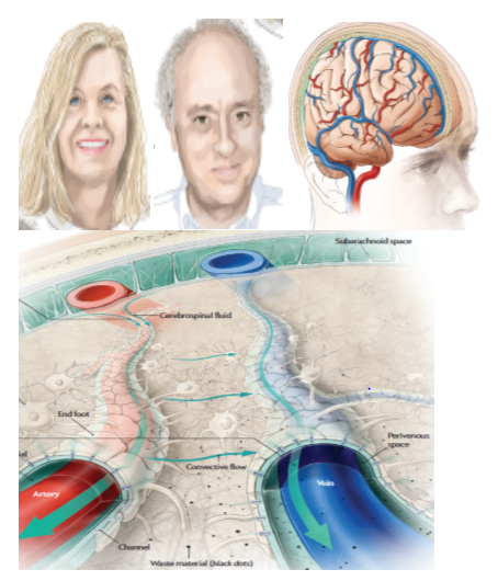
An intricate system of vessels—the Glymphatic system—snakes throughout the brain, carrying fluid that rids the organ of discarded proteins and other wastes that can clump together and turn toxic if left in place. The protein fragments known as beta-Amyloid peptides, which are present in Alzheimer’s disease, are examples of the cellular detritus cleared through the drainage system, mostly during sleep (Figure 1).2

Figure 1(a) (b) (c) Portraits of Maiken Nedergaard and Steven A. Goldman (taken from Ref 2) Glea-lymphc system surround the blood capillary without use another lymph. Both artery and vein in the brain are surrounded by glia cells. Outside the closed blood capillary are those leaky tubes surrounded by all the end feet of Astrocytes glia cells, surrounding both the vein and the Artery.2 That’s why we need 100 billion of Astrocytes glia cells to connect among 10 billion neurons. The authors spent 5 years time over hundreds mice have identified detail chemical molecular mechanism for the cleansing mechanism during the sleep.2
In the recent publication “how to avoid Dementia Alzheimer Disease.3 we itemized 3 physical dimensions: “Exercise Daily; Eat Right; Sleep Tight;” and 3 mental dimension: “Social Often; Stimuli Daily; Relax Spirit (without Alcohol Cigar)... to grow new neuron interconnections.
Moreover, we must dispel the erroneous notion that the EEG is a traveling transversal electromagnetic (EM) wave. If EEG were the EM wave, according to the Einstein hypothesis, the EM wave must travel at the constant speed of light in the vacuum. But in the dense medium, the light must be reduced by a thousand folds, we will have 2πν=30Hz corresponding to hundred kilo meter.
Q.E.D.
Additionally, it cannot be the longitudinal acoustic wave either simply because the speed of sound is increased tenfold.
Prof:
Q.E.D.
Since EEG has never been implied as a wave of any kind, it is conceivable as a weak magnetic modulation because various circular solenoid magnetic fields are random mixed around at 100Hz ion per second, or 50Hz ion per sec, as the case may be. Here comes the D.O. Hebb learning rule: “Fire Together, Wire Together” to organize local groups by connecting neurons of similar firing rates of Calcium ions. Thus, the mixture of solenoids magnetic fields is also gathered and modulated in random groups.
"Further, let's give a paragraphic review of Clark Maxwell EM theory as follows:
Two Sources: (1) Gauss Law: the electric charge produces the electric field; and
2Biot-Savart Law: the magnet produces the magnetic dipole field".
Two crossover Couplings (when the source moves, it produces the other field):
1Ampere Law (Motors: current moves generating the magnetic fields): the electric charge current generates the Solenoid magnetic field around it (Right Hand Rule: right thumb follows the ion current, all fingers follow the magnetic field). Brain EEG consists of all direction solenoid fields by Ampere’s law of neuronal axon currents; and
2Faraday Law (Generator: magnetic field move generating the current): the magnetic flux induces the electric motive force that dives the current in a conductor. The pickup of leakage brain magnetic fields are by means of Faraday law of axial currents in mini-coils
Maxwell Law indicates the inconsistency in the capacitor that there is only charged flows but nothing inside the capacitor, thus James Maxwell postulated the displacement current. Now all those sources and couplings are consistent in the Albert Einstein’s space-time relativistic format (Figure 2). According to Faraday’s induction law, modulation of magnetic fields will generate an induction currents at the scalp to be picked up by the EEG electrodes. Faraday observed and predicted how a magnetic field will interact with an electric circuit to produce an Electromotive Force (EMF) In order to elucidate the fluctuation EEG, we need to identify mathematically the existence of glia cells from neurophysiology and to determine how to mathematically prove and furthermore define the missing half of Einstein’s brain besides neurons (Figure 3). Elucidating EEG brainwaves at the fast onset of Epileptic Seizure may shed some light on biologically realistic neural network (BNN) due to neuron firing spiking population and local field potential for the 2nd order phase transition of Helmholtz Free Energy wearing f-EEG hat Szu et al.,35 SPIE News 2015). This is for always-on mental health, either a fast epileptic seizure or a slow Alzheimer dementia without Astrocytes glial cells’ hard work during a good night sleep tight.

Figure 2 (a) James Maxwell’s Portrait, (b) Brain EEG picked up by tiny orthogonal coils, (c) Faraday Experiments.

Figure 3 (a), and (b) “The Missing Half of the Brain” by R. Douglas Fields. cf. Scientific Am: April 2004 (Thomas Harvey who performed the autopsy of Albert Einstein in 1955).
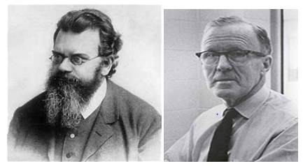
Donald O. Hebb, Professor of University of Toronto, famously said that “neurons that fire together, wire together” that is also known as Hebb’s rule as shown in the Figure 2. By a subsequent resistive measurement of Albert Einstein’s brain, we surprisingly know that he may have more glial cells relative to neurons. Then, the question remains why Albert Einstein seemed to be visually smarter and creative in imagination as a Homo sapiens. If we let the entropy for the degree of uniformity to represent the firing rate of neurons, we can link Boltzmann equilibrium 2nd law and in-equilibrium thermodynamics 3rd law to brain neuron firing rates and glia cells as follows (Figure 4) (Figure 5):
Given Homeostasis theory at isothermal equilibrium
(1)
We obtain from Boltzmann definition of Entropy as written on his headstone;
(2)
Where use is made of the 2nd Law of thermodynamics: the conservation of energy between the change of unknown brain energy and unknown reservoir entropy:
(3)
That allows us to replace the unknown reservoir entropy change with unknown brain energy
(4)
Then, Maxwell-Boltzmann canonic ensemble defines the Helmholtz free energy
(5)
Where we have derived the brain Helmholtz free energy
(6)
Based on the heat death of Boltzmann irreversible thermodynamics;
(7)
due to the incessant intermolecular collision toward the uniformity heat death.
We conclude
(8)
As a result, the dendrite input to a neuron is defined by the weighted synaptic matrix of all input neuron firing rates
(9)
Where use is made of by Einstein convention: Greek I,j,k, for individual and the repeated Latinα,β,γ are dummysummation over all the Greek indices. The chain rule allows us to define the glia cells
(10)
according to the Canadian neurologist Donald O Hebb observed 5 decades ago: that “linked together, fired together”, known as bi-linear Hebb rule from Newtonian dynamics of a gradient descent:
(11)
We have identified from Hebb rule the glia cell
(12)
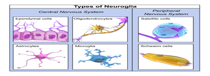
The different dendrite shape geometry defines one of 6 different glia cells (Figure 6) (Figure 7).

Figure 4 From (a) Ludwig Boltzmann to (b) Donald Hebb. (cf. Francisco Carlo Morabito: paper “ Entropy to EEG).

Figure 5 Glia cells surround the output axon to guide Calcium ions current flow within the tube (like 2 charge repulsive ducks(a)) force though the axon tube.(b); in a neuron (c).

Figure 7 (a) Typical Neuron in details and (b) different types (4+2) in the nervous systems ue to different form factors in the dendrite trees .
Given the local and thus irregular production of brain debris (e.g., beta-Amyloid) along with the cleansing dynamic by cerebrospinal fluid (CSF) outside the blood capillary in a leaky second tube, these two interactions following the sleep-wake cycle may generate a high-frequency fluctuation of EEG brain waves riding over the systematical low-frequency waves of Delta (5~10Hz), Theta (10~15Hz), Alpha (15~25), Beta (25~30Hz), as well as Gamma (30~100Hz). It is possible that the high-frequency modulation over D,T,A,B,G is real, the high-frequency albeit weak modulation would be a warning sign of potentially inefficient CSF cleansing of brain wastes. Our study goal is to help delay the onset of neurodegenerative diseases such as amyotrophic lateral sclerosis, Alzheimer's disease, Parkinson's disease and Huntington's disease through enhanced Glymphatic clearance system.
We propose a minimum invasive tool,4-9 namely a wireless EEG Head Mount to help monitor the effectiveness of Glymphatic clearance of brain wastes (Figure 8). The neuron density modulation of firing rates by means of Hebb’s rule: “fire together, wire together (FT,WT)” indicates that the dots density appears to be modulated from on 100Hz to off less than 50Hz. These shall not be confused as the usual travelling Electromagnetic wave, rather representing the neuronal firing population (of firing rates) density modulation. It behaves as, even if it were acoustic, it still has the transversal pointing vector waves but not acoustic longitudinal vector. The EEG consists of smooth DTABG group waves or wavelets10 allowing us to follow the over-complete ortho-normal bases and to expand the general EEG output.

Figure 8 (a) Wireless EEG head mount5; (b) measuring the Cortex 17 area in the back of the head, where the pin must be sharp enough to penetrate the hairs; (c) are typical correlation track of time correlation domain of firing rates where concentrate compressive sensing should be applied to reduce the band width of Smartphone.
Brain disorders include: (1) Multiple Sclerosis that is autoimmune disease when our own antibody is mistaking Myelin Sheath (made by glia cells) as other virus protein fat; (2) Epileptic Seizure that might be due to a suddenly feedback when axon wire crosses dendrite; (3) “.......(3) Dementia and Alzheimer’s Disease that are caused by toxic brain energy by-products not being removed by CSF from the synaptic junctions affecting electric, mechanical, and/or chemical signals; and (4) other bipolar Schizophrenia diseases that are caused more by genetic related factors than by beta-Amyloids. Although there are several medical reports about using continuously bricking LED light through pupils to reduce beta-Amyloid in mouse in the visual cortex 17 area, it is conceivably to be nuisance or impractical by driving the home alone seniors crazy.
In this paper, we elucidate how the EEG wireless head mount connected with a Smartphone can be used for distant nurse monitoring. We augment the electrode contact of wireless head mount (cf Fig. with a binary filter rect if otherwise zero frequency, where the band center frequency and the corresponding Fourier Transform is the sampling in the time domain: in their wireless EEG head mount of those high risk home alone seniors (HAS).
Since Adaptive Wavelet Transform is more efficient than Fourier Transform for feasibility studies, we shall make further remarks about several generation of wavelets as a group wave package base for the expansion kernel rather than less concise single sinusoidal wave base. The mathematical theorem for the completeness lossless proof has been given by 1st Gen Ingrid Daubechie wavelets. Thus, for compressive sensing, the useful tools for analyzing EEG fluctuation data are shown in Figure 9c that have been available over decades as follows:
(13a,b)
Note that the upper limit in Eq(13a) is the infinite term versus a finite term N in Eq(13b). The infinite term is the efficiency of compressive sensing that we are seeking for real time EEG offline reading on Smatphone (Figure 9). For those readers who are less familiar with the modern analysis tool, including a BNN adaptive kernel construction and adaptive wavelet expansion, we would make the following concluding remark for the edifice of biomedical wellness researchers. The community has extended the infinite terms of Fourier transform to the 1st Generation of wavelet by Ingrid Daubechies (so-called the “Mother Wavelets” by the general public due to her “Ten Lectures in Wavelets,” Book by SIAM), and then to the 2nd Generation of “lifting scheme” (Wikipedia) Discrete Wavelet Transform (DWT) of Wim Swelden, (www.wavelet.org). Now it has come to the Next 3rd Generations (NG) of Adaptive (Digital) Wavelet Transform (AWT), which preserves the statistical such as the 2nd moment variance.10 For those readers who are interested in the state of art review of the 1st Generation Daubechies mother wavelets, 2nd Generation Swelden lifting wavelets, as well as the 3rd Generation AWT statistics preserving wavelets by Szu et al., they shall consult reference,10–14 that have comprehensive review literatures. The compressive sensing is indicated by the a finite upper limit N, which could be few terms in the Least Means Square (LMS) error approximation sense to the original continuous function W(t) by taking away the DC component (to avoid the division by zero) for the matched filter of original wave package.12 The Shannon sampling theorem is given two discrete measurements within a period that can represent the continuous function. We can replace the Nyquist-Shannon-Whitaker kernel “sync function” with other band limited adaptive mother wavelet in order to be adaptively optimized to the local toxic deposition (to be experimentally determined).
The lossless NG_DWT accomplishes the data compression of "wellness baseline profiles (WBP)" of aging population at homes. For medical monitoring system at home fronts, we translate the military experience to dual usage of veterans and civilians alike with the following three requirements: (1) Data Compression: the necessary down sampling reduces the immense amount of data of individual WBP from hours to days and to weeks for primary caretakers in terms of moments, e.g., mean value, variance, etc., without the artifacts caused by FFT arbitrary windowing; (2) Lossless: our new NG_DWT must preserve the original data sets; and (3) Phase Transition: NG_DWT must capture the critical phase transition of the wellness toward the sickness with simultaneous display of local statistical moments. According to the Nyquist-Shannon-Whitaker sampling theory Eq(13b), assuming a band-limited wellness physiology, we must sample the WBP at least twice per day since it is changing diurnally and seasonally. Since NG_DWT, like the 1st Generation, 2nd Generation as well as the 3rd Generation are lossless in the Least Mean Square (LMS) error sense, we can reconstruct the original time series for the physicians' second looks. This technique of NG_DWT can also be adaptive to help monitoring the EEG without artificial horizon artifacts.10–13

Figure 9 EEG Brainwaves for an Epileptic Seizure Patience: (a) EEG average D,T,A,B waves Low Frequency composition under 30Hz cf. Figure 8; (b) Microscopic functional brain image reveals the neuron axon wire accidently cross backward and touched one the input dendrite tree, that might be the cause generates a feedback stability, Japan Kyushu Institute of Technology Prof. Takeshi Yamakawa et al; have applied the standard laser burning in a drilled hole in the brain scalp to remove the cross-over and turn a month long patient to a week patient; (c) indicating the sudden onset of phase transition of Epileptic seizure, furthermore the high frequency fluctuation might be due to irregular brain toxic production and irregular cleansing by CSF.
What’s the next?
Our suggestions for further Control Study following the NIH gold standard DC, NC, SS are as follows: anticipating as Principal Investigators close to NIH, we would like to collaborate with University of Rochester, Catholic University in DC, and other selected research groups at the tone of $10M over 5 years. The NIH, e.g., National Inst. of Mental Health Dr. Lalonta et. al.,9 requires a minimum invasive household tool with Gold Standard protocol such as our proposed wireless EEG head mount that can provide “Double Blinds (DB) to (both experimental PI and HAS volunteers (just a number ID))”; “Negative Control (NC) (e.g. insufficient sleep seniors)”; and “Sufficient Statistics (SS) (volunteers must be outside the sigmoid variance).” Brain biomedical wellness dial monitoring system is crucial to prevent various brain disorders for the high risk group of aging WWII baby boomers circa 72 years old. We have provided readers with Artificial Neural Network and Adaptive Wavelet Transform for Scale-Invariant Data Processing.10–13
Author declares that there is no conflict of interest.

©2018 Szu, et al. This is an open access article distributed under the terms of the, which permits unrestricted use, distribution, and build upon your work non-commercially.