Journal of
eISSN: 2379-6359


Case Report Volume 13 Issue 3
Department of ENT, Subbaiah Institute of Medical, India
Correspondence: Sphoorthi Basavannaiah, Associate Professor, Department of ENT, Subbaiah Institute of Medical Sciences, NH13, Purle, Holebenavalli Post, Shimoga- 577222, Karnataka, India, , Tel +91-7507491341
Received: August 08, 2021 | Published: August 23, 2021
Citation: Basavannaiah S. Deceiving & deceptive growth in the ear canal in an elderly female. J Otolaryngol ENT Res. 2021;13(3):74-77. DOI: 10.15406/joentr.2021.13.00494
Malignant neoplasms of the external auditory canal, middle and inner ear are rare. Squamous cell carcinoma (SCC) is the most common neoplasm to occur in this region. As such primary neoplasms of the external auditory canal and temporal bone are uncommon, as these structures more frequently are involved by the cutaneous squamous cell carcinoma (cSCC) of pinna or metastatic cSCC involving parotid gland or post auricular lymph nodes.
Keywords: external auditory canal, squamous cell carcinoma, pinna
Malignant neoplasms of the external auditory canal, middle and inner ear are uncommon. The most common neoplasm to occur in these sites is usually Squamous cell carcinoma followed by Basal cell carcinoma, Adenoid cystic carcinoma, Ceruminous adenocarcinoma and middle ear adenocarcinoma. As such primary neoplasms of the external auditory canal and temporal bone are unusual but it is very important to assess the growth clinically, analyse radiologically and then stage the disease accordingly based on which would be the further intervention. It is usually seen affecting fair skinned population and regions with high UV index.2,5 Here, is one such rare presentation of a suspicious, long-standing growth in the external auditory canal in a female patient.
62 year old elderly female patient comes to the ENT outpatient department with growth in the right external auditory canal since many years. She also gives history of occasional bleeding from the growth with occasional otalgia. She had consulted at various places for the same, with temporary management and no proper treatment advised anywhere. She gives no history of decreased hearing, otorrhoea, aural fullness, itching over the growth. She gives no history of increase in the size of the growth or any change in colour/discolouration of the growth over the years. No other previous medical or surgical history. Patient gives no history of any systemic illness or medications. On clinical examination, patient was stable and comfortable with vitals within normal limits. There were no abnormal findings detected on systemic examination.
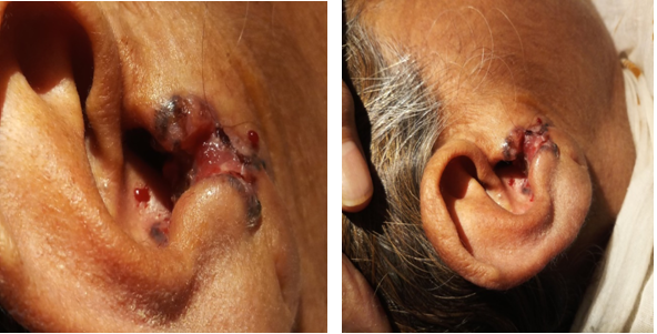
On local examination of the right ear as depicted in Figures 1 & 2, roughly 2X2 cm, irregular, hyper-pigmented growth involving the right tragus and cavum concha of the right ear. The growth is seen extending the right external auditory canal only involving nearly half of the cartilagenous portion of the canal. There is crusting with excoriation of skin over the growth along with ulcerative surface which bleeds on touch and is adhered to the skin of the auditory canal. The growth is not mobile and the base is ulcerative with areas of granulation seen with occasional serosanguinous discharge. On deep pressure, there is tenderness present over the growth. There is decreased sensation over and around the area of the growth with a fair amount of induration. Right tympanic membrane is well visualised and intact. Left ear, Nose and throat findings were within normal limits. The diagnostic work-up included clinical examination, imaging and biopsy. Hence, a biopsy was planned under local anaesthesia and sent for histopathological examination (HPE).

Figure 1 & 2 Growth in the right ear canal involving the tragus (in close up). Gross picture of the growth in the right ear canal.
On first opinion of HPE showed mucosa lined by stratified squamous epithelium with extensive areas of ulceration and granulation tissue formation. Focal areas in the mucosa shows hyperplasia with downward proliferation composed of dysplastic squamous cells with hyperchromatic nucleus, prominent nucleus and nucleoli. Mucosa shows areas of hyperkeratosis and acute inflammatory exudate. There is a dense mixed inflammatory infiltrate with few cholesterol clefts seen. Features are indicative of malignancy. But, was suggested follow up with Oncopathology opinion. Hence, a second opinion was planned as the first opinion was uncertain.
But second opinion of HPE showed keratinized stratified squamous epithelium with infected cholesteatoma like areas and focal suppuration. There is also invasion by small nests and irregular sheets of neoplastic basaloid cells with pleomorphic hyperchromatic nuclei and occasional mitosis that favours SCC (Basaloid type) with Cholesteatoma in the right ear as shown in Figures 5, 6 & 7. High Resolution CT temporal bone was proposed to look for features of any bone erosion by the growth, presence and involvement of cholesteatoma, status of middle ear structures, facial nerve, ossicular chain, labyrinth. The findings in this case of HRCT Temporal bone are mentioned as follows: Focal heterogeneously enhancing thickening in the outer part of the right external auditory canal and adjacent pinna. There was no adjacent bone erosion seen. Bilateral middle ear cleft appeared remarkable as shown in Figure 3 & 4.

Figure 3 & 4 Focal heterogeneously enhancing thickening in the outer part of the right external auditory canal and adjacent pinna. There was no adjacent bone erosion seen. Bilateral middle ear cleft appeared remarkable.

Figures 5, 6 & 7 5 - H & E stains showing the cholesteatoma area, 6 & 7 – both showing the tumor part with 5.
Later, this disease was staged as per Pittsburgh staging system modified by Hirsch for tumors arising from EAC which is mentioned below. Patient did not follow up to me after the biopsy and HRCT temporal bone. Patient was referred to higher centre for further management. As I was keen and followed up with patient’s relatives regarding management of this patient. Later, came to know that she underwent Wide local excision with Posterior auricular flap closure under General anaesthesia and was doing well presently.
Squamous cell carcinoma (SCC) can arise anywhere on the external ear and potentially involves middle ear, temporal bone, lateral skull base and surrounding sites. Periauricular soft tissues, parotid gland, temporomandibular joint and mastoid bone are the common sites for tumour progression. The carotid canal, jugular foramen, dura, middle and posterior cranial fossae are invaded in advanced stages of the disease.1-3 The tumor originates from the helix and antihelix margin where the skin receives greatest actinic exposure. Patients in 5th -6th decade of life show lesions that originate primarily from external auditory canal which is similar to this patient and present 10– 15 years earlier. Out of all, patients with SCC of the head and neck, 24% involve the ear and temporal bone. Association with HPV induced carcinogenesis, cold injury, chronic infection, fair complexion, radiation and sun exposure are among the most common predisposing factors.2,4
The tumor is a scaly, indurated, irregular maculo-papular lesion that shows an exo- or endophytic growth with hyperkeratotic or ulcerating surface accompanied by serosanguinous exudate which is similar to this patient. As it originates from the external auditory canal having hemorrhagic otorrhoea which is similar to this patient, it is falsely considered to be otitis externa due to which this patient was treated at many places on similar lines showing no response to treatment anywhere, until this visit to our ENT OPD setting. Suspicion arises when this condition fails to respond to conservative line of treatment and hence mandates for biopsy as in this patient. SCC lesions on nose and ear have highest tendency of recurrence as its close association with embryonic fusion planes and requires aggressive therapy.
Complete excision by micrographic surgery with tumor free margins is necessary for a successful outcome. Although this tumor grows in vertical fashion, it is less likely to respect the barriers of cartilage and bone when compared to Basal cell carcinoma (BCC). Therefore, intratemporal spread with involvement of external auditory canal is possible leading to conductive hearing loss. While further deep extension, the disease can involve and destroy vertical segment of facial nerve causing facial nerve palsy further advancing into internal auditory canal and cerebellopontine angle causing giddiness and/or sensorineural hearing loss.5,6
Once the tissue diagnosis and radiological investigations are known, it is important to stage the disease accordingly. It is important to provide apt treatment in any case of Carcinoma of EAC as it is said to recur soon although it is said to have a rare occurrence. Based on the diagnostic work-up, the patient was staged as per Pittsburgh staging system modified by Hirsch for tumours arising from EAC mentioned below as Table 1. In this case, it is T1N0 i.e (Stage I). There were no features suggestive of bone erosion, lymph node involvement and metastasis.7-10
|
T status |
Description External auditory canal (EAC) |
|
T1 |
Tumor limited to EAC without bony erosion or evidence of soft tissue extension. |
|
T2 |
Tumor with limited EAC bony erosion ( not full thickness) or radiographic finding consistent with limited (<0.5 cm) soft tissue involvement. |
|
T3 |
Tumor eroding the osseous EAC (full thickness) with limited (<0.5cm) soft tissue involvement or tumor involving middle ear and/or mastoid. |
|
T4 |
Tumor eroding the cochlear, petrous apex, medial wall of middle ear, carotid canal, jugular foramen or dura or with extensive (> 0.5cm) soft tissue involvement, where in patient presents with facial paralysis. |
|
N status |
Lymph node involvement is a poor prognostic sign and places the patient in advanced stage (i.e T1N1, Stage III) and T2,T3,T4N1 (Stage IV). |
|
M status |
M1 disease is a stage IV and is considered a very poor prognostic sign. |
Table 1 Pittsburgh staging system modified by Hirsch for Tumours arising from EAC9
Moreover, it is important to know the possible regional lymph node metastasis which signifies poor prognosis. There are locoregional metastasis that follow lymphatic drainage pattern which includes parotid and upper cervical nodes and undergoes regional lymphadenectomy (Neck-dissection level I-V), followed by postoperative irradiation. Nodal involvement is reported in 1–12.5% of all cases of SCC disease process. Any evidence of lymphovascular or perineural spread in the primary region, the adjacent "sentinel node" should be examined. In histologically aggressive malignancy, prophylactic lymphadenectomy and/or regional irradiation should be considered. Though large multi-institutional studies are missing, role of sentinel lymph node biopsy for SCC of the head and face region cannot be determined so far (Table 1).4,8
Squamous cell carcinoma of the external ear canal and middle ear are very rare malignancies which often present to the out-patient setting with long-standing chronic otitis media and also progressing to an advanced stage. The tissue diagnosis is fairly forthright; however, staging the disease has been a composite task that is best approached with consideration of clinical, radiological and pathological findings. Any intervention of these uncommon tumors is not well recognized and as such calibration of surgical and adjuvant treatment as well as irrational reporting will contribute to rather clear management pathways in the future.11
None.
None.
The Authors do not refer to Conflict of interest.

©2021 Basavannaiah. This is an open access article distributed under the terms of the, which permits unrestricted use, distribution, and build upon your work non-commercially.