Journal of
eISSN: 2379-6359


Case Report Volume 10 Issue 3
1Department of Otolaryngology, Kutahya Tavsanli State Hospital, Turkey
2Department of Patholology, Kutahya Tavsanli State Hospital, Turkey
Correspondence: Aysegul Sule Altindal, MD, Department of Otolaryngology, Kutahya Tavsanli State Hospital, Kutahya, Turkey, Tel 05079472488
Received: April 22, 2018 | Published: June 15, 2018
Citation: Altindal AS, Unal N. A case of unusual heteratopic salivary gland tissue mimicking tonsillar neoplasm and review of literature. J Otolaryngol ENT Res. 2018;10(3):157-160. DOI: 10.15406/joentr.2018.10.00336
Heterotopia or choristoma are characterized by a histologically normal tissue proliferation or nodule of a tissue type not normally found in anatomic site. Exact aetiopathogenesis of heterotopic salivary gland (HSG) is debatable. The histopathological finding of heterotopias is rare. In this article, we present a patient who underwent tonsillectomy for chronic recurrent tonsillitis with asymptomatic tonsillar mass and had chronic inflamed tonsillar tissue with HSG tissue in the palatine tonsil according to histological examination. Also we present a brief review about pathogenesis, pathologic features, tumorigenic potential, treatment and prognosis of HSGs. It could be one of the causes of recurrent tonsillitis. Also clinicans should be differentiate it from the tonsillar neoplasia due to similar appearance and should be aware of the fact that the neoplastic transformation of a HSG is a probablity.
Keywords: salivary gland; heterotopia; choristoma; ectopic; tonsil; neoplasia
HSG, heterotopic salivary gland; ENT, ear nose throat
Salivary gland tissue located in abnormal sites is variously defined as aberrant, accessory, ectopic, heterotopic, or salivary gland choristoma.1,2 Heterotopic salivary gland (HSG) is described as the presence of histologically normal mature salivary tissue that appears in an ectopic position due to placement defects during embryological development3 and HSG of the tonsil appears to be a developmental anomaly in the second pharyngeal arch. Furthermore, there is evidence of association of mucous glands with ducts in the palatine tonsils and these ducts scarcely open into the crypts but rarely does it manifest as visible mass with only isolated case reports having been found.4
Although HSGs are composed of lobules of normal salivary gland tissue histopathologically, they appear as soft tissue masses clinically and exhibit tumor like growth in otherwise normal salivary gland found in an abnormal location,5 even various neoplasms can arise from them.6,7 Thus, clinicans should consider HSGs in the differential diagnosis of soft tissue masses.
HSG has been reported in several areas of the body but rarely in the orofacial region;3 and the HSG tissue on tonsils are even rarer. Review of the literature published over the last 35 years showed only five reports of the presence of normal salivary gland tissue originating from it, on the tonsil region. In this article, we present a patient who underwent tonsillectomy for chronic recurrent tonsillitis with asymptomatic tonsillar mass and had chronic inflamed tonsillar tissue with HSG in the palatine tonsil according to histological examination. Also we present a brief review about pathogenesis, pathologic features, tumorigenic potential, treatment and prognosis of HSGs.
A 32-year-old male presented to our Ear, Nose, and Throat (ENT) Department with two years history of recurrent sore throat, pain, fever, painful swelling and a painless left tonsillar mass in April 2017. He also complained of snoring and halitosis, six month duration. Physical examination showed that tonsils were persistently enlarged (+3/+3 hypertrophic) and an irregular surfaced tissue tag on the middline of left palatine tonsil. The patient admitted smoking 20 to 30 cigarettes a day for more than 10 years. He was also an occasional drinker. He had no family history of cancer. Recurrent pain was related to chronic tonsillitis but presence of tonsillar mass and toxic habits raised suspicion of neoplasia.
Further examinations, including endoscopic inspection of the head and neck region, ultrasonography of the neck and neck magnetic resonance imaging, were performed to rule out a tonsillar neoplasia. None of the studies showed any pathology.
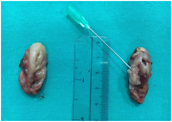
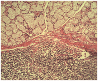
The patient was suggested to undergo tonsillectomy. Routine preoperative investigations were within normal limits and total surgical excision of the lesion through bilateral tonsillectomy was performed under general anesthesia. After completion of the tonsillectomy, both the specimens from the right and left tonsils measured approximately 3,5 cm x 2 cm x1.5 cm. Gross examination revealed a grey, irregular surfaced and 1x0,5 cm measured mass on the midline of left side tonsil (Figure 1). No other lesion was seen on the examination of the contralateral tonsil. Histopathologic examination of the left side revealed tonsilar tissue with chronic inflammation and lobules of mucous secreting salivary acini with ducts adjacent (HSG tissue) and musculary fibers to the surface squamous epithelium of the tonsilar tissue (Figures 2) (Figure 3). Histopathologic examination of the right side revealed only tonsilar tissue with chronic inflammation. A diagnosis of HSG in relation to tonsils was given. Postoperative period was uneventful and recovery was good. A year follow-up revealed no evidence of recurrence and no new relevant symptoms.

Figure 1 Gross specimen, right and left tonsils. Needle shows an exophytic grey, irregular surfaced tonsillar mass on the left tonsil.

Figure 2 Tonsillar tissue with adjacent islands of heterotopic mucinous salivary gland acini (Histopathologic examination, H&E stain, Magnifications: ×100).
Embryological anomalies are common in the neck region due to its complexity. HSG of head and neck with predilection to the oral cavity is described as the presence of histologically normal mature salivary tissue that appears in an ectopic position,3 with no tumoregenic features.8 Although the embryological placement defect is responsible for HSG formation, precise aetiopathogenesis of HSG tissue is debatable.
The palatine tonsils and tonsillar fossa are thought to be derived from the endodermal lining of the second pharyngeal pouch at the 9th week of fetal development Embryologically.9 The major salivary glands appear earlier, beginning as epithelial buds from the primitive oral cavity, between the sixth and eighth week.10 At last, minor intraoral salivary glands develop during the third month of intrauterine life and spread throughout the oral mucosa.10 So, it is first reasonable possibility these minor salivary glands as the mechanically displaced during prenatal development and have got implanted on palatine tonsillar walls and subsequently have developed into heterotopic tissue tags.3,8,10 In addition, there is evidence of association of mucous glands with ducts in the palatine tonsils and rarely opening these channels into the crypts is another probablity in the pathogenesis of HSG.11
A case of histologically diagnosed HSG tissue in the tonsillar fossa was first described by Neumann et al.,12 in 1928. In 1968, Samy et al.,13 reported that the ectopic salivary tissue, presented as a red mass at the lower pole of the tonsil in the Russian literature. A review of the literature over the last 35 years revealed only five isolated case reports in which ectopic salivary tissue was noted in the palatine tonsil. That makes the occurrence of HSG tissue on tonsil rare. Since 1982, the first such case reported by Banerjee et al.,14 which involved a newborn child with a first branchial arch defect in whom a large horseshoe-shaped, obstructing mass in the palatine tonsillar region revealed primarily salivary gland tissue on histopathologic examination. The second related case were presented by Shchepin et al.,15 involving ectopic salivary tissue identified incidentally in the tonsillar region during a resection for upper airway papillomas. The third case was reported by Wise et al.,16 which involved another child with a painless unilateral exophytic mass and turned out to be a salivary tissue. In the fourth case, Golan reported bilateral ectopic salivary gland tissue in the case of routine tonsillectomy.17 The last case with salivary gland tissue on palatine tonsillar wall, was presented by Shyamala et al.,18 in 2017. All these authors have mentioned that ectopic salivary gland tissue on tonsils is rare.
As sixth isolated case, we are reporting a HSG tissue seen on the left tonsil as an asymptomatic mass after tonsillectomy performed for chronic recurrent tonsilitis. Histopathological examination revealed ectopic salivary gland tissue with chronic inflammatoary tonsillar tissue on the left side. Tonsillar tissue on the right side revealed no evidence of the lesions. A year follow up revealed no evidence of recurrence and no new relevant symptoms.
The most frequent clinical presentation is a painless tumor and clinical presentation depends of location. HSG tissue has been reported in various body regions, including the tonsils, tongue, gingiva, oral palate, maxilla, mandible, buccinator muscle, optic nerve, lacrimal gland, frontal skin, middle and external ear, cervical lymph nodes, bilateral cervical skin, posterior triangle of the neck, thyroid parathyroid gland, anterior chest wall, bronchus, mediastinum, pituitary, cerebellopontine angle, pterygopalatine fossa, breast, stomach, vulva, vagina, prostate and colon.13–21 HSG tissue was revealed by a patient who underwent tonsillectomy for chronic recurrent tonsillitis with asymptomatic tonsillar mass in present case.
Normal salivary gland tissue such mucous and mixed acini predilection that may be came along with fibrosis have been shown in histology of HSG. Some of the variation seen such as associated ducts and areas of squamous metaplasia, may be related to chronic inflammation and lymph infiltration secondary to physical pressure of retained mucus.22 The present case showed these histological features probably due to mucosal secretion and chronic swelling.
Athough HSGs are composed of lobules of normal salivary gland tissue histopathologically, they appear as soft tissue masses clinically. So, ENT Physicians should be aware that such an entity as salivary gland heterotopias existance and should be considered as one of the differential diagnoses of soft tissue masses in the oral cavity. Clinically, heterotopia rests can be confused with true neoplasms, when they are sufficiently large. Moreover, it is possible that in rare cases this “heterotopic” salivary tissue undergoes neoplastic transformation, giving rise to a spectrum of neoplasms similar to those found in salivary glands, as reported in the literature.6,7 The neoplasms originating from the salivary gland have many histological types, ranging from benign to high-grade malignancy. Previous reports also suggest tumours in HSGs are rare and 80% of them are benign.22,23 Whartin’s tumour is the most frequent benign neoplasm which is followed by pleomorphic adenoma. Mucoepidermoid carcinoma is the most malignant neoplasm described.20 Hun-Soo Kim et al.,6 reported a case of acinic cell carcinoma of ectopic salivary tissue located in the tonsil. Adenocarcinoma, and adenoid cystic carcinoma are also seen to arise from this tissue.7 Chang et al.,24 noted that HSG tissue was likely to be under diagnosed before it underwent any neoplastic change. They also noted that when the lesion was suspected clinically, it should be totally excised to prevent further neoplastic changes, and all the sections should be cautiously checked. It is important to consider the possibility of it being a metastasis of a primary salivary gland tumour.22,23,25 Thus, there is a need for a proper diagnostic approach and the role of biopsy and histopathology in the diagnosis of such potentially tumorigenic lesions.
The treatment is still debatable as HSG tissue gives rise to neoplasm. Some authors believe that heterotopias are immature in nature, which theoretically increase the malignization potential26 so they prefer the complete excision of the mass.3,22,25 Some authors believe that salival heterotopia is a normal tissue and doesn’t require complete excision. Surgical removal is only necessary when there are signs of infection or neoplasia.3,27,28 But the general approach of the treatment is that simple excision or tonsillectomy is sufficient for non-neoplastic heterotopic mass as the present case.
Recurrences have not been documented in head and neck heterotopias, but some oral cases have been reported to be recurrent. In the present case, a year follow-up revealed no evidence of recurrence.
To conclude HSG tissue is rare histopathologic finding in the post tonsillectomy specimen. Only six isolated case reports have been reported in literature since 1982. Although it is rare occurrence in association with tonsils, it’s possible causing recurrent tonsilitis or tumorigenic potential cannot be ignored. If they get sufficiently large diameters clinically, diagnose can be confused with true neoplasms. Thus, ENT Physicians should be aware of the properties of such lesions to avoid false diagnosis or misdiagnosis.
Nil.
There are no conflicts of interest.
Obtained.

©2018 Altindal, et al. This is an open access article distributed under the terms of the, which permits unrestricted use, distribution, and build upon your work non-commercially.