Journal of
eISSN: 2379-6359


Case Report Volume 10 Issue 2
1Otorhinolaryngology and Head Neck Surgery Department Mater Dei Hospital, Malta
2Department of Sugery Mater Dei Hospital, Malta
Correspondence: Marija Agius Spiteri, Otorhinolaryngology and Head Neck Surgery Department, Mater Dei Hospital 47, Hompesch Road Fgura, Malta, Tel +35699245613
Received: February 06, 2018 | Published: March 7, 2018
Citation: Spiteri MA, Grech G, Bartolo A. Obstructive sleep apnoea caused by a pleomorphic adenoma: a case report. J Otolaryngol ENT Res. 2018;10(2):75–77. DOI: 10.15406/joentr.2018.10.00315
Background: Sleep apnoea syndrome is a condition characterised by excessive daytime somnolence, hyp-nagogic hallucinations, personality and sexual behaviour changes and hypertension. Most commonly, sleep apnoea syndrome is secondary to upper airway obstruction as a result of intermittent hypotonia of the soft palate and tongue musculature. In rare occasions, obstructive sleep apnoea can result from anatomical abnormalities such as a mass in the upper aerodigestive tract.1
Case summary: A 40 year old gentleman was referred in view of obstructive sleep apnoea symptoms. On ex-amination, a large right sided lesion was seen in the oropharynx. Further investigations with flexible nasoendoscopy, computed tomography (CT) and magnetic resonance imagings (MRI) of the neck were carried out which confirmed a large mass most likely arising from the right tonsillar bed. Surgical excision was carried out via a transoral approach. Histology was re-ported as pleomorphic adenoma.
Conclusion: It is widely accepted that management obstructive sleep apnoea involves treating the cause together with conservative and lifestyle measures are adopted initially. The majority of patients do not require surgery but a thorough assessment, including the upper aerodigestive tract, should be carried out to exclude rare causes of obstructive sleep apnoea.2
Keywords: Obstructive sleep apnoea, pleomorphic adenoma
CT, Computed tomography; MRI, Magnetic resonance imaging; OSA, Obstructive sleep apnoea
Obstructive sleep apnoea is a condition involving intermittent collapse of the pharynx, usually at the level of soft palate, causing episodes of apnoea during sleep1. Muscle tone declines during sleep and thus, upper airway dilating muscles are unable to maintain pharyngeal pa-tency. In severe cases, the patient wakes up briefly multiple times during the night to allow dilating muscles to re-open the airway.2 This results in severe sleep fragmentation leading to symptoms such as daytime somnolence (assessed by Epworth sleepiness scale), morning headaches and decreased cognitive performance. Polysomnography is the gold standard in-vestigation to diagnose Obstructive sleep apnoea. This involves monitoring of oxygen satu-ration, airflow at the nose and mouth ECG, EMG, chest and abdominal movement during sleep1. The occurrence of 15 or more episodes of apnoea or hypopnoea during 1 hour of sleep indicates significant sleep apnoea.3
Pleomorphic adenoma is a benign salivary gland tumour known for its pleomorphic appear-ance under light microscopy. It is the most common tumour of the salivary glands with 80-90% which arise from the parotid gland.4 Spiro RH in his study of 2078 patients with salivary gland neoplasia reported that 20-40% of all salivary gland tumours arise from minor salivary glands, mostly from the palate (42.63%), lip (10%), buccal mucosa (5.5%), retromolar area (0.7%) as well as floor of the mouth, tongue, tonsil, pharynx and nasal cavity.5–9
In rare occasions, obstructive sleep apnoea is a result of anatomical abnormalities such as a mass in the upper aerodigestive tract. Apart from obstructive sleep apnoea, tumours of this location may also cause dyspnoea, dysphagia and airway obstruction.10 The first case reported, was in 1981 where a patient was diagnosed with obstructive sleep apnoea secondary to nasopharyngeal carcinoma3. Up to 2005 only 30 cases of obstructive sleep ap-noea caused by head and neck tumours have been reported.11 In the English language literature, 9 pleomorphic adenomas were found to cause OSA.12
Pleomorphic adenomata are more common in patients between 40 to 60 years of age with a male to female ratio of 2:3 4. However case reports involving a 7-year old patient and an 82 year old patient are also seen.13 Such tumours are usually asymptomatic and thus they can grow a long time before being diagnosed. The potential risk of malignant transformation in-creases over the years with an incidence of 1 to 7%.14 The smaller the salivary gland that is affected, the more likely it is to trigger a malignant tumour.10
The diagnostic work-up begins with haematologic and serologic tests followed by imaging6. MRI provides precise information about tumour margins as well as the relationship with the surrounding structures. Fine Needle Aspiration Cytology (FNAC) can also be used to aid with cytological diagnosis. There are different approaches that can be used in pleomorphic adenomata of the palate. It is of utmost importance to have adequate exposure to identify and protect vital structures and ensure complete removal. Intra-oral and extra-oral interventions can be used. Transparotid, transcervical and transpharyngeal approaches are the extra-oral approaches which are commonly used. Mandibulotomy can be performed to improve exposure.4
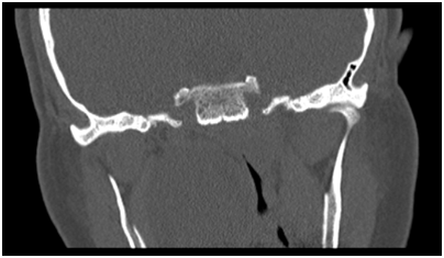
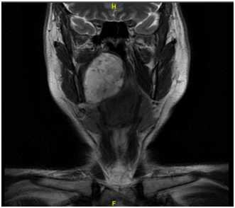
A 42 year old male, with no previous medical history was referred to the Otorhinolaryngology and Head & Neck surgery outpatients’ department at Mater Dei Hospital with Obstructive Sleep Apnoea (OSA) symptoms. The patient admitted to a few year history of such symptoms. During examination, a large right sided lesion was seen in the oropharynx which seemed to be arising from the soft palate. This lesion was seen to be crossing the midline and most likely contributing to his obstructive symptoms. The patient underwent a flexible nasoendoscopy which showed a large lesion extending to the lateral nasal cavity. Subsequently the patient underwent urgent imaging with computed tomography (CT) (Figure 1) of the neck and thorax which showed a large mass with the most likely site of origin being in the right tonsillar bed. A dedicated magnetic resonance imaging (MRI) (Figure 2) (Figure 3) of the neck was recommended for staging, to better assess the extent of the lesion. Two hypoechoic intrahe-patic liver lesions were discovered were also noted. A biopsy of the lesion arising from soft palate was taken under local anaesthesia which was reported as adenomatoid acinar hyper-plasia.


The patient underwent a MRI of the liver and spleen which showed signs of iron overload and reported the liver lesions as benign haemangiomas. Long discussions were undertaken with the patient and his wife with the proposed surgery with particular focus on the eventuality of tracheostomy as well as open approach.
A transoral approach was used using a Boyle Davies gag and cheek retractors to aid with ac-cess and exposure. Cheek retractors proved to be handy for better exposure. The lesion was 4 x 5 cm in diameter, partly cystic and unfortunately the capsule was breached during the ma-nipulation of the tumour. After resection, the defect was closed primarily with absorbable su-ture in an interrupted fashion (Figure 4) (Figure 5).
The patient was sent to Intensive Care Unit for overnight monitoring and was discharged on second postoperative day. The final histology was reported as pleomorphic adenoma.
The patient is being followed up regularly at the outpatients’ clinic.
None declared.

©2018 Spiteri, et al. This is an open access article distributed under the terms of the, which permits unrestricted use, distribution, and build upon your work non-commercially.