Journal of
eISSN: 2379-6359


Editorial Volume 6 Issue 4
Department of Special Surgery Jordan University of Science Technology, Jordan
Correspondence: Mohannad Al Qudah Professor of Otolaryngology Department of Special Surgery Jordan University of Science Technology P O Box 3030 Irbed 22110, Jordan, Tel 0096 2 79 9630066
Received: April 03, 2016 | Published: April 7, 2017
Citation: Al-Qudah M (2017) Endoscopic Sinus and Skull base Surgery: The Compartment Concept. J Otolaryngol ENT Res 6(4): 00173. DOI: 10.15406/joentr.2017.06.00173
Functional Endoscopic Sinus Surgery (FESS) is indicated for medically resistant sinusitis management. The procedure becomes one of the most common otolaryngology surgeries and replaces other surgical procedures designed to treat sinonasal disorders.
The rational of this minimally invasive technique is based on our modern understanding of Chronic Rhino Sinusitis (CRS) pathophysiology. Opening the natural ostium of infected sinus and removed severely infected tissues, will allow re-ventilation and regeneration of healthy functioning mucosa and cilia.
FESS is a broad term that involved stepwise dissection of consecutive lamellae with mucosa preservation. DeConde et al.,1 conducted prospective study to evaluate the outcome difference between complete versus targeted approaches in endoscopic sinus surgery. Patients undergoing bilateral frontal sinusotomy, ethmoidectomy, maxillary antrostomy, and sphenoidotomy were considered to have undergone “complete” surgery, whereas all other participants were categorized as receiving “targeted” surgery. Improvement was evaluated between surgical subgroups with at least 6-month follow up using the 22-item Sino-Nasal Outcome Test and the Brief Smell Inventory Test. The authors didn’t find a significant difference in the results between both groups.
These findings raise a big question of the beneficial value of opening all the sinuses' ostia and performing complete ethmoidectomy in every single sinus surgery. Similar to the term FESS, CRS is also a wide spectrum of heterogeneous disease. The disease can be classified based on microbiology etiological factors, the presence of polyp, microscopic presence of certain cells and markers.2,3
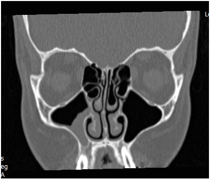
All these classifications are of value in the complex disease pathogenesis and prognosis understanding, but they don’t reflect on the disease extension and surgical steps and so don’t help in the preoperative planning, estimation of the duration of surgery or prediction of surgery related complications. For example, FESS is the term of procedure used to describe surgical management of a case with nasal polyposis and pansinusitis and it’s the same term used to describe surgical treatment of a case with unilateral maxillary sinusitis Figure 1. It is obvious that the first procedure has more serious complications, requires different instruments and experience, and indeed needs longer surgical and anesthesia time.
The increased costs of health service worldwide and providing a stander clinical quality require having obvious plan and protocol for any medical procedure. In our practice we divide FESS into compartments. When we approach the frontal sinus to treat frontal sinus pathology this will be the superior compartment, whereas treating maxillary disorders is the inferior compartment of FESS, (Figure 1). The anterior and posterior compartments are used to describe resection of diseased ethmoid cells and opening sphenoid sinus respectively.

Figure 1 CT scan for patient with unilateral maxillary sinus opacification demonstrating the Inferior compartment of FESS.
Recently nasal and sinus surgery have a tremendous expansion in the surgical filed and indications. Advances in image quality and reconstruction, better understanding of the anatomy, the introduction of navigation system and the availability of power instruments have allowed surgeons to broken the borders of the paranasal sinuses to perform complex surgery in the skull base and orbit.4
The compartment concept can also be applied in endoscopic skull base and orbit surgery. Because these cases always start with opening one or more sinus and then use the new created space as a corridor entry, the term extension can be added to describe them, so Endoscopic Endonasal Transsphenoidal Approach to skull base
considered as extended posterior compartment whereas endoscopic orbital or optic nerve decompression are an extended anterior approach.
We believe adapting this concept will make surgery more obvious to the whole operative team, open research studies for a new method of sinusitis classification and can be used to compare the outcome of different surgical techniques based on real surgical steps instead of patient’s demographic date and preoperative CT scan evaluation.
None.
Author declares there are no conflicts of interest.
None.

©2017 Al-Qudah. This is an open access article distributed under the terms of the, which permits unrestricted use, distribution, and build upon your work non-commercially.