Journal of
eISSN: 2377-4282


Review Article Volume 7 Issue 2
1School of Chemical Engineering and Technology, Tianjin University, China
2Key Laboratory of Systems Bioengineering of Ministry of Education, Tianjin University, China
Correspondence: Hao Qi, Key Laboratory of Systems Bioengineering of Ministry of Education, Tianjin University, Tianjin 300072, China
Received: February 11, 2018 | Published: March 21, 2018
Citation: Yasai H. Challenges in circulating tumor DNA analysis for cancer diagnosis. J Nanomed Res. 2018;972):76 – 80. DOI: 10.15406/jnmr.2018.07.00180
Correlation between circulating tumor DNA (ctDNA) and cancers have been investigated and reported in clinical studies. Due to the ease in collection of blood samples, ctDNA analysis with advanced molecular analysis technologies is becoming an alternative to current solid tissue biopsies in clinical diagnosis. However, there is still obscure issues resulting the short of consistency in clinical analysis of ctDNA. Here, we discussed the crucial issues including concentration and size profile of ctDNA which have led to contradictory results in different clinical studies. Then, the recent efforts being made to improve ctDNA analysis as a robust point-of-care application were review.
Keywords: cancer diagnosis, circulating tumor DNA, liquid biopsy, cell-free DNA, tumor
People knew that the DNA molecule existed outside of cell even before finding out its famous double helix structure. Mandel and colleagues identified DNA molecule, termed as cell-free nucleic acids (cfDNA) later, in human bloodstream in as early as 1948.1 However, at that time no people realized how these DNA molecules associate with human diseases. Thing started turning around until 1964, DNA was found being released into sera for certain systemic lupus erythematosus patients.2 Since then, many clinical studies were carried out, more evidences demonstrated the strong correlation between cell free DNA and human diseases, especially for cancer.3,4 It was observed that even DNA could be isolated from blood of healthy people, but the amount of DNA significantly increased in the blood sample from patients with serious tumor. Particularly, as the earliest research, DNA fragments from mutant K- ras gene were found in blood of pancreatic carcinoma patients5 and mutant N-ras gene fragment for myelodysplastic syndrome patients.6 These studies successfully demonstrated the direct correlation between circulating DNA and tumor. Recently it has been widely accepted that the levels of circulating nucleic acids strongly connected with tumor burden and malignant progression.7–11 For not being confused with cell-free DNA in healthy people, tumor cell related DNA circulating in human cardiovascular system were specially termed as circulating tumor DNA, ctDNA. Generally, it is widely considered that most DNA in circulation system is the debris of dead tumor cells. However, due to the complexity of cancer development, more fundamental studies are required to investigate questions, such as which processes contribute to ctDNA release from tumor cells12 and how the release process change the state of ctDNA in the circulation system. Besides being the debris left behind by dead cells, DNA is the key component of neutrophil extracellular traps (NETs), a host immune defense system against invading pathogens. Recently, increasing studies have demonstrated that NETs got involved in cancer development at every stages.13–15 With the development of molecular oncology, more and more tumor specific gene mutations were identified,16 and detail information about relevant tumor specific mutation could be found in systematically organized database, such as My Cancer Genome(www.mycancergenome.org). Up to now, ctDNA has been investigated with numerous of prevalent tumors, including Breast,17,18 Colorectal,7,19 Hepatocellular carcinoma,20,21 lung,22–24 Melanoma,25,26 Ovarian,27 Pancreatic28 and so on. In comparison with other biomarkers, e.g. protein, ctDNA is more informative with more precise analysis methods.29 Due to its nature, ctDNA is becoming a remarkable clinical tool. Especially, the convenience in collecting blood sample grant the liquid biopsy application great potential through ctDNA analysis in cancer diagnosis. However, precise analysis of ctDNA is still a challenge for some technique and biophysical reasons. For becoming a solid tool for tumor diagnosis, more clinical researches are still necessary to address some crucial questions about the physiological mechanism and analysis technical issues. In this review, we put the spotlight on the crucial issues in ctDNA based cancer diagnosis, especially the experimental issues which have led to contradictory results in different studies.
Analysis of circulating DNA
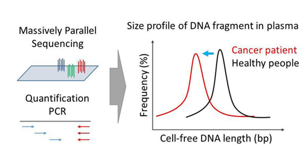
The concentration of ctDNA varies from person to person, also depending on the type and status of the tumor30–32 Generally, ctDNA is presence in the circulation system at a very low concentration. Based on estimation, it may up to 3.3% of DNA in tumor cells, which will be released into circulation system every day.33 If calculated as about 3x10¹⁰ tumor cells included in tumor tissue of 100g weight, there will be 105 copy genome DNA in 1 ml blood. High concentration of DNA in clinical blood samples of about 80 ng/ml, roughly equivalent to 104 copy genome DNA in 1 ml blood, has been reported.34 In comparison, even for same lung cancer 12.8 ng/ml of DNA in circulating plasma was reported in another study.35 The concentration varied between patients even with same type of tumor from a few nanogram to around 1000 nanogram in one milliliter blood plasma sample.36 Unfortunately, there is still no well optimized clinical standard for ctDNA isolation. Tumor progression is extremely complicate process, so many parameters including sample type and experimental protocol will significantly affected the final output of isolated ctDNA. The size of the ctDNA fragment is another key factor for further precise analysis as well (Figure 1). As crucial part of programmed cell death, fragmentation occurs before DNA released from cells undergoing apoptosis.37 Due to the highly ordered structure of DNA in nucleosomes, most part of the genome DNA was cut into fragments of roughly 180 base pairs or integer multiples in length via activation of endogenous endonucleases.12 Evidence has been found that ctDNA arose from necrosis was in larger size up to a few thousand base pairs.12 Jiang and colleagues investigated the plasma DNA from hepatocellular carcinoma patients using a massively parallel sequencing platform and found that the size of DNA fragment had a distribution with a prominent peak around 166 base pairs.38 In compared with the non-tumor cfDNA, a larger proportion of tumor derived ctDNA were of the length shorter than 166 base pairs. It was a little bit shorter than the 188 base pairs of cfDNA from healthy person reported in previous studies. Moreover, shortening in size was also observed in cell-free fetal DNA in comparison with the maternal cfDNA.39 In addition, it was found that the extent of DNA fragmentation increased, while the size of ctDNA decreased, with the tumor progression.41,42 There should be a reason behind the observed shortening of ctDNA42and further study definitely will improve the understanding of ctDNA biology. The size of ctDNA has been taken into account to improve the specificity and sensitivity of ctDNA analysis. Due to the shortening of ctDNA in cancer patients, it strongly indicated that for PCR based method optimized amplicon design is crucial. Florent Mouliere and colleagues thoroughly investigated the effect of amplicon size on ctDNA quantification.41 For specific gene target, a set of PCR primers were designed for amplicon of size 60-100, 100-150, 150-400 and larger than 400 base pairs respectively. DNA from metastatic colorectal cancer patients were analyzed following a general quantification PCR method. Interestingly, even for the same DNA sample, concentration quantified from larger amplicon was lower. For the DNA sample from big tumor, the ctDNA concentration quantified using smallest amplicon was over 1000 ng/ml, but only a few ng/ml using large amplicon. Moreover, Rikke and colleagues compared the detection of KRAS gene from blood sample using amplicon of 120 and 85 base pairs respectively. For specifically distinguishing the single base mutant target gene, ARMS-PCR, which is well designed for detection of single nucleotide base mutant, was used for quantification of target gene. Surprisingly, the mutant KRAS gene quantified from short amplicon was even three times higher than that quantified from long amplicon.43 These results strongly indicate that well designed short amplicon is preferred for ctDNA analysis, especially for tumor at late stage. Besides the biochemical features, the basic experiment methods have great impact on the ctDNA analysis as well (Figure 2). Good status of blood samples and appropriate sample handling including storage and DNA extraction are necessary for precise ctDNA analysis. As discussed, not like genome DNA directly from cell with compact structure, ctDNA is highly fragmental DNA molecule with exposed terminus, which is subject to degradation. Dozens of nucleotides, the major size of ctDNA, is a piece of cake for nucleases to degrade. The valuable sample will be lost in the twinkling of an eye without appropriate handling. In addition, it is crucial to avoid lysis of cells in blood sample during sample collection and DNA extraction, which will lead to high background from non-tumor cells. Patricia and colleagues took an investigation on the tubes used for blood collection and storage.44 Several commercially available tubes were compared. Blood of metastatic breast cancer patients were collected and stored in different tubes for a while and then DNA was extracted and analyzed by droplet digital PCR. It was found that the ctDNA integrity was well protected for up to a week at room temperature in Cell-free DNA BCT tubes manufactured by Streck (La Vista, NE), which has been shown being cable to stabilize cell membranes even in whole blood sample.45 However, the EDTA tubes, which could be considered as the golden standard used in hospital for blood sample collection, were only able to protect the ctDNA for just 2 hours. Qing and colleagues have obtained the similar result with comparing tubes of different brands in protecting the abundance of ctDNA.46 In circulation systems, low concentration and small length make the efficient extraction of DNA difficult. Therefore, Alison and colleagues investigated another crucial issue, the reagents of ctDNA extraction. Three commercially available DNA extraction kits on the market were compared thoroughly. The parameters including extraction efficiency, linearity of the extraction yield, inhibit contaminates and bias of fragment size were investigated and shown that the outcome of ctDNA extraction was dramatically different between these reagents.47 Similarly, other methods have been evaluated for ctDNA extraction as well respectively.48,49Based on these studies, it is very urgent to well standardize the basic method for ctDNA sample preparation.


Liquid biopsy
Due to the ease of collecting blood sample comparing with accessing solid tissue sample, ctDNA is widely considered as a promising alternative for invasive biopsy, termed as liquid biopsy. In 1997, research carried out by Dennis Lo at Chinese University of Hong Kong firstly demonstrated that DNA isolated from circulation system could be used as a powerful tool for prenatal diagnosis.50,51 Since then, liquid biopsy caught many attentions and has been expanded for diagnosis of various diseases, especially for early-stage cance.17,22,52–56 It has been reported that in Hong Kong liquid-biopsybased cancer screening has been applied on a large population including over twenty-thousand peoples and similar researches have been reported as well.57 With further combined with advanced DNA analysis assay,8,24,58–63 for example fast DNA sequencing ,64–66 isothermal DNA amplification,67,68 practical and precise point-of care cancer diagnosis assay could be achieved very soon. More detail related information could be found in recently published review articles with specific focus on this
liquid biopsy.69–71 With the significant potential in cancer treatments, liquid biopsy has become a fast growing market around the world. FDA approvals for non-invasive diagnostic tests for cancer detection drives more players with various technology platforms entering this market, part of them are listed in Table 1. Many biotechnology giant companies such as Illuminua, QIAGEN and Roche, have started putting focus on developing liquid biopsy ctDNA analysis technology or already have products on the market. However, there is still no systematic study demonstrating the consistency between these commercial products in analysis of ctDNA.
Company |
Product |
Analysis Technology |
Bio-Rad |
Cancer genomic alterations of BRAF V600E, |
Droplet digital PCR |
QIAGEN |
Reagents covering multiple cancers |
Digital DNA sequencing |
Roche (Cobas) |
Genomic alterations of EGFR, Exon 19 |
Real-time PCR |
deletion, L858R, T790M |
||
Guardant |
Panel of 73 cancer genomic alterations |
Digital DNA sequencing |
Health |
||
Genomic |
Panel of select 17 genes |
NGS |
Health |
||
Biocept |
Service for ctDNA analysis with Target |
SNV specific real-time |
Selector technology |
PCR |
|
Exosome Dx |
Specific cfDNA extraction technology |
qPCR or NGS |
Agena |
Panel of multiple cancer genomic alterations |
Massarray platform |
bioscience |
with specific extraction reagents |
|
Table 1 Commercialized liquid biopsy solutions for ctDNA test
It is predicted that cancer is becoming the leading causes of death for human. Very recently, an interesting research has been published, which demonstrated that most of the cancer related genomic mutation arose from random DNA replication.72 The authors investigated correlation between cancer risk and the stem cell divisions with a huge data from65 countries throughout the world. Surprisingly, they found that environment factors and inheritance may not contribute that much to cancer mutations not as people previously thought. The most of cancer mutations may arose inside but not being induced from outside. Random mistakes during DNA replication could be blamed as the real carcinogen. If correct, it will overturn the way how people deal with cancer. Cancer mutations are not preventable and occurrence of tumor is just a matter of time. Therefore, it became very crucial for preventing cancer by early-stage detection as argued by authors. Precise diagnosis based on analysis of DNA from circulation system is very attracting idea, especially for cancer. Since the concept has been proved, liquid biopsy assays of ctDNA have been developed for numerous types of cancers. Based on the huge clinical data so far, early detection significantly increases the success cancer treatment and cancer survival rate.73 Even though the future is promising, key challenges still lie ahead. More basic clinical researches are required to uncover the detail mechanism, by which ctDNA correlated with tumor development. Without these knowledge, only analysis of ctDNA could not lead to improved cancer therapy. Unfortunately, the lack of standardization is another crucial issue in ctDNA related research,74,75 which lead to contradictory results from different studies. As majorly discussed here, lack of standardization in ctDNA experiment including blood sample collection, and cfDNA extraction will hurt the development of this field. However, it will take time to reach a consensus for ctDNA related analysis method due to the short of understating about some key issues of ctDNA. For example, how the fragmentation of DNA occur in tumor cell and how long is the lifetime of ctDNA in circulation system. Based on these detail understanding, current method could be improved and standardized, therefore ctDNA could become a robust analyte and a golden standard for cancer diagnosis.
This work was supported by National Natural Science Foundation of China (Project No. 21476167). H.Q. was also supported by the Recruitment Program for Young Professionals.
None.

©2018 Yasai, et al. This is an open access article distributed under the terms of the, which permits unrestricted use, distribution, and build upon your work non-commercially.