Journal of
eISSN: 2373-437X


Research Article Volume 3 Issue 4
1Thomas Jefferson University, Philadelphia, PA, USA
2Rowan University, Glassboro, NJ, USA
Correspondence: Sheila Wood, Rowan University, Glassboro, NJ, USA
Received: March 22, 2016 | Published: July 6, 2016
Citation: Eckard A, Wood S (2016) In-Vitro Susceptibility of Different Morphological Forms of Borrelia burgdorferi to Common Lyme Antibiotics in Combination with Antimicrobial Peptides. J Microbiol Exp 3(4): 00097. DOI: 10.15406/jmen.2016.03.00097
Antimicrobial susceptibility testing was conducted with three antibiotics commonly used to treat Lyme disease (doxycycline, erythromycin, and tinidazole) and three AMPs (cecropin P1, magainin 1, and LL–37) against one strain of Borrelia burgdorferi. The susceptibility of spirochete and spheroplast (cyst) forms was determined by direct cell counting using dark field microscopy. Significant synergy was seen when erythromycin was combined with cecropin P1. Spirochete growth decreased (P=0.0015) and spheroplast (cyst) growth decreased (P=0.001) when compared to the growth control. Synergy was also seen when tinidazole was combined with LL–37. Spheroplast growth decreased (P=0.015). Combining antimicrobial peptides with commonly prescribed antibiotics to treat Lyme disease may provide a new approach to the treatment of chronic infections due to the significant synergistic effect of a combination therapy.
Keywords: antimicrobial susceptibility, borrelia burgdorferi, b. burgdorferi, lyme disease, doxycycline, erythromycin, tinidazole, spheroplast; antimicrobial peptides; antibiotics; synergistic effect
Combination therapies involving commonly prescribed antibiotics and antimicrobial peptides may provide a new approaches to the treatment of Lyme disease. Although early treatment of Lyme disease is generally successful, studies have shown that some patients who received antibiotic treatment develop persistent or recurrent Lyme disease symptoms. Possible explanations for the persistent symptoms may be the presence of different morphological forms of Borrelia burgdorferi, spirochetes and spheroplasts (cysts) and/or suboptimal treatment regimens. It is hypothesized that antimicrobial peptides (AMPs) will have a synergistic effect on the different morphological forms of B. burgdorferi when combined with current antibiotic therapies.
Lyme disease is a tick–borne illness that was first recognized in Lyme, CT in 1975 and subsequently shown to be caused by the spirochete Borrelia burgdorferi.1 Ticks of the genus Ixodes1 transmit the disease. Currently, Lyme disease is the most commonly reported vector–borne illness in the United States, and in 2013 it was the fifth most common Nationally Notifiable disease.2 Each year more than 30,000 cases of Lyme disease are reported to the CDC; however, the CDC estimates around 300,000 Americans are diagnosed each year.3 This estimate supports previously published studies indicating that the true number of cases is between 3– and 12–fold higher than the number of reported cases. Most cases of Lyme disease can be treated successfully with a few weeks of antibiotics2. However, approximately 10 to 20% of patients have symptoms that last after treatment, and in a small percentage of cases the symptoms last for more than 6 months.2 Sometimes called “chronic Lyme disease”, this condition is properly known as “Post–treatment Lyme Disease Syndrome” (PTLDS). The exact cause of PTLDS is not yet known, but possible explanations for this clinical observation are the spirochete’s ability to evade the human immune response and suboptimal treatment regimens.4–8 New approaches to the treatment of Lyme disease are necessary for the prevention, and possible treatment, of persistent or recurrent symptoms. The overall objective of this study is to use an established in–vitro system.8 to mimic the in–vivo effects of currently prescribed antibiotics and to combine them with antimicrobial peptides in order to enhance the treatment approaches for Lyme disease.
Clinical aspects of lyme disease
Once an infected tick has bitten an individual he may start exhibiting symptoms anywhere from three to 30 days after being bitten. Typical symptoms of early Lyme disease include general flu–like symptoms such as fatigue, chills, fever, headache, muscle and joint aches, and swollen lymph nodes. A characteristic skin rash called erythema migrans develops at the site of the tick bite in approximately 70 to 80% of infected individuals after a delay of three to 30 days. The rash will gradually expand over a period of several days, and can reach up to 30cm in diameter.2 As the rash expands, parts of it may clear resulting in a “bull’s eye” appearance.
If left untreated, the infection may spread from the site of the bite to other parts of the body and produce a range of specific symptoms including additional EM rashes, facial or bell’s palsy, severe headaches and neck stiffness due to meningitis, shooting pains that may interfere with sleep, and heart palpitations and dizziness due to changes in heartbeat. Although many of these symptoms may resolve over a period of weeks to months without treatment.7 lack of treatment can result in additional complications. Approximately 60% of patients with untreated infection may begin to have intermittent arthritis with severe joint pain and swelling mostly affecting the large joints like the knees.9 Up to 5% of untreated patients may develop chronic neurological symptoms months to years after infection. These symptoms may include shooting pains, numbness or tingling in the hands or feet, and problems with short–term memory.10 Due to the severity of the symptoms that may develop without treatment, it is extremely important to diagnose and treat Lyme disease as early and as effectively as possible.
Diagnosis is made based on symptoms, physical findings, and the possibility of exposure to an infected tick. If Lyme disease is suspected based on these criteria, a two–step laboratory testing process including an enzyme immunoassay (EIA) and immunoblot is recommended by the CDC. Results are only considered positive if both the EIA and the immunoblot are positive. Most cases of Lyme disease can be treated successfully with a few weeks of antibiotics. Antibiotics commonly used for oral treatment include doxycycline, amoxicillin, or cefuroxime acetil.2 Macrolide antibiotics such as erythromycin are not recommended as first–line therapy for early Lyme disease because those that have been compared to other antimicrobials in clinical trials have been found to be less effective. Macrolide antibiotics should be reserved for patients who are intolerant of, or should not take, doxycycline, amoxicillin, or cefuroxime axetil.11
Difficulties in diagnosis and treatment of lyme disease
As previously stated, diagnosis of Lyme disease is based on symptoms, physical findings, and the possibility of exposure to an infected tick followed by a positive two–tier testing protocol. However, if the classic Lyme disease symptoms are absent, the diagnosis may be missed. These classic symptoms may be absent due to the bacteria’s ability to evade the human immune response by use of “stealth pathology”4. Stealth pathology involves 4 basic strategies: immunosuppression; genetic, phase, and antigenic variation; physical seclusion; and secreted factors.4
Immunosuppression begins before the spirochete even enters the host. A tick’s saliva contains an analgesic, anticoagulant, and immunosuppressive factors that are injected into the wound. These components allow the spirochete to penetrate the skin while evading the local immune response. Once inside the host, B. burgdorferi induces immunosuppression through complement inhibition, induction of inhibitory cytokines, and induction of monocyte and lymphocyte toleration and antibody sequestration in immune complexes.4 Another aspect of immunosuppression employed by B. burgdorferi is the placement of the bacteria’s flagella. The flagella are located in the periplasmic space beneath the outer membrane, and they resemble the flagella of gram–positive bacteria. B. burgdorferi has 7–11 of these periplasmic flagella attached at each end that overlap in the center of the cell.5 Hiding their flagella, which are normally antigenic, helps the spirochetes to hide from host immune defenses.6
Borrelia burgdorferi has the ability to vary its morphology and assume a reversible, dormant cystic formation in response to unfavorable conditions. This dormant state allows the bacteria to persist in tissues without replicating, and provides the means to avoid antibiotics.4 Once conditions become more favorable for growth, ie antibiotics are removed, the cyst can transform back into the vegetative spirochete form. Recent studies have also demonstrated that gene mutations in B. burgdorferi provide resistance to various antibiotics in vitro.12,13
Another aspect of stealth pathology is physical seclusion. The bacteria can evade the immune system by physical seclusion at intracellular sites including synovial cells, endothelial cells, fibroblasts, macrophages, Kupffer cells, and neuronal cells. Physical seclusion at extracellular sites including the joints, eyes, and central nervous system may also occur.4
The final strategy of the spirochete’s stealth pathology is secreted factors. There are a number of factors secreted either by the bacteria itself, or in response to infection with the spirochete, including hemolysin, porin, adhesin, pheromones, and aggrecanase. Hemolysin causes the lysis of red blood cells by disrupting their cell membranes, however it is unclear how this function relates to the spirochete.4 Porin and adhesin are two proteins that allow the bacteria to adhere to cells and gain entry by piercing the cell wall.14 The pheromones secreted (including AI–2), act as autoinducers, and enable autoresuscitation like other dormant organisms.4 Aggrecanase is an enzyme that damages cartilage, and may be the mechanism by which the bacterium induces damage and inflammation in the joints.15
The bacteria’s stealth pathology may also be one of the reasons that treatment is not always successful, and patients develop post–treatment Lyme disease syndrome (PTLDS). Cases of persistent infection have been documented in patients who were previously diagnosed and treated for Lyme disease. Positive culture and PCR results were obtained from synovial fluid specimens of a patient seven years after treatment.16 This suggests that short–term antibiotic therapy may suppress the Lyme spirochete but not eradicate it. In another case, a patient’s condition deteriorated despite receiving repeated courses of antibiotic treatment over a period of two years. The patient was then treated with a year of IV antibiotics, followed by 11 months of oral antibiotics, and her condition significantly improved.17
Currently, there is much controversy over the treatment of post–treatment Lyme disease syndrome. Some previous studies have suggested that continued antibiotic treatment is beneficial for patients suffering from prolonged symptoms while other studies suggest that further antibiotic treatment is not recommended for PTLDS. The largest study supporting continued therapy included 277 patients who were treated with tetracycline for 1–11 months. After two months of therapy, 33% of patients had improvement in symptoms, and after three months of treatment, 61% of patients had decreased symptoms.18 At the Columbia/Lyme Disease Association’s annual meeting in 2005, two placebo–controlled studies were presented in support of prolonged antibiotic treatment for persisting symptoms. One study tested oral amoxicillin for 3 months versus a placebo given to previously treated patients. Re–treatment was successful for two–thirds of patients with the best initial quality of life.19 A second study involved IV ceftriaxone administered for 10 weeks to patients with persistent neurologic symptoms of Lyme disease.20 These patients showed significant cognitive improvement.
The National Institute of Allergy and Infectious Diseases, part of the National Institutes of Health (NIH), funded four placebo–controlled studies evaluating the effectiveness of prolonged antibiotic treatment on patients with persistent symptoms following recommended treatment regimens.21–23 These studies examined the susceptibility of B. burgdorferi to the antibiotics used and evaluated the ability of the antibiotics to remain at effective levels, to cross the blood–brain barrier, to access the central nervous system, and to kill bacteria living both inside and outside mammalian cells. This study evaluated the safety and welfare of patients enrolled in the trials. Overall, patients receiving antibiotics had some improvements in fatigue, but there were no significant improvements in cognitive abilities. In addition, patients in two of the studies experienced adverse effects associated with the studies including IV line infections, blood clots, allergic reactions, and even gallbladder removal. The overall conclusion in each of these studies was that further antibiotic treatment for PTLDS was not recommended due to the increased risk of further treatment. In order to combat post–treatment Lyme disease syndrome and the bacteria’s ability to evade the human immune response, new approaches to the treatment of Lyme disease should be developed.
Rational for progressive studies
This approach to evaluating the susceptibility of Borrelia burgdorferi to antibiotics was conceived as a step–wise discovery effort. While in vitro methods to test the susceptibility of Borrelia are evidenced in the literature, antibiotics in conjunction with antimicrobial peptides have not been tested. Antimicrobial peptides are able to penetrate membrane surfaces of bacteria selectively over mammalian cell membranes. Since Borrelia has two growth forms, the spirochete and the spheroplast (cyst), and treatment with antibiotics can fail; the ability to treat (perhaps by penetration) both forms of the organism becomes critical. Therefore, the membrane penetrating antimicrobial peptides could allow superior efficacy in treatment schemes.
The foundation experiment involved the comparative effects of doxycycline and erythromycin on growth of the organism. Both of these antibiotics disallow proper protein formation on the ribosome. The difference is that erythromycin is active against intracellular pathogens such as Mycoplasma that may give it an advantage with spirochetes and/or cysts. Concentrations of antibiotic were chosen to mimic achievable levels in serum, 16, 64, and 128μg/ml. Doxycycline and erythromycin were combined with 3μM cecropin P1 and spirochete counts were obtained after 24 hours and 2 weeks. The shorter time frame would be an indication of antimicrobial efficacy and the 2–week timeframe would measure peptide and/or combination effectiveness. Both of these assumptions are based on the half–lives of the components. Cecropin P1 was isolated from bovine cells and would not be ideal for use in the human. Due to its ease of procurement, low cost, and benchmark MIC of 3μM for E. coli, it was chosen for use in the initial feasibility phase of these experiments.
Because erythromycin plus cecropin P1 decreased spirochete growth more than doxycycline, the experiment was repeated using erythromycin at 128μg/ml and cecropin P1 at 1,3,6,and 9μM. The tests were performed in triplicate and both spirochetes and cysts were counted.
The next compounds chosen for use were tinidazole at 128μg/ml plus antimicrobial peptides magainin–1 and LL–37 at 3 and 6μM. This phase of experimentation sought to mimic what could possibly be used safely in the human as both magainin–1 and LL–37 are human–derived peptides. These were tested for efficacy against both spirochetes and cysts in triplicate. Tinidazole has been used as one of the components of combination therapy for treatment of Borrelia and reports have shown its effectiveness against cysts.
Antibiotics
Doxycycline is a synthetic derivative of tetracycline with a half–life of 18–22 hours.24 It has a bioavailability of 100%, being fully absorbed following oral administration, and making it a good choice in antibiotic treatment24. Doxycycline is lipophilic, and can pass through the lipid bilayer of bacteria. Once in the cell, the antibiotic disrupts protein synthesis by reversibly binding to the 30s subunit of the microbial ribosome. The ribosome has three active sites: the A site, the P site, and the E site. The A site is the point of entry for aminoacyl tRNA except for the first aminoacyl tRNA which enters at the P site. The P site is where the peptidyl RNA is formed, and the E site is the exit site of the uncharged tRNA after it gives it amino acid to the growing peptide chain. Doxycycline binds to the A site of the 30s subunit of the microbial ribosome, therefore blocking the binding of aminoacyl tRNA to mRNA, and inhibiting bacterial protein synthesis.24 (Figure 1). Multiple in vitro and clinical studies have been performed using doxycycline as a successful treatment for Lyme disease.8,25–29
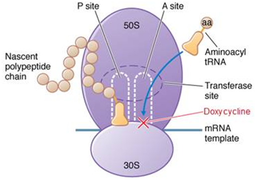
Figure 1 Mechanism of action of doxycycline: once inside the cell doxycycline binds to the A site on the 30S subunit of the microbial ribosome. During protein synthesis, a new tRNA with an amino acid attempts to bind to the A site of the ribosome, but cannot due to the site being blocked by doxycycline. Without the attachment of the aminoacyl tRNA to the A site protein synthesis is inhibited and cell death follows.
Source: National Information Program on Antibiotics.
Erythromycin is a macrolide antibiotic with a half–life of 0.8–3 hours. Erythromycin penetrates the bacterial cell membrane and inhibits bacterial protein synthesis by reversibly binding to the 50s subunit of the bacterial ribosome or near the P site (Figure 2). Binding to this site inhibits peptidyl transferase activity and interferes with translocation of amino acids and protein assembly. One disadvantage of erythromycin is that it is only effective against actively dividing organisms.30 Therefore, erythromycin alone will not be effective against the dormant stage of B. burgdorferi, but may be useful when combined with other antimicrobials. Although macrolide antibiotics are not recommended as first–line therapy for early Lyme disease, B. burgdorferi has been shown to be susceptible to erythromycin in in–vitro studies.28,29,31,32
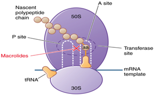
Figure 2 Mechanism of action of erythromycin: erythromycin penetrated the cell membrane and reversibly binds to the 50S subunit of the bacterial ribosome near the P site so than binding of tRNA to this site is blocked. Translocation of peptides from the A site to the P site is prevented, and subsequent protein synthesis is inhibited.
Source: quizlet.com.
Tinidazole is a nitroimidazole with a half–life of 12–14 hours.33 After oral administration, tinidazole is rapidly and completely absorbed.34. It is then distributed into virtually all tissues and body fluids, and is also capable of crossing the blood–brain barrier.34 Tinidazole impairs bacterial enzymes and destabilizes bacterial DNA, but the exact mechanism against B. burgdorferi is unknown. When used to treat Trichomonas, tinidazole is reduced, and the free nitro radical produced as a result of this reduction is believed to be responsible for antiprotozoal activity. Chemically reduced tinidazole has been shown to release nitrites and cause damage to purified bacterial DNA in vitro.34 Previous studies have demonstrated that tinidazole can be affective against mobile spirochetes at higher concentrations, but the main purpose for choosing the antibiotic for this study was the evidence that tinidazole significantly reduces the number of cystic forms of B. burgdorferi.8,35
Antimicrobial peptides
Cecropins are a group of antimicrobial peptides that act on the cell membrane of bacterial cells.36–40 Most of the antimicrobial peptides (AMPs) in this group have been isolated from insects, but cecropin P1, isolated from a pig intestine, is the only cecropin isolated from a mammal.36 Cecropin P1 consists of 31 amino acids, lacks hemolytic activity, and is not lethal to sheep red blood cells, human erythrocytes, or normal lymphocytes. This makes it a prime candidate for antimicrobial susceptibility studies and antimicrobial therapy.37 Gazit et al.36 determined the secondary structure, mechanism of membrane permeation, and orientation within the phospholipid membrane of cecropin P1 using attenuated total reflectance Fourier–transform infrared (ATR–FTIR) spectroscopy and molecular dynamics simulations. Cecropins are linear, cationic peptides that exhibit random coil structure in solution, but found to form α–helical structures consisting of an amphipathic N–terminal helix and a hydrophobic C–terminal helix upon interaction with a cell surface.36,37,40 (Figure 3A). Gazit et al.36 suggested that cecropin P1 exerts its antimicrobial activity by a “carpet–like” mechanism including preferential binding of the peptide monomer to negatively charged phospholipids in the membrane, alignment of amphipathic α–helical monomers on the surface of the membrane, rotation of the peptide leading to reorientation of the membrane, and finally disintegration of the membrane by disruption of lipid packing within the bilayer (Figure 3B). Currently no studies have been published on the susceptibility of Borrelia burgdorferi to cecropin P1, but a study was published that explored the effect of cecropin P1 on Escherichia coli. E. coli D21 exhibited instantaneous lysis and killing when cecropin P1 was added to growing cells.40 E. coli and B. burgdorferi have some similar cell wall components that may allow the peptide to exhibit similar activity on both species. Based on previous studies regarding cecropin P1’s mechanism of action, it was hypothesized that cecropin P1 would disrupt the membrane of not only the motile spirochete form of B. burgdorferi, allowing easier access for antibiotics, but that it would also act on the cyst form which is not easily treated with antibiotics.
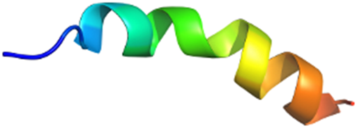
Figure 3a Example of α-helical antimicrobial peptide.
Source: Structural Classification of Proteins.
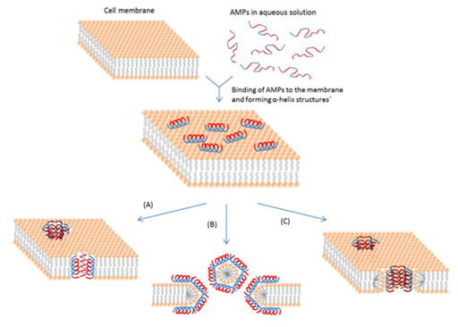
Figure 3b Mechanisms of membrane pore formation by antimicrobial peptides.
(A) demonstrates the barrel-stave mechanism: peptides associate to form a bundle of α-helices embedded in the membrane to form a membrane pore.
(B) demonstrates the “carpet” mechanism: peptides bind to the membrane surface until a threshold concentration is reached. α-helices associated with the lipids insert into the bilayer and rotate leading to reorientation and finally disintegration of the membrane.
(C) demonstrates the toroidal pore mechanism: peptide helices insert into the membrane and induce the lipid monolayers to bend continuously through the pore so that both the inserted peptides and the lipid head groups line the core.
Source: Bahar A, Ren D. Antimicrobial Peptides. Pharmaceuticals. 2013; 6(12):1543-1575. doi: 10.3390/ph6121543
Magainins are a class of AMPs first discovered in the skin of the African clawed frog Xenopus laevis.41,42 Magainins consist of 23 amino acids, are water soluble and non–hemolytic (at low concentrations), and do not exhibit significant toxicity to mammalian cells.41–45 This selective toxicity is believed to be due to the preferential interactions of magainins with anionic phospholipids, which are abundant in bacterial membranes.43 Magainins are similar to cecropins in that they form an amphipathic α–helix when associated with membranes and kill bacteria by increasing membrane permeability.45 The magainin used in this study was magainin 1. One of the only differences between magainin and cecropin P1 is the mechanism by which the peptide disrupts the membrane. Instead of the “carpet–like” mechanism of disruption seen with cecropin P1, magainin 1 has demonstrated toroidal pore formation within the membrane.44,45 (Figure 3B). At low concentrations, magainin 1 orients itself parallel to the membrane surface similar to cecropin P1, however at higher concentrations the peptide is driven into an inserted state within the membrane.45 In toroidal pore formation, the peptide helices insert into the membrane and induce the lipid monolayers to bend continuously through the pore so that the core is lined by both the inserted peptides and the lipid head groups. The lipids in these openings then tilt and connect the two leaflets of membrane forming a continuous bend from top to bottom.46 Although cecropin P1 and magainin 1 are similar in multiple ways, the slight differences in membrane permeability may make B. burgdorferi more susceptible to one antimicrobial peptide over the other.
LL–37 is a member of the antimicrobial peptide class cathelcidins. Similar to the other peptides in this study, LL–37 is an amphipathic α–helical cationic peptide that kills pathogens by forming pores in membranes.47,48 LL–37 is a 37 amino acid peptide, and is the only peptide in this study to be isolated in humans. LL–37 is produced in bone marrow and is present in epithelial cells of the testis, skin, the gastrointestinal tract, the respiratory tract, and in leukocytes such as monocytes, neutrophils, T cells, and B cells.48–50 It has been found to have defensive roles such as regulating the inflammatory response; chemo–attracting cells of the adaptive immune system to wound or infection sites, binding and neutralizing lipopolysaccharides, and promoting wound closure.48,49 The exact mechanism by which LL–37 destabilizes the membrane is unknown, but studies have ruled out the barrel–stave mechanism, leaving either the carpet model or toroidal model (Figure 3B). Sambri et al.50 presented a comparison of LL–37 and four other cathelicidin–derived AMPs against three pathogenic species of spirochetes including Borrelia. LL–37 was less active than the other cathelicidin–derived AMPs. Although LL–37 has lower activity against spirochetes than both cecropin and magainin, it would not induce an immune response if added to current treatment regimens due to its origin in the human body. There is currently no data on the immunogenicity of cecropin or magainin, but the fact that these peptides are produced in non–human mammals increases the chances of an immune reaction if they were to be administered as treatment in vivo.
Culturing B. burgdorferi
Borrelia burgdorferi N40 D10/E9 was isolated from a human and obtained from Tufts University, Medford, MA. B. burgdorferi was cultured in Barbour–Stoner–Kelley H (BSK–H) media (Sigma–Aldrich, St. Louis, MO, #B3528) modified with the addition of 6% rabbit serum (Pel–Freeze Biologicals, Rogers, AR, #31126–5). The cultures were stored in a sterile 15ml glass tube without antibiotics and incubated at 34 °C with 5% CO2.
Preparation of antimicrobial agents
Doxycycline (Fisher Scientific, Pittsburgh, PA, #NC0424034) was prepared at a concentration of 10mg/ml in sterile water. A 1:10 dilution of the stock solution was made in sterile water to prepare a 1mg/ml working stock. Erythromycin (Fisher Scientific, Pittsburgh, PA, #BP920–25) was prepared at a concentration of 10mg/ml in dimethylsulfoxide (DMSO). A 1:10 dilution of the stock solution was made in sterile water to prepare a 1mg/ml working stock. Tinidazole (Sigma–Aldrich, St. Louis, MO, #32553) was reconstituted in 60% acetonitrile/40% sterile water according to manufacturer’s specifications to produce a 10mg/ml stock. A 1:10 dilution of the stock solution was made in sterile water to produce a working stock with a final concentration of 1mg/ml. Cecropin P1 (American Peptide Company, Sunnyvale, CA, #87–9–61B) was reconstituted in sterile water to produce a 1mg/ml working stock. Magainin 1 (Sigma–Aldrich, St. Louis, MO, #M7152) was reconstituted in sterile water to produce a 0.5mg/ml working stock. LL–37 (AnaSpec, Fremont, CA, #AS–61302) was reconstituted in sterile water to produce a 1mg/ml working stock.
In vitro testing of antimicrobial agents
Experiment 1– Doxycycline, Erythromycin, and Cecropin P1: Doxycycline, erythromycin, and cecropin P1 were added to a set of 2 ml culture tubes containing spirochetes at a concentration of 1 x 10⁵ cells/ml, loosely capped, and incubated at 34 °C with 5% CO2. Concentrations of doxycycline, erythromycin, and cecropin P1 were chosen based on previously published minimum inhibitory concentrations (MICs) and minimum bactericidal concentrations (MBCs),8,32,51,52 (Table 1). Table 2 summarizes the different treatments.
|
Antimicrobial |
MIC |
MBC |
|
Doxycycline |
0.4µg/ml |
25µg/ml |
|
Erythromycin |
10µg/ml |
>500µg/ml |
|
Cecropin P1 |
3µM |
Undetermined |
Table 1 MIC and MBC from published data
Note: MIC/MBC for doxycycline was determined by microdilution method.8 MIC/MBC for erythromycin was determined by turbidity, change in the color of the medium, and microscopy.32 MIC for cecropin P1 was determined by growth inhibition assays performed with E. coli.52
|
Tube # |
Treatment |
|
1 |
No Treatment (Control) |
|
2 |
3µM Cecropin P1 |
|
3 |
16µg/ml Doxycycline |
|
4 |
64µg/ml Doxycycline |
|
5 |
128µg/ml Doxycycline |
|
6 |
16µg/ml Erythromycin |
|
7 |
64µg/ml Erythromycin |
|
8 |
128µg/ml Erythromycin |
|
9 |
3µM Cecropin P1 + 16µg/ml Doxycycline |
|
10 |
3µM Cecropin P1 + 64µg/ml Doxycycline |
|
11 |
3µM Cecropin P1 + 128µg/ml Doxycycline |
|
12 |
3µM Cecropin P1 + 16µg/ml Erythromycin |
|
13 |
3µM Cecropin P1 + 64µg/ml Erythromycin |
|
14 |
3µM Cecropin P1 + 128µg/ml Erythromycin |
Table 2 Experiment 1 Treatments
After 24 hours, the culture tubes were removed from the incubator, and the spirochete concentration was determined by directly counting the spirochetes using a bacterial counting chamber (Hausser Scientific, Horsham, PA, #3900) on an Olympus BX40 under dark field microscopy. The culture tubes were then returned to the incubator and kept for 2 weeks at 34 °C with 5% CO2. After the 2–week incubation period the culture tubes were again counted and the spirochete concentration was calculated to determine the effects of the treatments. These specific time points were chosen due to the half–life of the antimicrobials tested.
Experiment 2– Erythromycin and Cecropin P1: Overall, erythromycin demonstrated greater activity than doxycycline against B. burgdorferi (see Results). Erythromycin combined with 3μM cecropin P1 had a greater effect than any other treatment after 24 hours, but after 2 weeks the combination therapy showed little difference compared to erythromycin alone. Due to this finding, and the fact that the established MIC for cecropin P1 was determined using E. coli, it was decided to test different concentrations of cecropin P1 in order to find the optimal concentration for use with B. burgdorferi. The treatments for experiment 2 are summarized in Table 3. Experiment 2 followed the same procedure as experiment 1 with a few exceptions: the starting concentration of spirochetes was increased to 5 x 10⁵ cells/ml, treatments were performed in triplicate, and both the spirochete and cyst morphology were counted at each time point.
|
Tube # |
Treatment |
|
1(A-C) |
No treatment (Control) |
|
2(A-C) |
128µg/ml Erythromycin |
|
3(A-C) |
1µM Cecropin P1 |
|
4(A-C) |
3µM Cecropin P1 |
|
5(A-C) |
6µM Cecropin P1 |
|
6(A-C) |
9µM Cecropin P1 |
|
7(A-C) |
128µg/ml Erythromycin + 1µM Cecropin P1 |
|
8(A-C) |
128µg/ml Erythromycin + 3µM Cecropin P1 |
|
9(A-C) |
128µg/ml Erythromycin + 6µM Cecropin P1 |
|
10(A-C) |
128µg/ml Erythromycin + 9µM Cecropin P1 |
Table 3 Experiment 2 Treatments
Experiment 3– Tinidazole, Magainin 1, and LL–37: Tinidazole, magainin 1, and LL–37 were added to sets of 2ml culture tubes containing spirochetes at a concentration of 5 x 10⁵ cells/ml, loosely capped, and incubated at 34 °C with 5% CO2. The concentration of tinidazole was chosen based on previously published data which stated that the MBC of tinidazole against motile spirochetes was >128µg/ml.35 The concentrations of the two peptides, magainin 1 and LL–37, were based on the cecropin P1 data gathered in experiments 1 and 2. Experiment 3 treatments are summarized in Table 4. The culture tubes were removed from the incubator after 24 hours, and bacterial concentrations of both spirochetes and cysts were determined as previously described. The same tubes were then incubated for 2 weeks at 34 °C with 5% CO2. After the 2–week incubation period the culture tubes were again evaluated for growth, or inhibition of growth, of spirochete and cyst forms.
|
Tube # |
Treatment |
|
1(A-C) |
No treatment (Control) |
|
2(A-C) |
128µg/ml Tinidazole |
|
3(A-C) |
3µM Magainin 1 |
|
4(A-C) |
6µM Magainin 1 |
|
5(A-C) |
3µM LL-37 |
|
6(A-C) |
6µM LL-37 |
|
7(A-C) |
128µg/ml Tinidazole + 3µM Magainin 1 |
|
8(A-C) |
128µg/ml Tinidazole + 6µM Magainin 1 |
|
9(A-C) |
128µg/ml Tinidazole + 3µM LL-37 |
|
10(A-C) |
128µg/ml Tinidazole + 6µM LL-37 |
Table 4 Experiment 3 Treatments
Experiment 4– Tinidazole at high concentrations: Although the authors were looking for synergistic effects at therapeutically viable antimicrobial concentrations, the academic question arose concerning higher concentrations of tinidazole and the effect it may have on spirochetes and cysts. Concentrations of 250 and 500μg.ml tinidazole alone were used and some effect was seen at 500μg/ml. Even so, this concentration is unlikely to be achievable in the serum (Table 5).
|
Tube # |
Treatment |
|
1(A-C) |
No Treatment (Control) |
|
2(A-C) |
250µg/ml Tinidazole |
|
3(A-C) |
500µg/ml Tinidazole |
Table 5 Experiment 4 Treatments
Statistical analysis used in the studies
In the evaluation for statistical significance, the mean of raw triplicate values per treatment was compared to the mean of triplicate counts of the growth control in that run using the Student’s T in a 2–tailed evaluation. For determination of synergy, the mean of the combined therapy raw values was compared to the mean of the antimicrobial alone using Student’s T.
To evaluate in vitro antimicrobial sensitivity of spirochetes and cyst, and in vitro persistence of spirochetes and cyst morphologies, B. burgdorferi was incubated for 24 hours and 2 weeks after treatment with different antimicrobials at varying concentrations surrounding previously published MIC and MBC values. Susceptibility was evaluated using direct cell counting and dark field microscopy.
Results of Experiment 1: Doxycycline, Erythromycin, and Cecropin P1
The results of experiment 1 are summarized in Table 6. Growth occurred in every tube after 24 hours, but most tubes that were given antimicrobial treatments had less growth than the tube with no treatment. The greatest effect on spirochete growth was observed with the combination treatment of 3µM cecropin P1 and 128µg/ml erythromycin. The combination treatment had less spirochete growth than the control and less growth than the culture tubes containing 3µM cecropin P1 or 128µg/ml erythromycin alone (Figure 4).
|
Treatment |
Spirochete Concentration after 24 hours in cells/ml |
Spirochete Concentration after 2 weeks in cells/ml |
|
No Treatment |
1.00 x 10⁶ |
1.28 x 10⁸ |
|
3µM Cecropin P1 |
5.00 x 10⁵ |
1.03 x 10⁸ |
|
16µg/ml Doxycycline |
1.00 x 10⁶ |
4.00 x 10⁶ |
|
64µg/ml Doxycycline |
5.00 x 10⁵ |
1.00 x 10⁶ |
|
128µg/ml Doxycycline |
5.00 x 10⁵ |
2.00 x 10⁶ |
|
16µg/ml Erythromycin |
5.00 x 10⁵ |
5.00 x 10⁵ |
|
64µg/ml Erythromycin |
7.50 x 10⁵ |
2.50 x 10⁵ |
|
128µg/ml Erythromycin |
5.00 x 10⁵ |
7.50 x 10⁵ |
|
3µM Cecropin P1 + 16µg/ml Doxycycline |
5.00 x 10⁵ |
7.50 x 10⁵ |
|
3µM Cecropin P1 + 64µg/ml Doxycycline |
5.00 x 10⁵ |
5.00 x 10⁵ |
|
3µM Cecropin P1 + 128µg/ml Doxycycline |
5.00 x 10⁵ |
2.50 x 10⁵ |
|
3µM Cecropin P1 + 16µg/ml Erythromycin |
5.00 x 10⁵ |
5.00 x 10⁵ |
|
3µM Cecropin P1 + 64µg/ml Erythromycin |
5.00 x 10⁵ |
2.50 x 10⁵ |
|
3µM Cecropin P1 + 128µg/ml Erythromycin |
2.50 x 10⁵ |
2.50 x 10⁵ |
Table 6 Results of Experiment 1
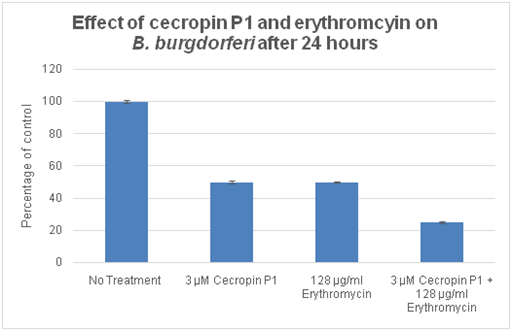
Figure 4 Comparison of the susceptibility of the spirochete B. burgdorferi to the antimicrobial peptide cecropin P1 and erythromycin alone and in combination after 24 hours.
After two weeks, half of the tubes exhibited additional spirochete growth. Of the other half of the tubes, four exhibited no additional growth after two weeks, and three had a decrease in spirochete concentration (Figure 5). All tested concentrations of doxycycline exhibited continual spirochete growth throughout the incubations, but the data shows that the antibiotic had some effect on B. burgdorferi. The lowest concentration of spirochetes in tubes treated with doxycycline was observed at a concentration of 64µg/ml. Erythromycin had a significantly greater inhibitory effect on spirochete growth than doxycycline at all tested concentrations (Figure 6A). The greatest inhibitory effect was seen in the tube treated with 64µg/ml erythromycin. Erythromycin combination therapies had a greater effect on spirochete concentration than combination therapies using doxycycline (Figure 6B). An interesting observation was that cecropin P1 alone had little effect on the spirochete concentration after two weeks, but combination therapies generally had greater effects on spirochete concentration than either cecropin P1 or the antibiotic alone suggesting a possible synergistic effect (Figure 6C).
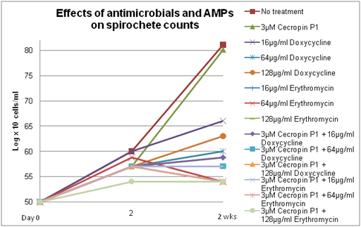
Figure 5 Comparison of spirochete concentration, in log x 10 cells/ml, of B. burgdorferi over a period of 2 weeks when treated with a variety of antimicrobial therapies.
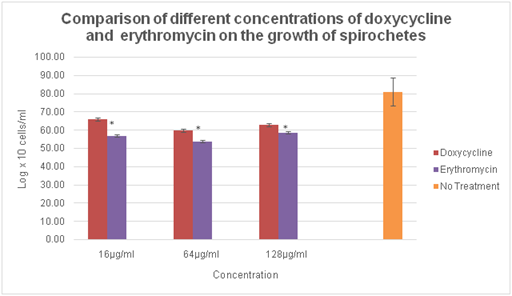
Figure 6a Comparison of the spirochete concentration, in log x 10 cells/ml, of B. burgdorferi 2 weeks after treatment with different concentrations of doxycycline and erythromycin.
Note: *P values <0.05 indicates statistical significance compared with equivalent concentration of doxycycline

Figure 6b Comparison of the spirochete concentration, in log x 10 cells/ml, of B. burgdorferi 2 weeks after treatment with different concentrations of doxycycline and erythromycin in combination with cecropin P1.
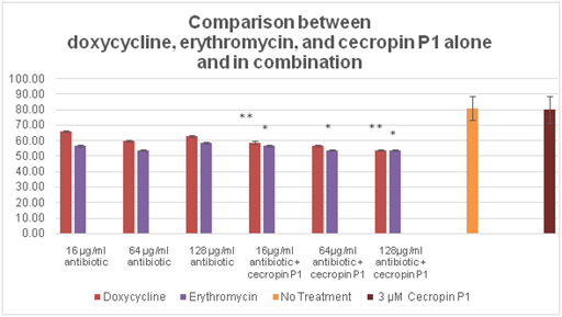
Figure 6c Comparison of the spirochete concentration, in log x 10 cells/ml, of B. burgdorferi 2 weeks after treatments consisting of different concentrations of doxycycline and erythromycin alone, cecropin P1 alone, and cecropin P1 in combination with concentrations of doxycycline and erythromycin.
Note: *P values <0.05 indicates statistical significance compared to cecropin P1
**P values <0.05 indicates statistical significance compared to antibiotic and cecropin P1 individual treatments.
Results of Experiment 2: Erythromycin and Cecropin P1
Results of experiment 2 are summarized in Table 7. After 24 hours, the spirochete concentration in every tube increased, however multiple effects on the amount of spirochete growth were observed. The greatest effect on spirochete growth after 24 hours was seen with the combination treatment of 3µM cecropin P1 and 128µg/ml erythromycin. This experiment confirmed the results of experiment 1 that the combination therapy was more successful inhibiting bacterial growth after 24 hours than either cecropin P1 or erythromycin alone at these concentrations. The other combination treatments had less of an effect on spirochete growth than the equivalent treatments of cecropin P1 and erythromycin alone after 24 hours. The cyst morphology was also counted in experiment 2. After 24 hours, the lowest concentrations of cysts were seen in the tubes treated with 128µg/ml erythromycin and 1µM cecropin P1 alone; all other treatments had similar cyst concentrations (Figure 7).
|
Treatment |
Avg. spirochetes/ml after 24 hrs. |
Avg. cysts/ml after 24 hrs. |
Avg. spirochetes/ml after 2 wks. |
Avg. cysts/ml after 2 wks. |
|
No Treatment |
1.25 X 10⁶ |
1.48 X 10⁷ |
6.56 X 10⁷ |
1.02 X 10⁷ |
|
128µg/ml Erythromycin |
6.67 X 10⁵ |
7.58 X 10⁶ |
2.50 X 10⁵ |
5.25 X 10⁶ |
|
1µM cecropin P1 |
9.17 X 10⁵ |
6.50 X 10⁶ |
7.28 X 10⁷ |
6.92 X 10⁶ |
|
3µM cecropin P1 |
1.00 X 10⁶ |
1.13 X 10⁷ |
7.01 X 10⁷ |
1.04 X 10⁷ |
|
6µM cecropin P1 |
1.00 X 10⁶ |
1.44 X 10⁷ |
7.52 X 10⁷ |
8.92 X 10⁶ |
|
9µM cecropin P1 |
8.33 X 10⁵ |
1.28 X 10⁷ |
8.62 X 10⁷ |
1.30 X 10⁷ |
|
128µg/ml erythromycin + 1µM cecropin P1 |
8.33 X 10⁵ |
1.06 X 10⁷ |
3.33 X 10⁵ |
5.42 X 10⁶ |
|
128µg/ml erythromycin + 3µM cecropin P1 |
5.83 X 10⁵ |
1.07 X 10⁷ |
1.67 X 10⁵ |
4.67 X 10⁶ |
|
128µg/ml erythromycin + 6µM cecropin P1 |
1.17 X 10⁶ |
1.61 X 10⁷ |
8.33 X 10⁴ |
4.42 X 10⁶ |
|
128µg/ml erythromycin + 9µM cecropin P1 |
1.00 X 10⁶ |
1.63 X 10⁷ |
1.67 X 10⁵ |
4.00 X 10⁶ |
Table 7 Results of Experiment 2 – Erythromycin and Cecropin P1
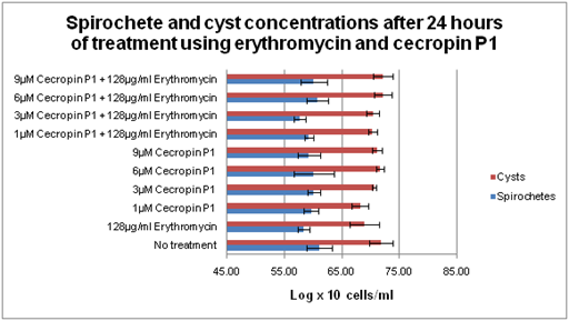
Figure 7 Spirochete and cyst concentrations, in log x 10 cells/ml, of B. burgdorferi after 24 hours of treatment.
After 2 weeks, all tubes containing erythromycin had a significant decrease in spirochete concentration (p<0.01). Tubes containing individual doses of cecropin P1 had increased spirochete counts at the 2–week time point. Tubes containing combination treatments had lower spirochete counts than the erythromycin treatment alone with the exception of erythromycin plus 1µM cecropin P1. (Figure 8). A statistically significant synergistic effect was seen when 6µM cecropin P1 was combined with 128µg/ml erythromycin (p≤0.05). This treatment was the most effective treatment against spirochetes overall, and significantly reduced spirochete growth compared to the control (p<0.01).
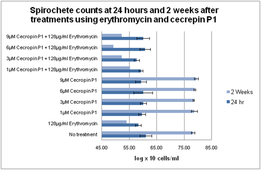
Figure 8 Comparison of spirochete concentrations, in log x 10 cells/ml, of B. burgdoferi at 24 hours, and 2 weeks after being treated with erythromycin alone and different concentrations of cecropin P1 alone and in combination with erythromycin.
Most tubes displayed a decrease in cyst concentration at the 24–hour time point with the exception of 1µM cecropin P1 and 9µM cecropin P1. It is of note that the cyst concentration in tubes treated with 1µM cecropin P1 was still lower than the cyst concentration in the control. This was not true for 9µM cecropin P1. Erythromycin/cecropin combination treatments had significantly lower cyst concentrations than cecropin P1 alone at concentrations of 6µM and 9µM (P<0.01). These combination treatments also had lower cyst concentrations than erythromycin alone, but this observation was not statistically significant. The greatest decrease in cyst concentration was seen with the combination treatments involving higher concentrations of cecropin P1 (Figure 9). Erythromycin alone reduced cyst concentration by ~49% while 6µM cecropin P1 alone reduced cyst concentration by ~13%. However, when the two therapies were combined they reduced cyst concentration by ~57%.
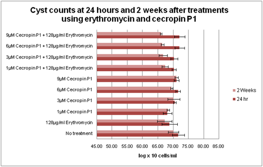
Figure 9 Comparison of cyst concentrations, in log x 10 cells/ml, of B. burgdorferi 24 hours and 2 weeks after treatment with erythromycin and different concentrations of cecropin P1 alone and in combination.
When comparing the spirochete and cyst concentrations in the combination treatments it became apparent that there was a concentration–dependency of cecropin P1 (Figure 10). As the concentration of cecropin increases the spirochete concentration decreases until a threshold is reached at 6µM cecropin P1. If a concentration higher than 6µM cecropin P1 is used, it is no longer as effective against spirochete growth. A similar trend is seen when comparing cyst concentrations. As the concentration of cecropin P1 increases the cyst concentration decreases. However, no threshold is observed in this experiment that may suggest that either a threshold does not exist against cysts or that it exists at a concentration greater than 9µM cecropin P1.
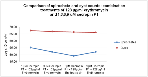
Figure 10 Comparison of spirochete and cyst concentrations, in log x 10 cells/ml, of B. burgdorferi treated with therapies involving 128µg/ml erythromycin combined with various concentrations of cecropin P1.
Results of experiment 3: treatment with tinidazole plus magainin and tinidazole plus LL–37
The results of experiment 3 are summarized in Table 8. Unlike the treatments in experiments 1 and 2, a couple of the treatments in experiment 3 did not exhibit an increase in spirochete concentration after 24 hours. The combination of 3µM magainin 1 and 128µg/ml tinidazole showed no change in spirochete concentration after 24 hours. Another combination therapy consisting of 3µM LL–37 and 128µg/ml tinidazole showed a decrease in spirochete concentration after only 24 hours (Figure 11). The 24–hour cyst concentration in tubes treated with 3µM magainin–1 and 128µg/ml tinidazole was similar to the concentrations in the other tubes. However, the 24 hour cyst concentration in tubes treated with 3µM LL–37 and 128µg/ml tinidazole was the highest of all treatments. A comparison of the spirochete and cyst concentrations shows that combination treatments involving 3µM AMP+128µg/ml tinidazole resulted in a decrease in spirochete concentration combined with a high concentration of cysts. This trend is also seen in tubes treated with 128µg/ml tinidazole and 3µM magainin –1. The trend may suggest that these therapies cause the spirochetes to transform into cysts within the first 24 hours of treatment (Figure 12).
|
Tube # |
Treatment |
Avg. spirochetes/ml after 24 hrs. |
Avg. cysts/ml after 24 hrs. |
Avg. spirochetes/ml after 2 weeks |
Avg. cysts/ml after 2 weeks |
|
1 (A-C) |
No treatment |
9.17 x 10⁵ |
1.55 x 10⁷ |
6.35 x 10⁷ |
1.33 x 10⁷ |
|
2 (A-C) |
128µg/ml tinidazole |
5.83 x 10⁵ |
1.58 x 10⁷ |
6.06 x 10⁷ |
1.28 x 10⁷ |
|
3 (A-C) |
3µM magainin 1 |
5.83 x 10⁵ |
1.53 x 10⁷ |
3.83 x 10⁷ |
1.33 x 10⁷ |
|
4 (A-C) |
6µM magainin 1 |
7.50 x 10⁵ |
1.68 x 10⁷ |
5.31 x 10⁷ |
1.48 x 10⁷ |
|
5 (A-C) |
3µM LL-37 |
1.00 x 10⁶ |
1.66 x 10⁷ |
4.46 x 10⁷ |
1.03 x 10⁷ |
|
6 (A-C) |
6µM LL-37 |
1.50 x 10⁶ |
1.84 x 10⁷ |
8.21 x 10⁷ |
9.58 x 10⁶ |
|
7 (A-C) |
128µg/ml tinidazole + 3µM magainin 1 |
5.00 x 10⁵ |
1.63 x 10⁷ |
6.12 x 10⁷ |
1.44 x 10⁷ |
|
8 (A-C) |
128µg/ml tinidazole + 6µM magainin 1 |
1.08 x 10⁶ |
1.94 x 10⁷ |
5.13 x 10⁷ |
1.40 x 10⁷ |
|
9 (A-C) |
128µg/ml + 3µM LL-37 |
3.33 x 10⁵ |
2.06 x 10⁷ |
5.78 x 10⁷ |
9.83 x 10⁶ |
|
10 (A-C) |
128µg/ml tinidazole + 6µM LL-37 |
5.83 x 10⁵ |
1.93 x 10⁷ |
5.93 x 10⁷ |
7.83 x 10⁶ |
Table 8 Results of Experiment 3: treatment with tinidazole, magainin, and LL-37
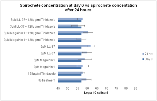
Figure 11 Comparison of spirochete concentration, in log x 10 cells/ml, of B. burgdoferi at day 0 and 24 hours after treatment with tinidazole, magainin 1, and LL-37 alone and in combination therapies.
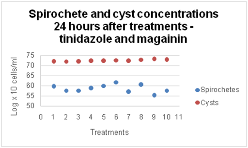
Figure 12 Comparison of spirochete and cyst concentrations, in log x 10 cells/ml, of B. burgdorferi 24 hours after treatment with tinidazole, magainin 1, and/or LL-37. Treatments are as follows: 1) No treatment, 2) 128µg/ml tinidazole, 3) 3µM magainin 1, 4) 6µM magainin 1, 5) 3µM LL-37, 6) 6µM LL-37, 7) 128µg/ml tinidazole + 3µM magainin 1, 8) 128µg/ml tinidazole + 6µM magainin, 9) 128µg/ml tinidazole + 3µM LL-37, 10) 128µg/ml tinidazole + 6µM LL-37.
After 2 weeks, the spirochete concentration increased in every tube, and the concentrations in treated tubes were very similar to the concentration in the untreated tubes (Figure 13). The greatest effect on spirochete concentration was seen with 3µM magainin –1 treatment. At the 2–week time point, this treatment had less spirochetes than the control, and displayed the least amount of growth from the 24–hour time point. These observations suggest that the treatments did not shift the spirochete morphology. However, effects on cyst concentration were detected. The cyst concentration at the 2–week time point was slightly decreased in each of the tubes compared to the 24–hour concentrations (Figure 14) but still had greater cyst concentrations than the control.
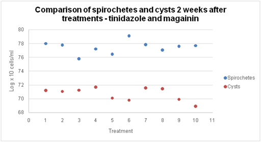
Figure 13 1) No treatment, 2) 128µg/ml tinidazole, 3) 3µM magainin 1, 4) 6µM magainin 1, 5) 3µM LL-37, 6) 6µM LL-37, 7) 128µg/ml tinidazole + 3µM magainin 1, 8) 128µg/ml tinidazole + 6µM magainin, 9) 128µg/ml tinidazole + 3µM LL-37, 10) 128µg/ml tinidazole + 6µM LL-37.
The greatest changes in cyst concentration from the 24–hour time point to the 2–week time point occurred in tubes treated with LL–37 alone and in combination with tinidazole. Cyst concentrations were slightly lower in the combination treatments compared to individual treatments (Figure 15). Statistical analysis indicated that the combination of 128µg/ml tinidazole + 6µM LL–37 demonstrated a synergistic effect against the cyst form of B. burgdorferi compared to tinidazole alone (p<0.02) (Figure 16). This treatment decreased cyst concentration compared to the control. Individual treatments of 128µg/ml tinidazole and 6µM LL–37 decreased cyst concentrations but the decreases were not statistically significant. While the combination of 128µg/ml tinidazole + 3µM LL–37 had a lower cyst concentration than the individual treatments, this observation was not statistically significant.
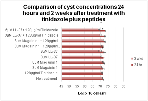
Figure 15 Comparison of cyst concentrations, in log x 10 cells/ml, of B. burgdorferi 24 hours and 2 weeks after treatment with tinidazole, magainin 1, and/or LL-37.
Note: *P values <0.05 indicates statistical significance compared to control
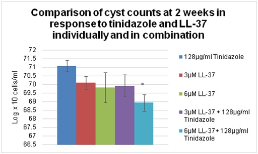
Figure 16 Comparison of cyst concentrations, in log x 10 cells/ml, of B. burgdorferi 2 weeks after treatment with tinidazole and LL-37 individually and in combination.
Note: *P values <0.05 indicates statistical significance compared to tinidazole.
A comparison of spirochete and cyst concentrations was performed using the 2–week data (Figure 17). Cysts were decreased in number and spirochetes had increased counts at two weeks. The 24–hour data (Figure 18) had the opposite showing higher cysts than spirochete counts. In order to determine if changes in concentration were due to antimicrobial treatment or transformation from spirochetes to cysts or vice versa, a comparison of rates of change of spirochete and cyst concentrations was performed (Figure 19). This comparison showed that the rate of spirochete growth exceeded the rate of cyst decrease. This implies that changes in spirochete and cyst concentrations may not solely be due to changes in morphological types.
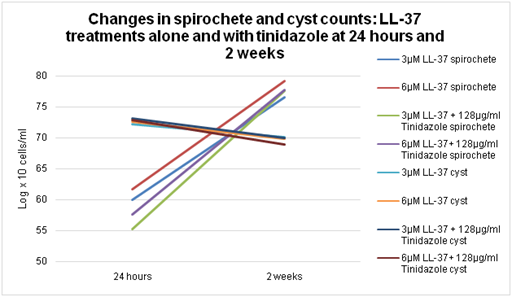
Figure 19 Changes in spirochete and cyst concentrations of B. burgdorferi 24 hours and 2 weeks after LL-37 treatments alone and in combination with tinidazole.
Experiment 4 Results: Treatment with high levels of tinidazole
Results of experiment 4 are summarized in Table 9. After 24 hours, all tubes exhibited spirochete growth from day 0, but tubes treated with tinidazole demonstrated an inhibitory effect on the growth of spirochetes after 24 hours as evidence by lower spirochete concentrations than the control. Cyst concentrations were exactly the opposite after 24 hours; both treated tubes had higher cyst concentrations than the control. The lower spirochete concentrations and increased cyst concentrations in treated tubes may again suggest a transformation of spirochetes into cysts during this 24–hour period.
|
Tube # |
Treatment |
Avg. spirochetes/ml after 24 hrs. |
Avg. cysts/ml after 24 hrs. |
Avg. spirochetes/ml after 2 wks. |
Avg. cysts/ml after 2 wks. |
|
1 (A-C) |
No treatment |
1.17 x 10⁶ |
5.00 x 10⁶ |
6.58 x 10⁷ |
9.08 x 10⁶ |
|
2 (A-C) |
250µg/ml tinidazole |
6.67 x 10⁵ |
5.92 x 10⁶ |
1.07 x 10⁸ |
7.50 x 10⁶ |
|
3 (A-C) |
500µg/ml tinidazole |
1.00 x 10⁶ |
6.33 x 10⁶ |
2.37 x 10⁷ |
4.00 x 10⁶ |
Table 9 Results of Experiment 4 – treatment with high levels of tinidazole. Spirochete and cyst counts after 24 hours and again at 2 weeks
After 2 weeks, the spirochete concentration in each tube increased. Interestingly, tubes treated with 250µg/ml tinidazole had higher concentrations of spirochetes than untreated tubes. Tubes treated with 500µg/ml tinidazole demonstrated a decrease in spirochete concentration compared to the control, but this observation was not statistically significant. Cyst concentrations were increased in both the control and 250µg/ml tinidazole treatment, although tubes containing treatment had a lower concentration of cysts than the control. A decrease in concentration was seen in tubes treated with 500µg/ml tinidazole, but again this finding was not statistically significant. Based on these results, tinidazole is slightly effective against this strain of B. burgdorferi at a concentration of 500µg/ml.
The goal of this study was to demonstrate the in vitro susceptibility of different morphological forms of B. burgdorferi to various antimicrobials in order to determine if new combinations of antimicrobial therapies could be used successfully to treat all forms of B. burgdorferi that may cause disease. B. burgdorferi has a number of mechanisms to avoid the immune response and remain undetected including immunosuppression, genetic, phase, and antigenic variation, physical seclusion, and secreted factors.4 One of the most difficult mechanisms to combat is the bacteria’s ability to vary its morphology and assume a reversible, dormant cystic formation when under unfavorable conditions such as change in temperature, pH, lack of nutrients, and antibiotic exposure.4,8 Several studies have demonstrated that B. burgdorferi can convert from the spirochete form to the cyst form in vitro when presented with an unfavorable environment, and the organisms can then revert back to the spirochete form when conditions are again favorable for growth.51–54 Although the cause of persistent and post–treatment infection is unknown, a possible explanation could be that certain antibiotics cause this conversion from spirochete to cyst. Once converted, the cyst is able to lie dormant, and possibly unaffected by the antibiotic. Then, once the antibiotic has cleared the body, the cyst can revert back to the spirochete form and continue to cause symptoms. For this reason it is necessary to develop treatments which are effective against both morphological forms of Borrelia burgdorferi.
In this study, an established method of in vitro antimicrobial susceptibility evaluation was used. This method included optimal culture conditions such as limited oxygen exposure and constant incubation at a steady temperature and CO2 level. An adequate amount of inoculum was determined by trial–and–error. If the inoculum was too low, ie 1 x 10⁵ cells/ml, bacterial counts were more difficult due to the lower number of cells. If the inoculum was too high, ie 8 x 10⁷ cells/ml, the spirochete growth at the 2–week time point was so great that the ability to get a correct count was hindered.
Doxycycline decreased spirochete growth at the optimal concentration of 64µg/ml, but the same concentration of erythromycin was significantly more effective. The antimicrobial peptide cecropin P1 had little effect on spirochete growth when used individually, but when combined with antibiotics produced greater inhibitory effects than the antibiotics alone. A statistically significant synergistic effect was seen when 6µM cecropin P1 was combined with 128µg/ml erythromycin (P<0.05). This treatment was the most effective treatment against spirochetes among all of the testing conditions (P<0.01). This treatment also significantly reduced cyst concentrations (P<0.01).
A concentration–dependency of cecropin P1 was also determined from the data. As the concentration of cecropin P1 increased, the spirochete concentration decreased until a threshold was reached at 6µM cecropin P1. At concentrations higher than 6µM, cecropin P1 was not as effective against spirochetes. This dependence was not as apparent against cyst forms. As the concentration of cecropin P1 increased, the cyst concentration decreased, but no threshold was observed suggesting that either a threshold does not exist or it exists at a concentration greater than 9µM cecropin P1. This phenomenon supports the experimentally defined activity of AMPs on the cell membrane. Because of the planar effect of the peptide initially lying on the surface in a parallel fashion and then bending into the membrane, concentration dependency plays a part in causing disruption of the membrane. Since the composition and arrangement of spirochete and cyst membranes are very different, our data further supports the concept that the membrane composition and AMP concentration are important factors in cell lysis and most be approached with detailed experimental design. Further detailed studies are important in this realm of study.
The antimicrobial peptide magainin –1 had little effect on spirochete or cyst concentrations of B. burgdorferi. The peptide LL–37 was not effective against spirochetes, but was effective against cysts. In addition, combination treatments involving tinidazole and LL–37 had slightly lower cyst concentrations than either individual treatment. A significant synergistic effect was demonstrated when 6µM LL–37 was combined with 128µg/ml tinidazole (P<0.02).
As more knowledge is gained regarding antimicrobial peptides, we will learn how feasible it may be to use them as treatment for multiple conditions including Lyme disease. Cecropin P1 demonstrated a synergistic effect on B. burgdorferi when combined with erythromycin, but it is not yet clear whether introduction of cecropin P1 into the body would cause an immune response or harmful side effects. LL–37 may be a better option because it is synthesized in humans. LL–37 was also effective against cysts when it was given as the only treatment, unlike cecropin P1. Further testing needs to be performed to determine the most efficacious combination of antimicrobial and peptide against the two forms of Borrelia.
These results demonstrate antimicrobial concentrations that are effective in vitro. Whether equivalent effectiveness can be attained in vivo still remains to be determined. Although more studies are necessary to explore the use of these antimicrobials as a treatment for Lyme disease, this study provided an introduction to new possibilities in combination therapy that may prove useful in the treatment and prevention of persistent symptoms or post–treatment Lyme disease syndrome.
The research was supported by Advanced Laboratory Services, Inc., Sharon Hill, Pennsylvania, USA. The authors would like to thank Kishore Alugupalli, Namrata Pabbati, and Akshita Datar for valuable input during the studies.
Authors declare that there is no conflict of interest.

©2016 Eckard, et al. This is an open access article distributed under the terms of the, which permits unrestricted use, distribution, and build upon your work non-commercially.