Journal of
eISSN: 2471-1381


Case Report Volume 4 Issue 2
1Department of Internal Medicine, Guilan University of Medical Sciences, Iran
2Razi Clinical Research Development Center, Guilan University of Medical Sciences, Iran
Correspondence: Manouchehr Aghajanzadeh, Inflammatory Lung Disease Research Center, Department of Internal Medicine, Razi Hospital, School of Medicine, Guilan University of Medical Sciences, Rasht, Iran, Tel 9891 1131 1711
Received: February 28, 2018 | Published: March 27, 2018
Citation: Aghajanzadeh M, Asgary MR, Hemmati H, et al. Multiple and bilaterally pulmonary hydatid cystic and liver mimicking metastatic lesion from ovarian malignancy. J Liver Res Disord Ther. 2018;4(2):76 – 78. DOI: 10.15406/jlrdt.2018.04.00103
Hydatid cyst is a parasitic infections of the humans that caused by echinococcus infection. This infection is considerable problem of public health. The most common locations in human body of a hydatid cyst are the liver (75%) and the lung tissue (15%) in the review of medical literature there was a few reports from bilaterally pulmonary hydatidosis which clinically mimics metastatic malignant tumors at initial clinical finding. Because of unusual presentation, the diagnosis with clinical presentations and CT-scan and CXR may easily be missed. For of this missed diagnosis problem, multiple and bilaterally hydatid cyst of lung should be in difrienciated diagnosis of others pulmonary diseases.
Case report: A 23 year-Old Iranian female patient presented with a six months history of fever, chest and abdominal pain, loss of appetite, weight loss, cough, night sweating and dyspnea was referred to our hospital. Chest X-Ray and Computed tomography of the chest shows multiple nodules in both lungs. Computed tomography of the abdomen and pelvic shows multiple cystic and solid lesions. Laparotomy allowed diagnosis. When the lesion of pelvic was incidentally opened, laminated membrane and daughter cysts were seen in two of pelvic lesions. The histological diagnosed also was hydatid cyst. Postoperatively, Albendazol (800mg daily) was administered for three cores of 28 days. The patient was discharged after five days post operatively with well conditions. In six months postoperatively follow up; there was regression of hydatid cysts in the lungs and others organs.
Conclusion: Multiple bilateral pulmonary hydatid cyst with cavitation and abdominopelvic mass is an extremely unusual condition and should be included in the differential diagnosis of multiple bilateral pulmonary hydatid cyst, especially in endemic areas.
Keywords: computed tomography, hydatid cyst, echinococcus infection, endobronchial lesion, albendazole, antihelminthic therapy, pulmonary infection,
Echnoccouse disease is a health problem in some countries like Iran, where it is endemic in Iran.1,2 Hydatid cysts are endemic in some areas in the world.1‒3 The endemic areas for hydatid cyst include the Mediterranean region, Africa, South America, Australia, Middle East and India.2,4‒6 Hydatid cysts can be found in almost any organ of the body but the most common sites are liver (50%-77%), lung (15%-47%), spleen (0.5%-8%), and kidney (2%-4%)1,4,5,7 The others organs which involved include, muscle, pancreas, peritoneum, heart bone and brain.2‒4,7 The rare localizations of hydatidosis such as heart, thyroid, spleen, pancreas and muscles lead to atypical clinical presentation causing difficulties in establishing the diagnosis.7 Definite diagnosis is mostly based on imaging techniques: ultrasound (US), computed tomography (CT) and magnetic resonance imaging (MRI). Patients who have an extrahepatic and intraabdominalhydatid cyst present mostly with abdominal pain and discomfort.5,8‒10 Bilateral pulmonary hydatidosis accounts for 4% to 26.7% in of all cases1,3 and multiple pulmonary hydatid cysts occur in 30% of cases.3,4 Bilaterally and multiple pulmonary hydatid cysts are very rare and in review of literature we found a few reports. Bilaterally and multiple pulmonaryhydatid cysts with ovarian and liver hydatidcysts are extremely rare and these cases may missed with pulmonary metastasis or tuberculosis or others granolomatosis disease. Our case was presented with fever, cough, abdominal pain, Wight loss and nonspecific chest symptom. Computed tomography (CT) of chest andabdominopelvic show multiple bilateral nodule some of nodule had cavitation and liver and pelvic solid-cystic masses and radiologist’s report from four center, was metastasis lesions of lung from ovarian malignancy. Definitive diagnosis obtained after laparotomy and evacuation of the abdominal lesions.
A 23 year-Old Iranian female patient presented with a six months history of fever, chest and abdominal pain, loss of appetite, weight loss, cough, night sweating and dyspnea was referred to our hospital. Physical examination revealed fever (39 centigrade), blood pressure 110/60 and coarse crackles at middle and lower area of both lungs. Abdomen was soft but a budging was seen in lower abdomen near the suprapubic region. Others organ, upper and lower extremity was normal. There was bilateral multiple nodular lesions at his chest X-ray. In hospital she takes ceftriaxone 2Gr and clindamycin 600mg twice daily. US of abdomen and pelvic was obtained and showed a cystic–solid lesion in the pelvic and in the liver. Computed tomography (CT) scan of the chest, abdomen and pelvic with IV contrast was obtained and showed multiple cavitary and solid-cystic mass lesions was showed in both lungs and the size of lesions were various in diameter. The lesions were located in the central and peripheral zone of both lungs (Figure 1). The abdomen and pelvic CT–scan showed solid-cystic mass in the pelvic and liver Radiologist finding and differential diagnosis was inflammatory lesion as Wegener granuloma, septic emboli, sarcoidosis and pulmonary metastasis from ovarian malignancy (Figure 2). Two days after admission, fever dropped and general condition of the patient becomes better. All lab date was normal except (ESR=40(0–20mm/h.CRP=22(0–5mg/L), WBC=16000). Fibroptic bronchoscopy was performed. Endobronchial lesion was not observed and bronchial lavage was obtained, Pathology examination of the bronchial lavage was normal, all other biochemical tests (CEA, ACE, RF, CA-125, hydatid tests and rheumatological tests) were normal. Laparatomy was performed ,the pelvic mass was excised and mass was opened, laminated membrane of hydatid cyst was seen, the lesion of liver was aspirated and fluid was clear, the mass was opened carefully and laminated membrane was seen (Figure 3) (Figure 4). With left mini anterolateral thoracotomy at 4 thintercostal space, chest wall was opened multiple nodules was palpable the big one was resected as wedge resection, the specimen was opened, laminated membrane was exposed (Figure 5). Chest tube was inserted and chest wall was closed in layers. Second post operative day, Albendazole was started at a dose of 10mg/kg/day for three cycle of 28days with 14days interval. Pathologist’s repot was hydatid cyst of lung in all three specimens (Figure 5). Patient was discharged in good condition 5days postoperative in the six months follow -up time there was no increased the size of both pulmonary nodules.
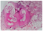
Hydatid cysts (HC) has a worldwide distribution and is a serious health problem in some area in the world such as: Mediterranean region, Asia, Newzealand, South Africa, Turkey, Greece, southern Europe and middle east.1,2,7 It is most prevalent in dog and cattle breeding countries.3,5,6 Humans may contract the infection either by direct contact with a dog or by ingestion of foods or fluids contaminated by the eggs.1,2,4 Dog is the definitive host.2,4,7 After ingestion in the GI tract, the eggs loss their coating and larvae penetrate the mucosa of the proximal portion of jejunum and after this stage the larvae enter the venous and lymphatic channels to every region of the body.2,4,7 Although any organ may involve, but liver and lungs most often affects.1,2,5,7 Liver involve in (75%) and the lungs tissue in (15%), and other organs only 10% .2,4,5,7 Clinical presentations in liver hydatid cysts depend on cyst status (intact or ruptured) and usually remain asymptomatic until the time of complication.8 Liver hydatid cyst can develop any of the complications which can be life threatening. These complications are: intrabiliary rupture and jaundice, intraperitoneal rupture and anaphylactic reaction, liver abscess, intera pleural and parenchymal rupture.1‒3 Rupture may occur during antihelminthic therapy or percutaneous aspiration and trauma can lead to severe complications. In our case, liver cyst was finding incidentally during U&S graph of abdomen. Primary pelvic hydatid cyst remains a very rare site of cyst involvement and leading to be a difficult pre-operative diagnosis.5,9,10Ovary seems to be the most frequently genital organ involved and constitutes 0.2% of different hydatid disease locations.5,9,10 Although the mechanism of primary pelvis involvement has not been clearly known, it has been suggested that larve access to the pelvis by the lymphatic or haematogenous way.9,10 Bilateral pulmonary (HC) accounts for 4% to 26.7%,1,3,4 and multiple cysts in 30% of cases.1,3,4 The hydatid cysts of organs may remain asymptomatic for a long time (Figure 6) (Figure 7).1,2,4

Figure 7 Pathology of lung and hydatid cyst, Microscopic examination shows inflamed pulmonary tissue with a hydatid cyst composed of a laminated.
During the growth , the cysts of lung may rupture in the tracheobronchial system or pleural space and patients complain of cough, expectoration of membranes, dyspnea, hemoptysis, and chest wall pain.1‒4 But in most uncomplicated cases of lung cysts are incidental finding or the patient may presents with dry cough, dyspnea, and chest pain.2‒4,8 The most common complication of pulmonary hydatid cyst is a secondary bacterial infection.2‒4,8 Infection resulting difficulty in differentiation it from an abscess or neoplasm lesions.3,4 Our case presented with fever, productive cough, dyspnea, chest pain and night sweating and this clinical presentation of our case mimicked a pulmonary infection such as tuberculosis or pulmonary abscess or pulmonary metastasis. Pelvic ultrasound has a low cost and a high sensitivity and constitutes the method of choice for pelvic and liver hydatid cysts.2,3,9 Pelvic and abdominal computed tomography allows to show the features, anatomic localization and extension of cystic masses. Pelvic magnetic resonance imaging (MRI) may be useful to recognize differential diagnosis of some tumor lesion in pelvic which including myxoid tumor such as myxoidneurofibroma and angiomyxoma(tun), Chest radiographs are useful to detect associated pulmonary hydatid cyst.9,10 In our case, pelvic hydatid cyst diagnosis was suggested by ultrasound CT-scan. In our case, hydatid serology test was not performed .The optimal treatment of pelvic hydatid cyst remains surgery. In our case we performed a diagnostic laparotomy, during exploration, we find a cystic–solid lesion of ovarian, after aspiration and evacuation, laminated membrane of hydatid cyst presented, lesion on the liver was aspirated, evacuated, hydatid cyst membrane was presented. With a mini thoracotomy a wedge resection of left lung was performed, daughter cyst and laminated membrane was presented from this lesion. The definitive diagnosis obtained from pathologist in all three specimens. Postoperatively, Albendazole was started at a dose of 10 mg/kg/day for three cycles of 28days with 14days interval. Patient was discharged in good condition 5days postoperative in the six months follow-up time there was no increased the size of both pulmonary nodules.
Multiple bilateral pulmonary hydatid cysts with cavitation and abdominopelvichydatid cysts is an extremely unusual condition and should be included in the differential diagnosis of multiple bilateral pulmonary mass or lesions and pulmonary metastasis especially in endemic areas as Iran. For definitive diagnosis VATS, laparascopy or open biopsy are choice.
All authors contributed toward data analysis, drafting and revising the paper and agree to be accountable for all aspects of the work.
There is no conflict of interest.

©2018 Aghajanzadeh, et al. This is an open access article distributed under the terms of the, which permits unrestricted use, distribution, and build upon your work non-commercially.