Journal of
eISSN: 2376-0060


Research Article Volume 3 Issue 4
1Department of Pathology, Gujarat University, India
2Department of Internal Medicine, Gujarat University, India
3Department of Orthopaedics, Gujarat University, India
Correspondence: Mitul B Modi, PG Resident, Department of Pathology, Gujarat Cancer Research Institute (GCRI), Gujarat University, Ahmedabad, Gujarat, India, Tel 919662000000
Received: June 10, 2016 | Published: June 28, 2016
Citation: Modi MB, Rathva MR, Shah NR, et al. Role of FNAC in lung carcinoma and its histo-cytological correlation. J Lung Plume Respir Res. 2016;3(4):109–112. DOI: 10.15406/jlprr.2016.03.00090
Aims and objective
Methods and material: We used Chest CT scan and Radiograph of patients for radiological data. We used H & E stain, Modiefied PAP stain and Modified Giemsa stain for slide preparation.
After cytological evaluation, We further divided positive cases into metastatic, Small cell carcinoma, Non small cell carcinoma and others category. Final diagnosis is based on HPE study, as we considered HPE study as GOLD standard. Immuno histochemistry was done in few cases where necessary.
Results: Diagnostic sensitivity and specificity of FNAC for Lung carcinoma was 91.5% and 72.5% respectively. Diagnostic Positive predictive value and Negative predictive value of FNAC for Lung carcinoma was 94.7% and 61.5% respectively.
Here we considered HPE study as GOLD standard. Accuracy of FNAC was 88.5% based on final HPE report.
Conclusion: FNAC is a simple, safe and reliable method with high diagnostic sensitivity for the diagnosis of lung cancer.
Keywords: FNAC, lung carcinoma, histo-cyto correlation
Lung cancer, also known as carcinoma of the lung or pulmonary carcinoma, is a malignant lung tumor characterized by uncontrolled cell growth in tissues of the lung. Worldwide, lung cancer is the most common cancer in terms of both incidence and mortality.
If left untreated, this growth can spread beyond the lung by process of metastasis into nearby tissue or other parts of the body. Most cancers that start in the lung, known as primary lung cancers, are carcinomas that derive from epithelial cells. The main primary types are small-cell lung carcinoma (SCLC) and non-small-cell lung carcinoma (NSCLC). Treatment and long-term outcomes depend on the type of cancer, the stage (degree of spread), and the person's overall health.
Fine needle aspiration cytology (FNAC) is an accurate and sensitive way for the diagnosis of Lung mass lesions.1,2
FNAC not only distinguishes between benign and malignant lesions but also helps in tumor typing of Lung cancer. So initiation of specific therapy like chemotherapy or surgery, is possible without delay.
The special advantage of FNAC includes detection of those tumor types like small cell carcinoma, lymphomas which are more appropriately treated by chemotherapy rather than surgery.
The study was taken in the Department of Pathology, Gujarat Cancer& Research Institute, during the period of July 2014 to November 2014.
There were consecutive cases over a period of 5 months. Retrospective study was undertaken patients having lung mass lesions suspected to be neoplastic by chest radiograph or CT scan were referred to our Institute.
A total 70 cases were included where we got adequate FNAC smear, histopathology reports and clinical history as sex, age and smoking history. Some cases were excluded, in which we did not get either FNAC or Biopsy report.
Air dried smears were stained with May-Grunwald-Giemsa (MGG) whereas alcohol fixed smear stained with modified Papanicolaou (PAP) stain for cytopathological evaluation of the lesions.
By cytological study we widely divided cases into Positive, Negative, suspicious and unsatisfactory groups. We further divided positive cases into metastatic, Small cell carcinoma, Non small cell carcinoma and others category.
Final diagnosis is based on HPE study, as we considered HPE study as GOLD standard. Immuno histochemistry was done in few cases where necessary.
IHC was performed on formalin fixed paraffin embedded tissue sections, using ABC technique with antigen epitope enhancement by heat. The Diaminibenzidine (DAB) reaction was used in final detection step. The slides were counterstained with Mayer’s hematoxyline. The method for epitope retrieval was overnight incubation at 60 C. After antigen epitope enhancement, the staining was performed by fully automated machine VENTANA BENCHMARK XT. Appropriate positive and negative controls were included in ll stains to ensure the quality and consistency of staining results.
Following set of antibodies were used wherever required:
A total 70 cases were included in the study where we got both FNAC smear and histopathology report of lung masses.
Out of 70 cases, 53 were male and 17 were female.
General demographic findings of the study and common disease pattern have been given in Tables 1–4.
Subject |
Total No |
Percentage |
Age |
||
<40 years |
04 |
5.7% |
40-49 years |
11 |
15.7% |
50-59 years |
26 |
37.2% |
60-69 years |
25 |
35.7% |
>70 years |
04 |
5.7% |
Sex |
||
Male |
53 |
75.7% |
Female |
17 |
24.3% |
Smoking |
||
Smoker |
45 |
64.2% |
Non smoker |
25 |
35.8% |
Table 1 Demographic description of the study
FNAC |
Male |
Female |
Smoker |
Non Smoker |
Positive |
41 |
11 |
37 |
15 |
Metastatic |
02 |
01 |
02 |
01 |
Small Cell Carcinoma |
03 |
00 |
01 |
02 |
Non Small Cell Carcinoma |
||||
Adenocarcinoma |
26 |
06 |
26 |
06 |
Squamous Cell Ca |
04 |
03 |
03 |
04 |
Others |
01 |
01 |
01 |
01 |
Poorly Differentiated Ca |
05 |
00 |
04 |
01 |
Suspicious |
02 |
02 |
02 |
02 |
Negative |
03 |
02 |
03 |
02 |
Unsatisfactory |
07 |
02 |
03 |
06 |
Table 2 Distribution of Lung lesions according to cytological study
Biopsy |
Male |
Female |
Smoker |
Non Smoker |
Positive |
47 |
12 |
41 |
18 |
Metastatic |
03 |
00 |
02 |
01 |
Small Cell Carcinoma |
04 |
00 |
03 |
01 |
Non Small Cell Carcinoma |
||||
Adenocarcinoma |
23 |
09 |
22 |
10 |
Squamous Cell Ca |
11 |
02 |
09 |
04 |
Others |
02 |
01 |
02 |
01 |
Poorly Differentiated Ca |
04 |
00 |
03 |
01 |
Negative |
07 |
04 |
04 |
07 |
Table 3 Distribution of Lung lesions according to histological study
Biopsy |
Positive |
Negative |
FNAC` |
||
Positive |
54 |
03 |
Negative |
05 |
08 |
Table 4 Cytological and histological correlation
Diagnostic sensitivity and specificity of FNAC for Lung carcinoma was 91.5% and 72.5%.1,2
Here we considered HPE study as GOLD standard.
Positive predictive value 94.7%, Negative predictive value 61.5%
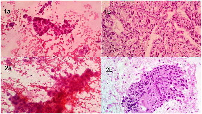
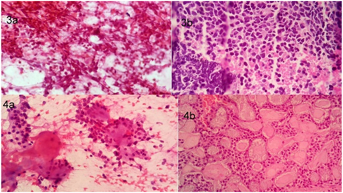
Accuracy of FNAC was 88.5% based on final HPE report Figures 1–4.

Figure 1 1a: Adeno carcinoma (PAP stain): Cells arranged in clusters & singly with eccentrically placed nuclei.
1b: Adeno carcinoma (HPE study): Malignant cells forming glandular pattern and showing intracytoplasmic mucin.
2a: Squamous cell carcinoma (PAP stain): Malignant squamous Cells arranged in clusters.
2b: Squamous cell carcinoma (HPE study): A cluster of malignant squamous cell showing cytoplasmic keratin and intercellular bridging s/o SCC.

Figure 2 3a: Small cell carcinoma (PAP stain): Small sized cells arranged in clusters with scant or no cytoplasm.
3b: Small cell carcinoma (HPE study): Section shows clusters of small cells with hyperchromasia & nuclear molding with cytoplasm.
4a: Adenoid Cystic carcinoma (PAP stain): Small round hyperchromatic cells with Prominent hyaline globules seen.
4b: Adenoid Cystic carcinoma (HPE study): Biopsy shows small round hyperchromatic cells arranged in cribriform pattern & containing hyaline basement membrane material.
FNAC is an accurate and safe method for evaluation of lung mass. It enable categorization of malignant lesion in majority of cases. All cases were adults. The peak age of incidence (50-59years) was the same as the documented in recent studies.3,4
The mean age in our study was 55.3years.4,5
The malignant cases formed largest category including Non small cell carcinoma, small cell carcinoma, metastatic carcinoma and poorly differentiated carcinoma. There was male predominance (75.7%) compared to female in our study. Among the patients (64.2%) was active smokers. Among the malignant lesion, the most common was Adenocarcinoma (47.14%) followed by squamous cell carcinoma (18.5%) and small cell carcinoma (5.71%).
The incidence of Adenocarcinoma was significantly higher than that of squamous cell carcinoma.
Adenocarcinoma, squamous cell carcinoma and small cell carcinoma can be effectively diagnosed by cytology. A high degree of accuracy in cytological typing can be great importance in these days where no confimatory histology is abailable. Maximum cases of lung malignancy were primary while 3 cases represented as metastatic carcinoma from different sites.
No major complication was found when FNAC was done in our study. In our study, FNAC showed almost perfect agreement with histological diagnosis. So FNAC was found to be highly accurate.
FNAC is an accurate and safe method for the evaluation of lung nodules and it enables sub classification of bronchogenic carcinomas in the vast majority of cases. It is also useful for the diagnosis of tuberculous pulmonary nodules.6
Hence FNAC diagnosis alone can be used with confidence to select treatment modalities and to avoid unnecessary surgeries in patients with lung malignancies. FNAC is a simple, safe and reliable method with high diagnostic sensitivity for the diagnosis of lung cancer.
FNAC is a simple, safe and reliable method with high diagnostic sensitivity for the diagnosis of lung.
We all would like to thank the Director of Gujarat University and our institutions’ for allowing us to publish this article.
The author declares no conflict of interest.

©2016 Modi, et al. This is an open access article distributed under the terms of the, which permits unrestricted use, distribution, and build upon your work non-commercially.