Journal of
eISSN: 2377-4312


Case Report Volume 12 Issue 1
1Sindh Agriculture University Tandojam, Pakistan
2PMAS-Arid Agriculture University Rawalpindi, Pakistan
Correspondence: Adnan Yousaf, Sindh Agriculture University Tandojam, Pakistan
Received: September 12, 2022 | Published: January 12, 2023
Citation: Kumar L, Mushtaque A,Yousaf A, et al. Therapeutic management of dermatitis in a female German shepherd bitch in Islamabad, Pakistan. J Dairy Vet Anim Res. 2023;12(1):1-3 DOI: 10.15406/jdvar.2023.12.00313
A female German shepherd bitch with baldness, widespread purulent lesions, hyperpigmentation, and acute itching was presented at the Ali veterinary clinic, Islamabad. The common reasons of the dermatitis problem are Mange/mite. According to history and current conditions of bitch mange/mite were ruled out as after a laboratory investigation. The condition has been identified as atopic dermatitis other bacterial and fungal infection agents also caused secondary lesion. Anti-allergic/antihistaminic drugs along with the administration of corticosteroids and nutritional supplement of omega fatty acid had noticeable marginal recovery in the bitch health.
Keywords: dermatitis, itching, diffuse purulent, canine atopic dermatitis
Canine atopic dermatitis (CAD) is a prevalent, multifaceted, pruritic skin disease with a complex etiopathogenesis. Canine atopic dermatitis (CAD) appears to be caused by a confluence of genetic and environmental variables that lead to impaired skin barrier function, immunological dysregulation, and skin microbial dysbiosis.1,2 Although immunoglobulin E (IgE) is an important component in atopic dermatitis (AD), recent studies have shown that AD is not always IgE-mediated.3 As in humans, a small subset of dogs with AD do not have detectable by either serology or intradermal testing allergen-specific IgE. This form of AD has been recently referred to as atopic-like dermatitis.4 According to Scott and Miller,5 almost 15% to 30% of dogs are thought to be affected with canine atopic dermatitis (CAD), which is typically a chronic condition. Immune dysregulation in the skin of atopic dogs leads to overproduction of pro-inflammatory and pruritogenic mediators, such as T-helper type-2 cytokines, including interleukins IL-4, IL-5, IL-10, and IL-13.4 More recently, IL-31 was shown to play a significant role in CAD, inducing inflammation and pruritus in atopic dogs via activation of Janus kinase (JK) signal transduction. This led to the development of the JK inhibitor, oclacitinib maleate (Apoquel, zoetisus.com)6 Although the precise etiology of CAD is not yet fully understood, it is believed to be caused by immediate and late-phase hypersensitivity reactions to environmental allergens that are mediated by immunoglobulin (Ig) E.7 A deficiency in their skin's natural protective barrier causes itching in atopic dermatitis-affected canines (Rebecca et al., 2021). The secondary source of infections that worsen the severity of CAD is this itching, which supports the skin microbiota.8 Staphylococcus pseudintermedius is the most prevalent bacteria on lesional skin of dogs with AD, while Malassezia pachydermatis is the primary fungus representative, according to canine microbial culture-based investigations.9 There is increasing evidence that dogs with AD have epidermal barrier abnormalities, including abnormal lipid and ceramide composition. Clinical symptoms typically start to show between the ages of 6 months and 3 years. The first sign is typically chronic itching, which is followed by "primary skin lesions" such as erythema, papules, and pustules.10–12 Alopecia, cutaneous lichenification, and frequently bacterial infections are examples of "secondary skin lesions" in chronic CAD.10,12
Aim of study
This new understanding has led to updated approaches for avoidance of allergen and microbial exposure, and development of novel therapies to restore or protect the skin barrier of atopic dogs to know the treatment and prevention measures.
Case history and observation
A female German shepherd bitch, age 3 years, who body weight 30 kg and had a history of persistent itching was presented at the Ali veterinary clinic, Islamabad-Pakistan (GPS Coordinates 33.6334773, 72.9186582) in August, 2022. The dog was raised (outdoors) and dog been in contact with different dogs. Beginning six months ago, scratching the region close to the neck and hind limb signaled the onset of pruritus.
General physical examination
The patient seemed illiterate yet was responsive. The rectal temperature was elevated (104.5°F), the respiratory rate was normal (22bpm), and the heart rate was normal (80.5bpm).
Dermatological examination
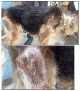
The patient periodically rubbed her ears while attempting to reach his paws and the theme-dial side of his thighs through his nose during the consultation. Both auricles exhibited no lesions upon otoscopic evaluation. The external auditory canals showed signs of light erythema. No indications of purulent otitis were present. Clinical examination of the lateral aspect of the thighs and neck revealed pyodermatitis over the skin with diffuse purulent lesions, lichenification, alopecia, and patchy hyperpigmentation. Ecto-parasites were not observed. On the rest of the body, there were no additional primary or secondary skin lesions.
Diagnosis
A diagnostic issue is identifying canine atopic dermatitis (CAD). There are no known pathognomonic symptoms or specific biomarkers. In general, the diagnosis is made to rule out other infections with comparable symptoms, such as ectoparasitic infestations.2,13 The two most common allergy tests are the Allergen-Specific IgE Serology (ASIS) test2,13 and Intra Dermal Testing (IDT), which was not done in this case (Figure 1).

Figure 1 Picture of German shepherd bitch having hyper pigmentation, alopecia and diffuse purulent lesions showing dermatitis.
Management
The prescription of topical or systemic glucocorticoids, antifungals, the implementation of a flea control program, nutritional supplementation with essential fatty acids, antibiotic treatment, and frequent shampooing comprise the symptomatic treatment for CAD.14–16 Due to the prevalence of the Staphylococcus genus in comparison to healthy controls on the skin of AD dogs, as well as the commensal yeast Malassezia pachydermatis (M. pachydermatis), which is often present on mammalian skin, and dermatitis in dogs.17,18 Therefore, to repair or reduce the detrimental effects of an excessively self-perpetuating inflammatory response, our remedies must simultaneously target multiple locations.
Treatment
The dog received treatment with intramuscular (I/M) injections of anti histaminics chlorpheniramine maleate for 5 days, Prednisolone for 5 days @ 1mg/kg of B.WT, Ketoconazole for 5 days @ 10mg/kg of B.WT, Ceftriaxone-tazobactum antibiotic for 5 days @ 25mg/kg of B.WT, Ivermectin @ 2mg/kg of B.WT once. Omega fatty acid-containing vitamin supplements were given orally for two weeks while receiving supportive care. The owner was told to maintain the body dry and free of moisture. After a week, the owner brought the dog back, who had made some minor progress. Even though there had been some improvement but not totally free of pruritus.
An allergic skin condition with genetic predisposition to inflammation and itching is called atopic dermatitis. The function of the epidermal barrier is frequently altered, which increases the risk of disease.19 The use of glucocorticoids, antihistamines, omega-6/omega-3 fatty acid supplements, topical antipruritic drugs, antibiotics, antifungal, and combinations there are among the standard therapy protocols for canine atopic dermatitis.7 Specific skin test could not be performed in this case and the specific immunotherapy was not tried. Antihistaminics were also used historically in conjunction with glucocorticoids as a synergistic way to lower glucocorticoid doses.20 Dogs with CAD frequently respond differently and inexplicably to antihistamines.5,7 In some dogs with CAD, chloropheniramine proved effective in controlling pruritus.5 In atopic dogs, a reduced diversity of the microbiome and an increase in Staphylococcus have been linked to clinical flare-ups of the condition.21 Antipruritic therapies restored biodiversity and stabilized skin barrier indices, according to longitudinal investigations in dogs with CAD.17 According to Chermprapai et al.,22 topical antibiotic therapy has also been shown to promote skin biodiversity in atopic dogs. Although the therapeutic responses were adequately represented, they were not successful in this case. In addition, Tarpatki N19 also reported that no single treatment is universally effective in treating canine Atopic Dermatitis.23,24
None.
Author declares there is no conflict of interest in publishing the article.
None.

©2023 Kumar, et al. This is an open access article distributed under the terms of the, which permits unrestricted use, distribution, and build upon your work non-commercially.