Journal of
eISSN: 2377-4312


Mini Review Volume 12 Issue 1
Clínica Veterinária Reino Animal, São Paulo, Brazil
Correspondence: Paolo Ruggero Errante, Clínica Veterinária Reino Animal, Avenida José Maria Whitaker, 1650. Planalto Paulista, São Paulo, SP, Brazil
Received: January 23, 2023 | Published: February 3, 2023
Citation: Errante PR. Extravasation injuries in the intravenous therapy with drugs with properties vesicants and irritants in the veterinary medicine of small animals. J Dairy Vet Anim Res. 2023;12(1):19-22 DOI: 10.15406/jdvar.2023.12.00317
The extravasation injuries are uncommon complications during the administration of intravenous therapy in the small animal veterinary clinic. However, the efflux of drugs with vesicant or irritant properties can cause severe and/or irreversible functional and aesthetic changes. The correct catheterization technique and vigilance during intravenous administration of drugs with vesicant or irritant properties are fundamental in preventing the appearance of iatrogenic injuries. The therapeutic approach to this iatrogenic complication involves the combination of non-pharmacological, pharmacological, and surgical measures. In small animal veterinary medicine, there are no uniform recommendations about the best strategy could be adopted, and the interventions described in the literature lack evidence of effectiveness based on scientific studies.
Keywords: extravasation injuries, vesicant drugs, irritant drugs, medicine veterinary, lesion
The extravasation injuries are produced by the efflux of drugs from the bloodstream into the perivascular spaces.1 The extravasation of intravenous therapy can occur in several clinical contexts, and the global incidence in small animal veterinary medicine is not known. The case reports focus mainly on cancer patients undergoing intravenous chemotherapy,2–4 and in a minor degree in cases of extravasation of parenteral contrast media during radiological examinations.5
All sick animals under intravenous pharmacotherapy are at risk of drug extravasation. The extravasation is predominantly associated with the use of peripheral venous catheters, being repeated catheterization and its performance by an inexperienced professional the main causes that contribute to the increased risk, as described in humans.6,7
Also in many cases, sick animals undergoing chemotherapy often have small-caliber, fragile veins that are difficult to catheterize. In obese animals, the visualization or palpation of the venous path may be difficult due to the thickening of the subcutaneous adipose tissue.8 The animals with generalized skin diseases, because the pathological changes, are also a risk population.9,10 The sick animals with sensory deficits (for example in Canine Cognitive Dysfunction Syndrome) may not complain of pain associated with extravasation, while patients with altered state of consciousness (for example coma) may not be able to manifest this symptom.11,12
The administration of chemotherapy drugs in an area previously submitted to radiotherapy can cause reactivation of local skin toxicity and, in sick animals with a history of extravasation resulting from previous pharmacological therapy, a remote inflammatory response can be induced in the older site of the extravasation lesion.3
Pathogenesis of extravasation injury
The tissue damage associated with extravasation of intravenous therapy depends on the volume and properties of the extravasated drug. These drugs can be classified according to their potential of local toxicity into irritants or vesicants.1,13,14 The irritants drugs start a local inflammatory response characterized by pain, heat, and erythema, but do not induce tissue necrosis. The inflammation is self-limiting and not associated with long-term sequelae. The vesicants drugs locally produce vesicles and bullae and can cause tissue necrosis with involvement of the skin, and, in severe extravasations, deep tissue involvement can occur.15 The cytotoxic agents are the best described vesicant drugs in the literature (Table 1), however, there are cytotoxic drugs with irritating properties (Table 2), and non-cytotoxic drugs associated with extravasation lesions.15,16
Alkylating agents |
Platinum compounds |
Bendamustine |
Cisplatin |
Carmustine |
|
Dacarbazine |
Vinca alkaloids |
Mechlorethamine |
Vinblastine |
Vincristine |
|
Anthracyclines |
Vindesine |
Daunorubicin |
Vinorelbine |
Doxorubicin |
|
Epirubicin |
Others |
Idarubicin |
Dactinomycin |
Mitomycin |
Table 1 Cytotoxic vesicant drugs associated with extravasation injuries
Alkylating agents |
Platinum compounds |
Busulfan |
Carboplatin |
Cyclophosphamide |
Cisplatin |
Dacarbazine |
Oxaliplatin |
Ifosfamide |
Taxanes |
Melphalan |
Docetaxel |
Tiotepa |
Paclitaxel |
Antimetabolites |
Others |
Cladribine |
Bleomycin |
Fluorouracil |
Etoposide |
Gemcitabine |
Irinotecan |
Ixabepilone |
|
Anthracyclines |
Mitoxantrone |
Daunorubicin (liposomal) |
Teniposide |
Doxorubicin (liposomal) |
Topotecan |
Table 2 Cytotoxic irritant drugs associated with extravasation injuries
In addition to the drug's intrinsic cytotoxicity, the severity of extravasation lesions also varies depending on its physicochemical properties such as DNA binding capacity, vasoconstrictor or vasodilator properties, pH outside the range 5.5-8.5, osmolarity higher than plasma (>290mOsmol/L), and presence of other compounds in the formulation, such as alcohol or polyethylene glycol (Table 3).13–15
Vasoactive drugs |
Drugs with pH extreme |
Dobutamine |
Acetazolamide |
Dopamine (vesicant) |
Aminophylline (vesicant) |
Epinephrine (vesicant) |
Amphotericin |
Norepinephrine (vesicant) |
Dantrolene (vesicant) |
Vasoepinephrine (vesicant) |
Phenytoin (vesicant) |
Vancomycin |
|
Hyperosmolar solutions |
|
Calcium chloride (vesicant) |
Others |
Calcium gluconate 10% (vesicant) |
Diazepam (contains propylene glycol) |
Dextrose > 10% (vesicant) |
Digoxin (vesicant) |
Mannitol > 10% |
Promethazine (vesicant) |
Potassium chloride ≥ 0.1mEq/mL |
|
Sodium bicarbonate ≥8.4% |
|
Sodium chloride ≥ 3.7% (vesicant) |
Table 3 Non-cytotoxic drugs associated with extravasation injuries
The extravasation of cytotoxic drugs can cause acute and persistent lesions, progressive or late. These characteristics are typical of DNA-binding cytotoxic drugs (for example alkylating agents and anthracyclines), that after being released from the necrotic cells into the extracellular space, are taken up by adjacent cells, initiating a new cycle of tissue destruction. The cytotoxic drugs that do not have DNA binding capacity (for example vinca alkaloids, taxanes) are rapidly metabolized and eliminated from tissues, causing lesions to disappear more quickly.1,13–15
The extravasation injuries associated with drugs with vasoconstrictor properties (for example dopamine, epinephrine) are caused by local ischemia followed by coagulative necrosis. The use of drugs with vasodilator properties can aggravate extravasation and increase the extent of associated lesions. The drugs with acidic pH (for example amphotericin and etomidate) can induce protein denaturation and coagulation necrosis. On the other hand, drugs with alkaline pH (for example phenytoin and thiopental) can cause liquefaction necrosis, with the presence of deeper lesions.14
The extravasated hyperosmolar solutions locally modify the oncotic pressure of the interstitial fluid. The increase of oncotic pressure associated with extravasation of hyperosmolar solutions can cause tissue necrosis by cellular dehydration, while the decrease in oncotic pressure by hyposmolar solutions can cause necrosis by cellular edema.15
Clinical manifestations
The extravasation of irritating drugs is usually manifested by discomfort or pain, accompanied by heat, redness, and local edema. The presence of redness and swelling around the infusion site favors the possibility of extravasation.1 Clinical manifestations associated with vesicant cytotoxic drugs extravasation injury are initially mild and like those described for irritant drugs, including localized pain, erythema, edema and pruritus.13 In dogs and cats, incessant licking of the area is common. In most cases, the onset of symptoms is immediate, but there may be a latency period of a few days to several weeks after extravasation. After 2 or 3 days, the progression of the injury can be emergence by the appearance of pain, increased erythema, discoloration, hardening, necrosis, desquamation, or skin blisters formation. 1,14,15
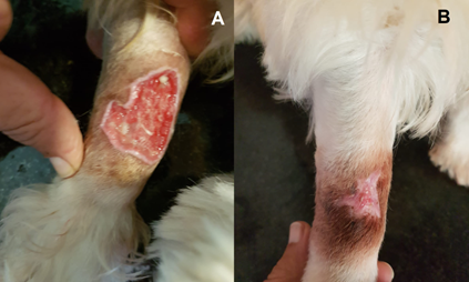
Lesions associated with extravasation may evolve over several weeks, with the formation of a dry eschar whose desquamation produces an ulcer at the site of extravasation. The ulcers formed are characterized by a yellowish necrotic central base and raised edges, erythematous, painful, and with the formation of granulation tissue (Figure 1).

Figure 1 Diazepam extravasation injury in a male dog breed West Highland White terrier. A. Deep skin ulceration due to extravasation of diazepam. B. Resolution of the lesion after 30 days of pharmacological treatment. Font: Errante, 2023.
The evolution of larger lesions is gradual and indolent expansion, and if the natural course is not altered, there may be involvement of joint capsules, tendons, nerves, and vessels, with potentially serious consequences (permanent joint stiffness, nerve compression syndromes, neurological deficits, residual sympathetic dystrophy).1
Prevention
The extravasation injuries can be prevented by installing simple measures. In pets (usually in dogs and cats), as well as in humans, preference should be given to the veins of the forearms (cephalic vein). The catheterization site should allow for easy access, secure attachment to the skin, and regular inspection. Joints and folds should be avoided, as they represent anatomical spaces containing tendons and nerves.17 The severity of extravasation lesions is greater in regions with reduced subcutaneous tissue,1 as the veins located on the anterior surface near the carpal joint, or laterally over the tarsal joint in dogs.
The peripheral venous access device, preferably a silicone catheter, must be fixed to the skin with adhesive tape, without occlusion of catheter entry site in the skin. Before starting the perfusion, check its permeability by the instillation of 5-10 mL of 0.9% NaCl solution. During administration of intravenous therapy, monitoring for the appearance of any symptoms of extravasation and regular inspection of the catheterized area should be performed.1
If an infusion pump is used, the decrease in the infusion rate can be a warning sign. After other causes of resistance to flow (change in body position, equipment tube compression) have been excluded and in the presence of characteristic clinical signs or symptoms, catheter migration and extravasation should be considered. The absence of blood return through the catheter, without other associated manifestations, does not always indicate extravasation. On the other hand, in some cases there may be blood return even after catheter migration.1,17
Treatment
The approach to the case of extravasation injury involves a combination of non-pharmacological measures to limit the injuries, neutralizing agents for the drugs involved and, when necessary, surgical intervention. The decision should be based on the properties of the drug, availability of antidotes and degree staging of the lesion. In view of the suspicion of intravenous therapy extravasation, the drug infusion should be interrupted immediately and the catheter should not be immediately removed, which should be aspirated and used to drain any subcutaneous collections.1,15–17
The intermittent topical application of heat or cold may be beneficial in extravasation of non-vesicant drugs. The vasodilation caused by heat increases local blood flow and enhances drug absorption into the systemic circulation. The vasoconstrictor action associated with cold promotes drug concentration and acceleration of tissue metabolism, in addition to reducing inflammation and producing pain relief. The topical application of cold compresses in periods of 20 minutes, 3 to 4 times a day, in the first 48-72 hours, is indicated in the extravasation of vesicant agents, with the exception of vinca-derived alkaloids (for example vinblastine, vincristine), epipodophyllotoxins (for example etoposide) and vasoconstrictor agents.1 Topical cold application should not be used concurrently with the administration of systemic antidotes, such as in the use of dexrazoxane in extravasation of anthracyclines.18
Different techniques for washing the subcutaneous tissues can be used through the percutaneous instillation and drainage of a 0.9% NaCl solution. The Gault flush-out technique initially consists of hyaluronidase infiltration in the region where the extravasation occurred. Four small peripheral incisions are made, through which 500 mL of 0.9% NaCl solution are injected consecutively into each of the incisions. A blunt catheter with side holes should be used to minimize tissue trauma. In immunosuppressed animals, prophylactic antibiotic therapy is recommended. Incisions should not be sutured and should be heal spontaneously.19
Despite several recommendations for the use of antidotes in the prevention of necrosis and ulceration in extravasation of cytotoxic and non-cytotoxic drugs, there are no controlled clinical trials that prove the full effectiveness in pets or humans. Interventions to neutralize the action of extravasated agents, as well as techniques for the physical elimination of drugs from perivascular tissues, are more effective when performed in the period between the onset of extravasation and appearance of structural changes in the tissues. In the literature based on observations in humans, this interval is defined as 4-6 hours for vasoconstrictor agents, 6 hours for radiological contrast media and 72 hours for cytotoxic drugs.1,16
The use of subcutaneous sodium thiosulphate in humans is recommended in cases of extravasation of dacarbazine, mechlorethamine and cisplatin, which promotes neutralization of active metabolites.20 In pets, its use has not yet been described in lesions caused by extravasation.
The hyaluronidase causes hydrolysis of hyaluronic acid, a constituent of the extracellular matrix, facilitating drug dispersion and dilution in perivascular tissues and increased systemic absorption. Subcutaneous administration of this agent is indicated for extravasation of vinca derived alkaloids, paclitaxel, epipodophyllotoxins (for example etoposide) and ifosfamide.1 The literature reports the beneficial use of hyaluronidase in dogs in cases of extravasation injury.21
The dimethyl sulfoxide, an oxygen free radical scavenger, is recommended topically in humans for anthracycline extravasation and can also be used, via subcutaneous injection, for mitomycin extravasation.20
The dexrazoxane is a useful drug in preventing the cardiotoxicity of anthracyclines and the only antidote for extravasation injuries in humans with proven efficacy in clinical trials,22 being successfully used in the treatment of extravasation injury caused by doxorubicin in dogs and cats.23–25 Dexrazoxane should be given intravenously, ideally within the first 6 hours after extravasation of anthracyclines. Its beneficial effect is mediated by iron chelation with a decrease in oxidative stress dependent of this metal.1,15,16,22
There is no consensus regarding recommendations for the use of glucocorticoids in the treatment of extravasation lesions in both humans and pets. However, this kind of therapy is contraindicated in the extravasation of alkaloids derived from vinca and etoposide, due to the possible aggravation of the lesions.1
In the event of failure of conservative therapy, onset of sepsis, deterioration of the clinical status or associated necrotic progression, debridement of all necrotic tissue and establishment of drainage become urgent.1,2 The surgery is also indicated in the presence of cutaneous ulcers or extensive necrosis of the subcutaneous tissues, pain, or inflammation refractory to the initial conservative approach. The excision of all necrotic tissue should be make, to alleviate the associated morbidity and reduce the interruption time of treatment aimed at the primary disease. The surgical reconstruction can be performed immediately or scheduled for later, usually involving a skin graft or cutaneous flap.1,2,3,16,17
The intravenous therapy extravasation injuries are uncommon in the small animal veterinary clinic, but they can determine serious and/or irreversible complications, and their prevention is fundamental. When extravasation occurs, correct diagnosis and treatment can prevent long-term sequelae. Although there are no uniform recommendations regarding the best strategy to be adopted, it is essential to know the general principles that guide the approach to injuries caused by drug extravasation.
None.
Author declares there is no conflict of interest in publishing the article.
None.

©2023 Errante. This is an open access article distributed under the terms of the, which permits unrestricted use, distribution, and build upon your work non-commercially.