Journal of
eISSN: 2377-4312


Case Report Volume 12 Issue 1
Clínica Veterinária Reino Animal, Avenida José Maria Whitaker, 1650, Planalto Paulista, São Paulo, CEP: 04057-000, Brazil
Correspondence: Paolo Ruggero Errante, Clínica Veterinária Reino Animal, Avenida José Maria Whitaker, 1650, Planalto Paulista, São Paulo, CEP: 04057-000, Brazil
Received: May 20, 2023 | Published: June 1, 2023
Citation: Errante PR. A case report of pulmonary edema associated with left cardiac failure secondary with mitral insufficiency in dog. J Dairy Vet Anim Res. 2023;12(1):66-68 DOI: 10.15406/jdvar.2023.12.00325
The cardiac left insufficiency with mitral dysfunction is a relevant cardiac pathology with importante repercussion in the lung, leads to pulmonary edema. Pulmonary edema corresponds to the accumulation of fluid in the lung parenchyma and alveoli, and can be classified as cardiogenic or non-cardiogenic. Clinical signs and symptoms include presence of dyspnea, cough, tachypnea, and alteration of normal sounds during lung auscultation. In this case report, a 16 year old female Lhasa Apso dog breed was presented to the clinical facility with signals of apathy, hyporexia, cough and dyspnea. After emergency treatment with oxygen therapy to stabilize the respiratory condition, imaging tests (radiography, Doppler echocardiography) were performed, confirming the suspicion of cardiogenic pulmonary edema due to left heart disease. The treatment of cardiac dysfunction was recommended through the use of pimobendan, which has a positive inotropic effect on the heart, and benzepril, an angiotensin-converting enzyme inhibitor. These drugs are important to control pulmonary vascular pressure, reducing the risk of developing pulmonary edema. Even though it is a veterinary clinical emergency, cardiogenic pulmonary edema when treated immediately and correctly tends to present a favorable prognosis in affected dogs.
Keywords: dog disease, heart disease, pulmonary edema, congestive heart failure, diagnostic imaging
The cardiogenic acute pulmonary edema in dogs can be caused by functional mitral valve insufficiency or dilated cardiomyopathy with severe involvement of the left side of the heart. Due to the increase in hydrostatic pressure in the pulmonary capillaries, there is extravasation of fluid into interstitium and alveoli, compromising the ventilation of pulmonary areas way, decreasing tissue perfusion and hypoxemia.1
During pulmonary auscultation, respiratory sounds may be loud and harsh due to inspiratory dyspnea, and during cardiac auscultation, and presence of a gallop rhythm accompanied by arrhythmia and heart murmur can be detected.2
The diagnosis is initially obtained through chest auscultation and its correlation with clinical signs and symptoms. The chest radiography and Doppler echocardiography should preferably be performed when the patient is stable and able to tolerate the procedures.3,4
The treatment of dogs should be symptomatic, seeking to relieve signs of congestion, improve cardiac function, promote tissue perfusion and reduce the dog's stress level.5
The oxygen therapy is indicated in dogs with respiratory distress,6 and diuretic drugs7 are used in an attempt to reduce circulating fluid volume and preload of heart. The use of vasodilators, such as nitroglycerin and nitroprusside is recommended with constant monitoring of blood pressure essential to avoid severe hypotension caused by the use of these drugs.8
In dogs with reduced cardiac output and blood pressure, the use of inotropic drugs such as dobutamine9 is recommended. The hydralazine10 promotes arteriolar vasodilation and is used to reduce the regurgitant fraction associated with myxomatous degeneration of mitral valve in cases of evident tissue hypoperfusion with a clinical picture of inspiratory dyspnea and cyanosis of the mucous membranes.
In this case report, a 16 year old female Lhasa Apso dog breed was presented to the clinical facility with signals of apathy, hyporexia, cough and dyspnea. The owner reported that the dog had progressive cough and dyspnea in the last 5 days. The clinical examination revealed that the dog showed apathy, slightly pale ocular mucous membranes and dyspnea; during pulmonary auscultation, characteristic sounds of pulmonary rales were detected. Support with oxygen therapy and administration of furosemide (4 mg/kg every 8 hours intravenously) was started. After stabilization of respiratory condition, imaging tests were performed. The chest radiology showed diffuse pulmonary opacification with an interstitial-alveolar pattern, global increase in the size of the cardiac silhouette and hepatomegaly (Figure 1).

Figure 1 Chest radiography. Diffuse pulmonary opacification of an interstitial-alveolar pattern in the perihilar.
Region of dorsocaudal lung fields (*). Global increase in the dimensions of the cardiac silhouette, more pronounced in the topography of the left chambers (**), dorsally displacing the tracheal path, carina and left main bronchus. Hepatomegaly characterized by hepatic silhouette surpassing the limits of the rib cage (***).
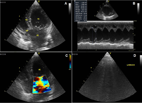
The Doppler echocardiography revealed the presence of mitral valve degeneration with prolapse of the septal leaflet and countless B lines in the right and left hemithorax (Figure 2A–2C).

Figure 2 Doppler echocardiography of heart. A. Degenerative disease of mitral valve. B. Echodopplercardiography and spectral doppler of mitral valve. Mitral valve showing a degenerative appearance with prolapse of septal leaflet. 2C. Doppler study and color flow mapping demonstrate significant insufficiency. 2D. Presence of countless B lines in the right and left hemithorax.
After confirmation of cardiogenic pulmonary edema due to left valve heart disease, benazepril (0.5 mg/kg every 12 hours) and pimobendan (0.25 mg/kg every 12 hours, one hour before meals) were prescribed orally. The owner was advised to have biannual consultations with a veterinary specialist in the area of cardiology.
The pulmonary edema is considered a clinical emergency that requires rapid care to stabilize the patient, and may be caused by a chronic degenerative valve disease (mitral regurgitation) or dilated cardiomyopathy with involvement of the left side of heart.1
The cardiogenic pulmonary edema presents on cardiac auscultation a gallop rhythm and heart murmur, whose clinical signs vary from tachypnea to severe dyspnea2, as observed in the patient in this case report.
In this case report, the patient had acute pulmonary edema with suspected valvular heart disease involving the left side of heart, which was subsequently confirmed by imaging tests, such as chest radiography.11,12 The radiographic findings of pulmonary edema were more evident in the caudal lung lobes with an alveolar pattern in the middle lobe, accompanied by a change in cardiac silhouette with increased dimensions and dorsal deviation of tracheal path.
The doppler echocardiography is essential for confirming structural and functional changes in the heart of dogs with suspected left heart disease.13
The presence of degenerative disease of mitral valve (valve prolapse) with significant hemodynamic repercussion was observed in the doppler echocardiogram. The B lines were also observed, characterized vertical artifacts, perpendicular to the pleural line that move along the pleural line, generally erasing the A lines. They reflect the filling of the inter and/or intralobular septa associated with pulmonary edema or interstitial lung disease.14
The initial approach of patients with cardiogenic pulmonary edema consists in the stabilizing respiratory condition through oxygen therapy 6,15 and intravenous diuretic administration, as described in this case report.
The drugs used to prevent non-cardiogenic pulmonary edema and stabilize left congestive heart failure include use of diuretics,7,16 angiotensin-converting enzyme inhibitors17 and drugs with cardiac positive inotropic effect,18 as described in this case report.
Finally, biannual monitoring by a qualified professional in the area of small animal veterinary cardiology is crucial to the prevention of complications resulting from left congestive heart failure in dogs.
Cardiogenic pulmonary edema is considered a veterinary clinical emergency, where the clinical stabilization of the patient's respiratory condition by the use of oxygen and diuretics is essential before performing complementary tests to determine the primary cause. After the diagnosis, treatment with drugs with a positive cardiac inotropic effect and angiotensin-converting enzyme inhibitors are essential to control congestive left heart failure and the risk of pulmonary edema development. Although an immediate and emergency approach is required, the clinical picture of acute cardiogenic pulmonary edema has a favorable prognosis when properly treated.
None.
Author declares there is no conflict of interest in publishing the article.
None.

©2023 Errante. This is an open access article distributed under the terms of the, which permits unrestricted use, distribution, and build upon your work non-commercially.