Journal of
eISSN: 2377-4312


Case Report Volume 13 Issue 1
1 Graduating in Veterinary Medicine. Nove de Julho University (UNINOVE), São Paulo, SP, Brazil
2Teacher of the Veterinary Medicine Course. Universidade Nove de Julho (UNINOVE), Brazil
Correspondence: Paolo Ruggero Errante, Teacher of the Veterinary Medicine Course, Nove de Julho University (UNINOVE), Santo Amaro Campus, Rua Amador Bueno, 389/491, Santo Amaro, São Paulo, SP, Brasil, Tel 55112633-9000
Received: May 10, 2024 | Published: May 27, 2024
Citation: Leon BM, Errante PR. A case report of canine kidney dysplasia. J Dairy Vet Anim Res. 2024;13(1):39-41. DOI: 10.15406/jdvar.2024.13.00346
The renal dysplasia consists of an abnormal development of the renal parenchyma and stroma, which gives it a whitish appearance and irregular surface, whitish color and firm consistency to the touch. Microscopically, the renal tubules have an adenomatous appearance, with the presence of immature or fetal glomeruli and tubules, primitive mesenchymal tissue with a myxomatous appearance and interstitial fibrosis. The renal dysplasia is considered a congenital and hereditary disease, mainly described in breeds such as Alaskan Malamute, Chow Chow, Golden Retriever, Lhasa Apso, Shih-Tzu, Miniature Schnauzer, Soft-Coated Wheaten Terrier and Standard Poodle. Dogs affected by the disease present polydipsia, polyuria, anorexia, vomiting, lethargy and weight loss. With disease progresses, secondary renal hyperparathyroidism and fibrous osteodystrophy may develop. In this case report, we describe a six-month-old Golden Retriever dog with a history of polydipsia, polyuria and growth retardation. A complete blood count, serum chemistry, urinalysis, abdominal ultrasound and ultrasound-guided percutaneous renal biopsy were requested, which were essential for establishing the diagnosis of renal dysplasia.
Keywords: kidneys, renal failure, renal dysplasia, congenital disease, dogs
Renal dysplasia is a congenital and hereditary disease that affects canine animals, where the disordered development of the renal parenchyma occurs during the embryonic phase.1 In dogs, tissue maturation of nephrogenesis normally occurs during the first ten weeks after birth. Causes associated with this renal tissue immaturity include intrauterine urethral obstruction, exposure to teratogenic substances and canine herpes virus infection.2 In renal dysplasia, nephrogenic tissue immaturity becomes persistent without the transformation of mesenchymal tissue into epithelial tissue.1,2 Renal dysplasia in dogs can occur unilaterally or bilaterally, being considered an important cause of the development of chronic kidney disease in dogs during the first and second year of life.3,4 This disease affects different breeds of dogs, such as Lhasa Apso and Shih-Tzu, Golden Retriever, Mastiff, Bernese, Boxer and Rhodesian.5 Clinical signs associated with disease include polydipsia, polyuria, growth retardation, anemia, dull coat, halitosis, diarrhea and renal osteodystrophy.6 Therefore, the clinical veterinarian for small animals should suspect a hereditary kidney disease in the presence of these signs in puppies or young adults.
In May 2024, an animal of the canine species, Golden Retriever, male, 6-month-old, was served with polydipsia and polyuria. The owner reported up-to-date immunization and deworming. During the clinical examination, the animal showed signs of growth retardation, pale mucous membranes, opaque and dry fur (Figure 1).

Figure 1 Clinical appearance of Golden Retriever puppies with renal dysplasia.
A six-month-old Golden Retriever puppy with growth retardation and dull coat.
A complete blood count, serum biochemistry, glucose, type I urinalysis, abdominal ultrasound were performed. In the blood count, a lower number of erythrocytes (3.15 million/mm3; normal values=3.5-6.0 million/mm3), hemoglobin (7,00 g/dL; normal values=8,5-13,0 g/dL), hematocrit (23; normal values=26-39%), HCM (21,53; normal values=22-25 pg), CHCM (26,9; normal values=31-35 g/dL), leukocytes (17.1 thousand/mm3; normal values=8,0-16,0 mil/mm3), platelets (525 mil/µL; normal values=200-500 mil/µL) was observed.
The dosage of aminotransferase (ALT) (32,7 U/L; normal values=10,0-88,0 U/L), aspartate aminotransferase (AST) (38 U/L; normal values=10,0-80,0 U/L), alkaline phosphatase (ALP) (21,0; normal values=10,0-156,0 U/L), glucose (81,0 mg/dL; normal=70,0-110,0 mg/ dL), cholesterol (122 mg/dL; normal=100,0-270,0 mg/dL), creatinine (3,25 mg/dL; normal values=0,50-1,40 mg/dL), urea (178,5 mg/dL; normal values=10,0-56,0 mg/dL), calcium (10,80 mg/dL; normal values= 9,0-11,3 mg/dL), phosphor (6,7 mg/dL; normal values=2,6-6,2 mg/dL), potassium (5,71; normal values=3,5-5,8 mmol/L), creatinine protein ratio (0,42; dog=0,2-0,5, equivalent to chronic kidney disease borderline proteinúria) was increased.
Type I urinalysis was performed by collecting urine through a cystocentesis, with a isostenuric urine, and the biochemical examination showed the absence of glucose, ketone bodies, occult blood. In sedimentoscopy, erythrocytes, leukocytes, cylinders, or crystals were not observed. However, presence of bactérias was confirmed, urine culture were performed, with the presence of Escherichia coli.
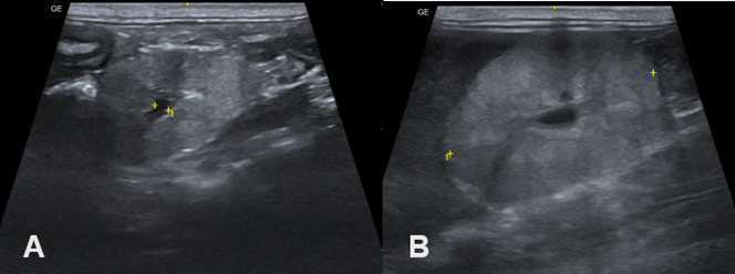
An abdominal ultrasound was performed which revealed reduced right and left kidneys, with loss of corticomedullary definition and coarse echotexture compatible with nephropathy indicative of renal dysplasia (Figure 2).

Figure 2 Abdominal ultrasound. A. Left kidney. B. Right kidney. Both kidneys showed a decrease in size, but there was a marked loss of corticomedullary relationship and coarse echotexture in the left kidney.
Due to the clinical signs and ultrasound findings, a ultrasound-guided percutaneous renal biopsy techniques was performed, which found the presence of immature glomeruli with persistent mesangial tissue, persistent metanephric ducts, and atypical tubular epithelium (Figure 3).
The renal dysplasia is a hereditary disease that affects dogs, characterized by the disorganized development of the renal parenchyma due to changes in nephrogenesis, with abnormal differentiation of the kidneys. In dogs, nephrogenesis is completed after birth, until the tenth week of age,1,2 and in renal dysplasia, the kidneys remain immature throughout life. In dogs, the disease is classified as a type of juvenile nephropathy that culminates in kidney failure between three months and three years of age. The severity of the disease depends on the proportion of immature nephrons and time course for the development of chronic renal failure.3,5
Renal dysplasia can be unilateral or bilateral, leading to the emergence of renal failure in young dogs, whose age at which clinical signs appear varies from 4 months to 2 years of age.3,4
Initial clinical signs include weight loss, polydipsia, polyuria, dehydration, and anemia,6 as was observed in the dog in this case report. Clinical signs associated with uremia include anorexia, vomiting, diarrhea, ulcerative stomatitis, lethargy and uremic breath.7
In severe cases, puppies after birth or between 3 to 6 months of age may die due to renal dysplasia. Clinical signs include excessive polydipsia and polyuria, poor growth, anemia, and renal osteodystrophy. Some dogs with moderate signs present with polyuria, growth retardation and azotemia. Most dogs with the moderate form live 1 to 2 years and gradually develop kidney failure.8,9 A less affected group, with mild dysplasia, may develop renal failure in adulthood or remain clinically normal and present only a clinical picture of moderate polyuria.3
The diagnosis of renal dysplasia is based on anamnesis, clinical signs, laboratory findings, diagnostic imaging and histopathology. In the blood count, non-regenerative anemia is often observed due to low renal production of erythropoietin,2 which is in agreement with this case report. With Hyperphosphatemia and hypocalcemia can also be observed, which can lead to the emergence of secondary renal hyperparathyroidism.10 In our case report, a small increase in serum phosphorus levels was observed. By measuring urea and creatinine levels, and creatinine protein ratio, it was possible to confirm renal failure, in accordance with the literature.2–4 In urinalysis, the presence or absence of proteins are described in the literature, while isosthenuria11–13 is a commonly described finding and is in agreement with our results, along with the presence of E. coli in the urine. The abdominal ultrasound examination is essential in detecting changes compatible with renal dysplasia, where the kidneys have reduced dimensions, loss of the corticomedullary relationship, and increased echotexture,2,14 fact described in our case report.
By performing a percutaneous biopsy guided by ultrasound, it was possible to define the diagnosis of renal dysplasia. Microscopic changes in renal dysplasia can be classified into three different groups: 1) primary lesions characterized by immature or fetal glomeruli, persistent mesangial tissue, persistent metanephric duct, atypical tubular epithelium, or dysontogenic metaplasia; 2) glomerular and tubular metaplasia or hypertrophy; and 3) degenerative lesions accompanied by interstitial fibrosis, tubulointerstitial nephritis/pyelonephritis, dystrophic mineralization, cystic glomerular atrophy and glomerular lipidosis.13,15 In our case report, morphological changes compatible with the presence of immature or fetal glomeruli and persistent mesangial tissue were observed. Due to the large number of affected breeds, including crossbred animals, and its hereditary nature, renal dysplasia must be suspected and investigated in detail in young animals with a racial predisposition that present clinical signs of chronic renal failure.
The renal dysplasia is a genetic disease that affects young animals and requires more detailed clinical and laboratory investigation for diagnostic confirmation.
None.
The authors declare that there are no conflicts of interest.
None.

©2024 Leon, et al. This is an open access article distributed under the terms of the, which permits unrestricted use, distribution, and build upon your work non-commercially.