Journal of
eISSN: 2373-4345


Case Report Volume 7 Issue 3
Department of Paediatric Dentistry, Population and Patient Health, King's College London, Dental Institute, UK
Correspondence: Mahshid Bagheri, Specialist Paediatric Dentist, Razavi International Hospital, Mashhad, Iran
Received: May 10, 2016 | Published: May 29, 2017
Citation: Bagheri M, Kabban M, Hosey MT. Sound levels in movie theaters: is there a potential for hearing loss? J Dent Health Oral Disord Ther. 2017;7(3):291-293. DOI: 10.15406/jdhodt.2017.07.00244
Hypodontia or missing teeth is a rare finding in the primary dentition. It could be due to genetic mutations, syndromes, trauma, infection, radiation, metabolic disease, infant oral mutilation or idiopathic reasons. The most common missing teeth in the primary dentition are maxillary lateral incisors and mandibular central and lateral incisors. Hypodontia of primary teeth may be an indicator of missing permanent successors. It may result in a lack of space and/or supporting bone for the successors when present. A 3 year old mixed non- syndromic White-Black Caribbean boy with missing upper and lower left primary canines the patient was referred from the GDP to The Department of Paediatric Dentistry, King's College Hospital, London was referred for diagnosis and possible dental treatment. There was no history of trauma, extraction or mutilation or hypodontia in the family. Intra-oral examination revealed a soft tissue notch and a tissue tag in upper left canine region, which raised concerns to the possibility of presence of a cleft associated with the missing teeth. Panoramic and upper standard radiographs indicated the presence of successor teeth and presence of a few carious lesions in other primary teeth. No evidence of a cleft was detected. Parents were reassured and no further treatment was carried out regarding the missing primary canines. The carious primary teeth were, however, restored. Hypodontia may present as an isolated finding without consequences on the permanent dentition. No specific treatment was required for this young patient with developmentally absent left primary canines as the permanent successors were present. However, maintenance of a caries-free dentition would remain paramount.
Keywords:hypodontia, primary dentition, canine
Hypodontia is a term used for missing teeth and is a rare finding in primary dentition.1 The reported prevalence of hypodontia in primary teeth differs according to geographic locations and the study methods used. Prevalence of 0.1 to 0.9% has been reported in Iceland, Scandinavia and the United Kingdom,2–4 while figures were as high as 5% in Japanese populations.5 The aetiology for hypodontia may be genetic mutations, syndromes,6 trauma, infection, radiation, metabolic disease, infant oral mutilation7 or idiopathic reasons. Genetic causes of hypodontia may be related to mutations in PAX9, MSX1 or AXIN2 genes.8 Genetic testing is a means of diagnosis,9 while gene mapping and karyotype is used for identification of genes responsible for dental anomalies. Underlying medical conditions such as Down syndrome, ectodermal dysplasia, cleft lip and/or palate, incontinentia pigmenti, Rieger syndrome, Ellis-van Crevald syndrome, Oro-facial digital syndrome and William syndrome may also be associated with missing teeth.10 Extirpation of the primary canine tooth follicles of infants is a type of infant oral mutilation which is mainly practiced in rural areas of Eastern Africa in order to treat high temperature, vomiting, loss of appetite and diarrhea.7 Therefore, precise history taking is of great importance in terms of diagnosis for missing primary canines. The most common missing teeth in the primary dentition are maxillary lateral incisors and mandibular central and lateral incisors.11 Hypodontia of primary teeth may be an indicator of missing permanent successors. It may result in a lack of space and/or supporting bone for the successors when present. This is a case report of missing upper and lower left primary canines. To date, there has not been a similar published report of a case with missing two teeth of the same type on only one side.
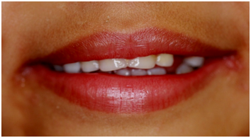
A 3 year old mixed White-Black Caribbean boy was referred to the Department of Paediatric Dentistry, King’s College Hospital, London with the chief complaint of missing upper and lower left primary canines. His mother gave a history of jaundice at birth and his medical history revealed eczema and sickle cell trait. The patient was born to non-consanguineous parents and mother reported an un-eventful pregnancy and normal birth conditions. The family history was not significant with six other siblings. Parents and siblings did not suffer from hypodontia or any other health issues. The child’s mother noticed a soft tissue notch on left maxillary canine area at birth. She reported to the Department of Paediatric dentistry when the tooth failed to erupt. She reported no history of previous trauma, exfoliation, or extraction of the missing teeth. The possibility of mutilation was also ruled out. Patient’s nails, skin, hair and sweating were also normal. On examination, no abnormality was detected extra-orally. Intra-orally, all soft tissues were normal, although a soft tissue tag could be detected in the upper left canine region. All primary teeth were present and erupted except for the upper and lower left primary canines which were missing. All teeth were normal in size and shape. Early dental caries was present in both upper central incisors (Figures 1–3). A panoramic and an upper standard occlusal radiograph were taken which revealed absence of upper and lower left primary canines. All permanent anteriors, canines and first molars were, however, present (Figures 4 & 5). The opinion of an Oral and Maxillofacial surgeon was sought in order to rule-out a sub-mucous cleft in the upper left primary canine area. We diagnosed this case as idiopathic hypodontia of upper and lower left primary canines. Mother was reassured regarding the presence of the successor teeth and no further treatment except 6-monthly reviews was scheduled for monitoring eruption of permanent teeth. The patient attended review appointments when right and left bimolar radiographs were taken and the presence of dental caries was confirmed in the upper first primary molars, lower right and left first primary molar and lower right primary canine. Comprehensive oral hygiene instructions, fluoride and diet advice were provided and further appointments were scheduled for the treatment of dental caries.

Figure 1 Extra-oral view showing spacing due to developmentally absent upper and lower left primary canines.
This case report is interesting due to the fact that missing teeth were only of one type, vertically in line, on the left side. Although a rare finding, there have been previous reports of hypodontia in the primary dentition. However, most are cases with oligodontia, missing six teeth or more.12–14 History taking revealed no previous oral mutilation, exfoliation or dental trauma. Although, dental trauma leading to the avulsion of upper and lower primary canines would be unlikely. Hypodontia in this case was not associated with any syndromes as patient’s facial features (eyes, nasal bridge, and frontal bone) as well as nails, skin and sweating were completely normal. However, presence of a sub-mucous cleft was also a possibility, as patients with cleft lip and/or palate may present missing teeth.15 This was also ruled out both clinically and radiographically. In summary, agenesis of primary teeth could be of genetic origin, associated with an underlying medical condition or a rare isolated finding without consequences on the permanent dentition.13 Gene mapping may be carried out as a diagnostic aid especially in cases where other family members report similar conditions or in patients who significantly benefit from the results of the genetic test in terms of treatment planning.
It was concluded that no specific treatment was required in the rare case of this young patient with developmentally absent left primary canines as the permanent successors were present. However, maintenance of a caries-free dentition would remain paramount.
None.
None.
The author declares that there is no conflict of interest.

©2017 Bagheri, et al. This is an open access article distributed under the terms of the, which permits unrestricted use, distribution, and build upon your work non-commercially.