Journal of
eISSN: 2373-4345


Research Article Volume 4 Issue 4
Department of Orthodontist, Aljouf University College of Dentistry, Aljouf University, Saudi Arabia
Correspondence: Ibadullah Kundi, Department of Orthodontist, Aljouf University College of Dentistry, Aljouf University, Saudi Arabia, Tel 00966 542048412, Fax 00923339172381
Received: February 09, 2016 | Published: March 8, 2016
Citation: Kundi I. Accuracy of assessment of gonial angle by both hemispheres of panoramic images and its comparison with lateral cephalometric radiographic measurements. J Dent Health Oral Disord Ther. 2016;4(4):97-99. DOI: 10.15406/jdhodt.2016.04.00116
Background: One of the most important values in cephalometric tracing is the gonial angle which is used to measure growth pattern of patients, teeth extraction pattern in Class II patients, surgical decision in class III skeletal base patients and age estimation in forensic medicine. Gonial angle measured from panoramic radiograph is found to be more reliable than lateral cephalometric radiograph. In the latter, superimposition of the left and right sides angle, makes it difficult to measure accurately gonial angle.
Aims: I aimed at testing the similarity of left and right sides of Orthopantomography in measuring the gonial angle vs. its measurement from lateral cephalometric radiograph.
Materials and methods: This cross-sectional hospital files-based study of patients visited the Staff Clinic of College of Dentistry, Aljouf University, Sakaka; Al-Jouf, Saudi Arabia was done in the period from 1st January 2015 to 30th September 2015. The prescribed panoramic and lateral cephalometric radiographs used and gonial angle were traced.
Results: Only measurements yielded from each of the panoramic images (right and left) vs. lateral cephalometric were statistically significant (p ˂0.030 and 0.001, respectively).
Conclusion: The gonial angles measured from each of left and right sides of panoramic images were equally reliable but when these measurements were compared to the gonial angles measured on the cephalometric radiographs, the measurements were found statistically different.
Keywords: gonial angle, panoramic radiography, lateral cephalometric radiography
Professor Yrjo Paatero, in 1961, first introduced the Orthopantomography (OPG).1 It has been extensively used in dentistry for analysing the number and type of teeth present, caries, impacted teeth, root resorption, ankylosis, shape of the condyles,2 temporomandibular joints, sinuses, fractures, cysts, tumours and alveolar bone level.3,4 Panoramic radiography is advised to all patients seeking orthodontic treatment; including Class I malocclusions.5
The gonial angle is crucial in determining growth pattern of patients,6 teeth extraction pattern in Class II patients,7 decision making whether to carry out surgery in class III skeletal base patients8 and age estimation in forensic medicine.9 Recent studies have highlighted that OPG can be used to calculate the gonial angle which is a key value in cephalometric diagnosis.10 Larheim & Svanaes11 in 1986 concluded that the gonial angle measured from a lateral cephalometric radiograph is not accurate and is difficult to measure because of right and left side superimposition. They also concluded that gonial angle measurements from panoramic radiographs are equal to those measured from dried human mandibles.11
There are two methods of constructing the gonial angle. The first is to draw a tangent on the posterior border of the ramus of the mandible and join it with another line passing through the points gonion and Gnathion.12 The second method is to draw a tangent on the lower border of the mandible and another tangent to the distal border of the ascending ramus and condyle and measure the angle in between them.13 Due to difficulty in reliably identifying gnathion on lateral cephalometric radiographs, I have used the later technique to determine the gonial angle.
The objective of this cross-sectional study was to test the reliability of measurements of the gonial angle yielded from left and right hemispheres of panoramic radiographs and compare them to the values obtained from lateral cephalograms. The rationale of performing the study is to check the possible application and reliability of panoramic radiographs for gonial angle determination by clarifying whether there is any significant difference between them and those from lateral cephalograms in an attempt to enhance the use of panoramic radiography for gonial angle measurements.
Records of patients who were enrolled for treatment at the Staff Clinic of the College of Dentistry, Aljouf University, Sakaka, Al-Jouf, Saudi Arabia were included in this cross-sectional study. Each patient was given a serial number to protect his/her confidentiality. The duration of this study was 9 months, i.e., from 1st January 2015 to 30th September, 2015. The sample size comprised 80 patients14 (50 male and 30 female patients). Radiographs selected were for patients with Class I malocclusion, taken from the same apparatus. The radiographs were analysed by two experienced orthodontists. 2H pencil was used to draw the tangents. Panoramic and cephalometric images were acquired with a Cranex D digital X-ray unit, Version 3 (Soredex Co., Tuusula, Finland). 85 KVP for panoramic radiograph and 73 KVP for lateral cephalometric radiograph were used. 20 seconds exposure time was selected through 2.7mm Al filtration for both types of radiographs. The two experienced orthodontists analysed clinically the radiographs for high quality and sharpness. Patients having history of trauma, previous facial/mandibular surgery, syndromes (affecting face/jaw) and incomplete records were excluded. Approval on the study design from the local ethical committee of the College of Dentistry, Aljouf University, Sakaka, Saudi Arabia, was obtained.
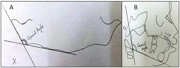
The gonial angle was measured between the two tangent lines; one to the distal border of the ascending ramus and condyle and the second line to the lower border of the mandible (Figure 1). The data were analyzed by SPSS 17. Paired Student’s t-test was used to compare the variables. The level of significance was set at p ≤0.05.

Figure 1 A) The approach used to measure gonial angle on a representative panoramic radiograph investigated.
B) The approach used to measure gonial angle on a representative lateral cephalogram investigated.
The radiographs were viewed and evaluated by two expert orthodontists. The gonial angle in the intersection of the ramal plane and mandibular plane was traced on tracing paper and measured using a protractor with 1 degree accuracy (Figure 2). Gonial angle measurement was done twice, with an interval of 1 month apart, and Dahlberg’s formula was used to determine intra observer reproducibility.

Figure 2 A) Example gonial angle tracing from a panoramic radiograph.
B) Example gonial angle tracing from on a lateral cephalogram.
The mean±standard deviations of gonial angle measured from lateral cephalometric radiographs vs. Right and left panoramic radiographs were 124.89±6.18, 123.06±6.39, and, 123.32±6.10, respectively (Table 1). No significant statistical difference was found when right and left side Panoramic measurements were compared (p = 0.129). A significant difference was found when each of the panoramic measurements (right and left) was compared to lateral cephalometric measurements (p ˂0.035 and 0.001, respectively) (Table 2).
|
Lateral Ceph |
Panoramic right |
Panoramic left |
Mean |
124.89 |
123.06 |
123.32 |
SD |
6.18 |
6.39 |
6.10 |
Table 1 Mean and standard deviations of gonial angle measured from lateral cephalometric radiographs as well as bilaterally from panoramic radiographs (right and left angles)
SD, standard deviation
|
|
Mean |
±SD |
P value |
Pair 1 |
Lateral Ceph - Panoramic Right |
2.212 |
4.277 |
0.035* |
Pair 2 |
Lateral Ceph - Panoramic Left |
1.600 |
3.723 |
0.001* |
Pair 3 |
Panoramic Right - Panoramic Left |
0.612 |
4.510 |
0.129 |
Table 2 Comparison of OPG and Lateral Ceph
*= Significant difference, paired Student's "t" test at P≤0.05
This study was undertaken to test the reliability of measurements of the gonial angle yielded from left and right hemispheres of panoramic radiographs as compared to the values obtained from lateral cephalograms in patients with ages ranging between 15-25 years having class I malocclusion. The gonial angle depicts the form and shape of the mandible, has a pivotal role in forecasting future mandibular growth and it also has certain effects on profile, changes, growth and the position of the mandibular anterior teeth.15 The basic reason of conducting the current study was to enhance the application of panoramic radiography in clinical practice to determine the gonial angle. A few previous studies done to investigate the same aim found no significant difference between gonial angle values measured from panoramic radiographs and those yielded from lateral cephalometric images.16 However, the current study revealed different results. This study is in agreement with a previous study done by Bhullar in 2014, which demonstrated equal values for measurements from both sides of panoramic radiographs.16
Clinical usefulness of panoramic radiography for determining the gonial angle is confirmed by previous studies. This is due to the facts that the left and right side gonial angles are not superimposed on panoramic radiographs, unlike lateral cephalograms which exhibit superimposition.10,17-19 Mattila et al.15 in an old study in 197l took measurements of gonial angle on cephalograms, panoramic radiographs and dried skulls. They showed that the measurements for right and left gonial angles from panoramic images were equal to the angles measured on dry skulls. They further reported that the means of the measurements made on cephalograms showed lower accuracy than those of the panoramic radiographs when compared to the dry mandibles’ measurements.
Nohadani et al.20 in 2008 compared vertical facial and dentoalveolar changes as measured on panoramic and lateral cephalometric radiographs. They stated that panoramic radiographs are not useful for measuring the vertical facial dimension changes. And, angular values from panoramic radiograph are more reliable than vertical values. This was because in the posterior and lateral aspects of the mandible, the angular values are not influenced by image distortion inherent with panoramic radiography. In a most recent study, Araki et al.21 in 2015 compared gonial angles measured from 49 panoramic radiographs with the gonial angle on lateral cephalometric radiographs taken for 2 dry mandibles. The gonial angle measurements were slightly smaller on the panoramic radiographs than on the lateral cephalometric radiographs. The mean gonial angle was 115.1±5.2°C on the panoramic radiographs and 122.2±6.4°C on lateral cephalometric radiographs.
No statistically significant difference was found when right and left side panoramic gonial angle measurements were compared. On the other hand, panoramic measurements (right and left) and lateral cephalometric measurements were not equal.
Limitations of the study
None.
None.
The author declares that there was no conflict of interest.

©2016 Kundi. This is an open access article distributed under the terms of the, which permits unrestricted use, distribution, and build upon your work non-commercially.