Journal of
eISSN: 2574-9943


Opinion Volume 2 Issue 3
Dermatology, STD?s & Leprosy, Ellahi Medicare, India
Correspondence: Tasleem Arif, MBBS, MD, Dermatology, STD & Leprosy, Ellahi Medicare, New colony Soura, near water supply control room, Srinagar, Kashmir, Pin 190011, India, Tel +91 9557 4803 45
Received: December 02, 2017 | Published: June 28, 2018
Citation: Arif T. Angiokeratoma of Fordyce associated with bilateral varicocele: look beyond the skin. J Dermat Cosmetol. 2018;2(3):159-161. DOI: 10.15406/jdc.2018.02.00067
Angiokeratomas are a group of disorders characterized by hyperkeratotic vascular lesions. They are broadly divided into two main types, localized and diffuse. The localized type includes angiokeratoma circumscriptum, angiokeratoma of Mibelli, solitary papular angiokeratoma and angiokeratoma of the scrotum/vulva (angiokeratoma of Fordyce). The diffuse type known as angiokeratoma corporis diffusum is a very rare disorder commonly found in association with metabolic disorders especially Fabry’s disease.1 In this article a case of angiokeratoma of scrotum with bilateral varicocele has been reported with an emphasis to look for a more sinister underlying pathology that is associated with angiokeratoma.
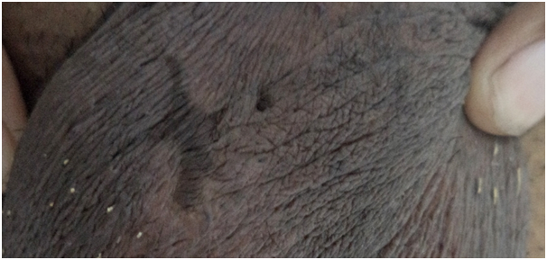
A 54year old male visited with a chief complaint of intermittent bleeding from the scrotal skin for the last 7-8years. He had visited many general physicians but with little relief. His past medical history was unremarkable except he was having seizures and was diagnosed as neurocysticercosis and was under the treatment of a neurologist. He denied any history of trauma to the affected site. He denied any urethral discharge, dysuria or sexual dysfunction. Examination revealed multiple 1-3mm skin colored to dark reddish brown papules (Figure 1A) present over scrotum. Many of the lesions were so small that can hardly be appreciated on naked eye examination. A superficial network of blood vessels could be easily seen around the skin lesions. Some lesions were covered with reddish brown blood crusts employing that the papular lesions were the source of blood. Some lesions have resolved leaving behind brownish-black pigmentation (Figure 1B). On further questioning, patient revealed that scratching in the scrotal area has been the source of bleeding on many occasions. There was a large star shaped atrophic scar over the scrotum measuring about 3cm × 2.5cm. There was no active bleeding. His abdomen was soft and non tender, with no palpable masses felt. With such a history and further supported by clinical examination, a diagnosis of angiokeratoma of scrotum (Fordyce) was made. Due to the chronicity of the condition and the age group affected, an Ultrasonography (USG) of the abdomen and colour doppler of the scrotum was advised. While USG of the abdomen was unremarkable, colour doppler of scrotum revealed grade 4 varicocele on left side (with maximum diameter of dilated veins being 5.1 mm) and grade 2 varicocele on right side (maximum diameter of dilated veins 2.5mm). With such an investigative report, the present case was diagnosed as angiokeratoma of Fordyce with bilateral varicocele. The lesions were treated with radiofrequency ablation. For varicocele, he was referred to surgery department.

Figure 1B Multiple brownish blackpapules of size13mm present overthe scrotum, many of which are covered with crusts. Some lesions have resolved leaving brownish black pigmentation.
Angiokeratomas are considered to be a vascular malformation comprising of ecstatic blood vessels in the papillary dermis, which may or may not be thrombosed. The histopathological features are characterized by marked ectasia of blood vessels within the papillary dermis. The overlying epidermis has acanthosis, papillomatosis and hyperkeratosis. The elongated rete ridges tend to encircle and enclose the vascular lacunae.2,3 However, in our case skin biopsy was not taken.
It was John Addison Fordyce in 1896 who first described a case of angiokeratomas of scrotum in a 60-year-oldman; hence the name angiokeratoma of Fordyce.4 The etiology of angiokeratoma of Fordyce is still not clear. However, many authors incriminate increased venous pressure to be the causative factor.5 Angiokeratomas have been described in association with varicocele and other conditions which are characterized by increased venous pressure viz., tumors of the urinary tract, hernias, trauma, tumors of epididymis and thrombophlebitis.5 According to a report, venous pressure conditions were seen in up to half of patients with angiokeratoma. 6 Agger et al have described a case where the surgical treatment of the varicocele was followed by the resolution of the angiokeratomas.5 Other factors which have been incriminated include vascular/nevoid malformations; and
acute or chronic trauma.7 Angiokeratoma has been reported in association with other dermatological conditions including human papilloma virus infection, papular xanthoma, angiokeratoma of the oral mucosa and nevus lipomatosus.8-11 Our case had bilateral varicocele for which he was referred to department of surgery. No other dermatological conditions were found in our patient.
Angiokeratomas are considered common with increasing age. Izaki reported a study from Japan which showed the prevalence of 0.6% in the age group of 16-20years to a prevalence of 16.6% in the age group of 70years or more.12 Angiokeratoma of Fordyce clinically presents as black-dark red to blue, dome-shaped hyperkeratotic vascular papules ranging in size from 1-6mm.13 While In younger patients, the lesions are more erythematous, smaller with less surface hyperkeratosis; older patients tend to have darker lesions which are larger with overlying scales.14The lesions can be solitary or multiple. Scrotum is considered the most common site. Other sites affected include penile shaft, corona of the glans penis, lower abdomen and the inner thighs.15,16 They can remain asymptomatic or symptoms like itching, soreness or bleeding can be the presenting complaints.13
Most of the times, diagnosis is straightforward. Dermoscopy can aid in the diagnosis. Angiokeratoma is characterized by well-demarcated lacunars red-to-black area round to oval in shape surrounded by a white veil which corresponds to hyperkeratotic and acanthotic epidermis17 In suspicious cases, a skin biopsy can be taken which is usually not the case. The treatment modalities include cryotherapy with liquid nitrogen, electrocautery and treatment with various lasers like 578-nm copper laser and the argon laser, 532-nm potassium-titanyl-phosphate (KTP) laser, long-pulse 1064-nm Nd:YAG laser, 595-nm variable-pulse pulsed dye laser, carbon dioxide and erbium-doped yttrium aluminum garnet lasers.13,18-22 Recently scerotherapy with repeated local injections of 0.5% ethanolamine oleate or 0.25% sodium tetradecyl sulphate have also been found effective.23
The author concludes that in a case of scrotal angiokeratoma, it is important to look for any cause of raised intra-abdominal pressure, especially if the patient is having associated systemic features. A thorough history and good physical examination of the abdomen for intra-abdominal masses may help to exclude any urinary tract tumours and hernias. The scrotum should be examined for evidence of varicocele which can be confirmed by advising a Doppler ultrasound. The rest of the cutaneous examination is a must to rule out other similar lesions which may be a part of angiokeratoma corporis diffusum or look for other associated dermatological conditions.
None
The authors declared that there are no conflicts of interest.

©2018 Arif. This is an open access article distributed under the terms of the, which permits unrestricted use, distribution, and build upon your work non-commercially.