Journal of
eISSN: 2574-9943


Mini Review Volume 2 Issue 2
1Assistant of the Dermatology Ambulatory of the Pontifical, Catholic University of Campinas, Brazil
1Resident of the second year of the Pontifical, Catholic University of Campinas, Brazil
2Resident of the third year of the Pontifical, Catholic University of Campinas, Brazil
Correspondence: Livia Matida Gontijo, Resident of the second year of the Pontificate, Catholic University of Campinas, Brazil
Received: September 26, 2017 | Published: March 19, 2018
Citation: Gontijo LM, Moreira MB, Mançano VS,et al. Dowling-Degos disease: a rare genodermatosis. J Dermat Cosmetol. 2018;2(2):104-105. DOI: 10.15406/jdc.2018.02.00053
Dowling Degos disease (DDD) is a rare, autosomal dominant genodermatosis, characterized by multiple small, reticular pigmented macules distributed in the flexural areas. In the present report, there is a case of the disease that affects 41-year-old female patients, with multiple clinical complications of the disease.
Keywords: genes, female, hyperpigmentation, facial scars, Dowling-Degos disease
Dowling-Degos disease (DDD) is a rare genetic disease of the skin (reticulate pigmented anomaly), clinically characterized by flexural brown pigmented reticulate macules, comedo-like papules on the back, neck and pitted perioral or facial scars. The diagnosis is established based on clinical and histopathological correlation 1,2. In this paper we report a patient of 41yearold females, with multiple clinical complications of DDD.
In 1978, Wilson Jones and Grice first described Dowling Disease (DDD).1,2 It is rare disease that affects women between the second and fourth decade of life. It presents a late onset with benign evolution; nevertheless, it is extremely unaesthetic. It is further characterized by its clinical picture and histopathology.3
Clinically, therefore, it presents with acquired hyper pigmentation of reticulated pattern predominantly in flexures that do not modify or suffer influence of the solar exposition. They are also verified lesions type cribriform scars and perioral acne, with no previous history of acne. In addition, DDD patients with comedo-like lesions on the face, dorsum and other areas mentioned above are observed in patients with DDD.
Histopathology of the skin showed presence of horny cysts and absence of melanocytes hyperplasia. In addition, there was acanthosis characterized by fine and irregular elongation of the inter papillary cones with hyper pigmentation at the extremities. The present case report is about a patient with typical DDD and aims to show the importance of histopathological evaluation, and especially the clinical examination for conclusive diagnosis.
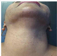
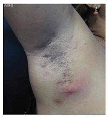
A 43-year-old female, from Campinas- Brazil, black, complained of acne form lesions in the face 30 years ago, and lesions in the armpits and intergluteal region of appearance 3 months ago. At the dermatological examination there were multiple atrophic scars and open comedo on the face, cervical and intermammary regions (Figure 1) (Figure 2). There were also erythematous nodules with exit of purulent secretion with open toes in areas of fibrosis and retractions in inframammary regions, armpits and inguinal region. Brownish hyper pigmentation of the reticular pattern located in the infra axillary and intergluteal region was also observed.

Figure 1 Black rhinoceros biological validation monitoring fecal hormone metabolite (FGM) concentrations before, during, and after transport to a new facility. Transport occurred on Day 29 (6/16/08) and levels began to increase on Day 31.

Figure 2 Open comets with brownish hyperpigmentation of reticular pattern and erythematous nodules with exit of purulent secretion in areas of fibrosis and retractions in right axilla region.
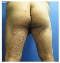
The histopathological finding was similar in the two biopsied samples (inframammary and left axilla) and presented extensions of the epithelial cones and hyper pigmentation of the basal. Sometimes the epidermis exhibited foci of atrophy and rectification. Confirming the diagnosis through clinical examination and histopathology. Patient, man, 61 years old, complained of asymptomatic lesions growing asymptomatic dark lesions growing in number and size that started 20 years ago. He also affirms acne form lesions with spontaneous improvement. Dermatological examination revealed brownish, clustered and confluent macules varying from 2-4mm in folds areas (inguinal region and armpits), Intergluteal cleft, posterior thigh root, pelvis and dorsum (Figure 3). It was also observed, atrophic scars in the bilateral and posterior cervical and dorsal. Histopathological study in two points showed in both samples acanthotic thickening of inters papillary ridges with hyper pigmentation of the same, with compatible Dowling Degos.

Figure 3 Confluent brownish-colored macules with a velvety aspect ranging from 24 mm in the region of intergluteal slit and distributing in both roots of posterior thigh.
DDD is a rare, autosomal dominant genodermatosis with variable penetrance, described by the first in 1938 by Dowling and Fredenthal.1 It is characterized by the dysfunction of keratin 5 (KRT5), present on chromosome 12q gene, which induces the proliferation of the abnormal pilosebaceous epithelium.4 Preferably affects women, in a ratio of 2:1 to men, in the third and fourth decade of life, and has no racial predilection.5,6 It is characterized clinically by acquired reticular hyper pigmentation, some of which are similar to lentigos, in areas of flexure, perioral acne form scars and hyperkeratotic comedo-like lesions in the cervical and perioral regions 1,2,3,4,5 Hyper pigmentation is symmetrical, asymptomatic and progressively increasing over time, appearing initially in the armpits and in the groin with late extension for the intergluteal folds, cervical and inframammary region and internal face of the arms and thighs.
Comedo-like lesions affecting the dorsal region, armpits and intermartial region, keratoacanthoma, epidermoid cysts, seborrheic keratosis and hidradenitis suppurativa represent the additional characteristics of the presentation of this disease in some patients, which corroborates the existence of a defect in the pilosebaceous epithelial proliferation in these patients.
Regarding histopathology, there was no melanocyte hyperplasia, presence of corneal cysts, with adenoid type seborrheic keratosis as the main histological differential diagnosis. In this, however, there is no infundibulum involvement and papillomatosis. In addition, acanthosis is characterized by fine and irregular elongation of the interpapillary cones with hyper pigmentation at the extremities, which also extends to the follicular infundibulum and there is sporadic follicle blockage.1 It is still necessary to advance in the treatment, all of which still have unsatisfactory results.1,2
These case reports, therefore, have the purpose of exposing that despite the rarity of DDD, this is a disease of easy and quick diagnosis, since it is established simply based on clinical and histopathological correlation. There is no need for another complementary examination for the diagnosis aid.
None.
The authors declared that there are no conflicts of interest.

©2018 Gontijo, et al. This is an open access article distributed under the terms of the, which permits unrestricted use, distribution, and build upon your work non-commercially.